

Transapical Access
Learn how transapical access enables minimally invasive heart valve surgery through a small chest incision, plus benefits, risks, and recovery.
What is transapical access
Transapical access is a specialized surgical technique used to reach the heart without performing a traditional, full open-chest operation. The term refers to making a small, precise entry through the apex—the very tip—of the left ventricle, which is the heart's main pumping chamber. Surgeons use this pathway to insert specialized tools and replacement valves directly into the heart.
Why the apex is chosen: The apex of the heart is located near the lower front portion of the chest, making it highly accessible through a minor incision between the ribs. This area provides a relatively straight, short route to the aortic valve. By entering here, the surgical team can bypass the need to navigate through the complex peripheral arteries in the groin or chest.
For patients facing cardiac surgery, understanding this approach can relieve significant anxiety. It allows your surgeon to perform complex procedures, such as replacing a narrowed aortic valve, using a minimally invasive method that prioritizes both precision and patient safety.
How transapical access works
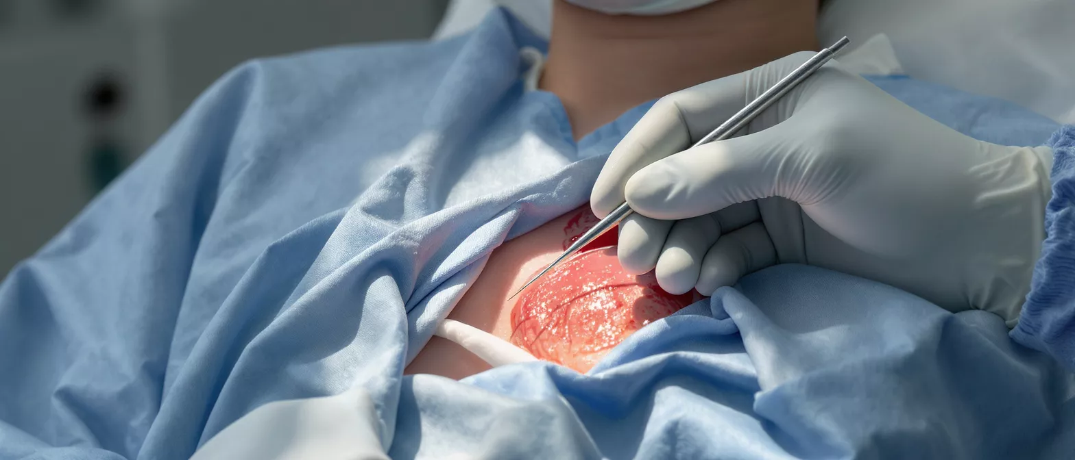
During a transapical access procedure, the surgical team makes a small incision—usually just a few centimeters long—between the ribs on the left side of the chest. The surgeon gently exposes the apex of the heart and places a series of specialized purse-string sutures around a tiny point on the heart muscle. These sutures act like a drawstring, ensuring the wound remains completely sealed around the instruments.
Inserting the delivery sheath: Once the sutures are secure, a narrow tube called a delivery sheath is carefully guided through the apex and into the left ventricle. The replacement heart valve, which is compressed onto a catheter, travels through this tube. Live X-ray imaging and echocardiography guide the surgeon as they advance the valve to the target position.
After the new valve is deployed and confirmed to be functioning properly, the delivery tools are gently removed. The surgeon tightens the purse-string sutures to close the puncture site in the heart muscle, ensuring there is no bleeding. A temporary pacemaker wire may be used during the deployment to safely regulate the heart rate, and the chest incision is then closed in layers.
Transapical access vs transfemoral approach
When planning a minimally invasive heart valve replacement, surgeons evaluate different routes to reach the heart. The transfemoral approach enters through a large artery in the groin, while transapical access enters through a small incision in the chest. The choice between them depends entirely on the patient's individual anatomy and overall health.
Navigating peripheral artery disease: Many patients requiring heart valve replacement also suffer from narrowed or calcified arteries in their legs. If the blood vessels in the groin are too small, heavily calcified, or severely tortuous, threading a catheter through them could cause vascular damage. In these cases, the transapical route offers a direct and safe alternative.
- Transfemoral path: Entirely needle-based access in the groin, requiring highly flexible arteries.
- Transapical path: Direct surgical access to the chest, completely bypassing the peripheral leg arteries.
Because it relies on anatomical pathways rather than pre-existing vessel health, transapical access provides a viable option for patients who are not candidates for the transfemoral method.
Benefits of transapical access heart surgery
The primary advantage of this technique is its ability to avoid a full median sternotomy, which is the traditional splitting of the breastbone. By using a smaller, intercostal incision between the ribs, patients often experience significantly less surgical trauma. This directly translates into a more comfortable postoperative experience.
Direct control for complex anatomy: The transapical approach allows the surgeon highly controlled, coaxial alignment with the aortic valve. This straight trajectory is particularly beneficial when dealing with heavily calcified valves or complex anatomical structures, as it reduces the risk of dislodging calcium fragments into the bloodstream.
Patients who undergo this method typically require less blood transfusions and experience a lower risk of major vascular complications compared to traditional open-heart surgery.
Who needs a transapical TAVR procedure
Transapical access is most commonly utilized for patients undergoing transcatheter aortic valve replacement (TAVR), particularly those diagnosed with severe aortic stenosis. Aortic stenosis is a condition where the heart's main valve becomes stiff and narrowed, restricting blood flow to the rest of the body and causing significant strain on the heart muscle.
High-risk surgical candidates: This approach is often recommended for older adults or individuals who have complex medical histories that make traditional open-heart surgery too dangerous. Conditions such as severe lung disease, previous heart surgeries, or advanced age frequently make patients better candidates for a catheter-based approach.
Your surgical team will conduct extensive imaging tests, including CT scans and echocardiograms, to map out your heart and vascular system. If your medical profile suggests that opening the chest or navigating from the groin carries undue risk, the transapical route becomes a life-saving alternative.
Transapical access incision and surgical details
Understanding the physical incision can help alleviate fear before surgery. The cut is typically two to three inches long, located on the left side of the chest just below the breast area. Because the ribs are spread apart only slightly, the structural integrity of the chest cage is largely preserved, which aids in faster respiratory recovery.
Anesthesia and monitoring: The procedure is performed under general anesthesia. Throughout the operation, advanced imaging provides real-time visualization of the heart. To safely place and deploy the replacement valve, the surgical team utilizes specialized cardiac surgery stabilization equipment to ensure absolute precision and prevent any movement.
Once the valve is securely placed within the diseased native valve, the delivery system is withdrawn. The surgeon closes the puncture in the heart muscle and places a temporary drain in the chest cavity to prevent fluid buildup. The skin is then sutured, leaving a relatively small scar compared to traditional bypass surgery.
Transapical access complications and risks
While transapical access is considered highly advanced and safe, every cardiac procedure carries some degree of risk. The surgical team takes extensive precautions to minimize these risks. Potential complications can include bleeding at the incision site, minor irregular heart rhythms, or the need for a permanent pacemaker if the heart's conduction system is disrupted.
Managing apical bleeding: Because the technique involves making a controlled puncture in the heart muscle, one specific risk is bleeding from the apex when the instruments are removed. Surgeons mitigate this through meticulous suture placement before the procedure begins, ensuring a tight seal the moment the tools are withdrawn.
Other rare risks include infection, stroke, or valve dysfunction. It is vital to discuss your specific risk profile with your cardiologist and surgeon. They will provide a balanced overview of the potential complications compared to the severe risks of leaving severe aortic stenosis untreated.
Transapical access recovery and hospital stay
Recovery from a transapical procedure is generally faster than recovery from a traditional open-chest operation. Most patients spend their first night in the intensive care unit (ICU) for close monitoring of their heart rhythm and chest drains. Once stable, they are transferred to a standard cardiac ward within a day or two.
Returning home and healing: Patients usually remain in the hospital for three to five days before being discharged. During this time, the medical team focuses on pain management, lung exercises to prevent pneumonia, and gentle mobility. The breastbone remains intact, so patients do not face the strict sternal precautions required after traditional surgery.
- Respiratory care: Deep breathing exercises are crucial to fully re-expand the lung near the incision site.
- Gradual movement: Walking with assistance begins within 24 hours to promote circulation.
- Wound care: Keeping the chest incision clean and dry prevents infection.
Full recovery takes several weeks, but many patients find they can return to light daily activities much sooner.
Is transapical access safe for older patients
Older adults are the most frequent candidates for transapical access heart surgery. As we age, the risk associated with major open-heart surgeries increases due to frailty and weakened lung or kidney function. This minimally invasive method is specifically designed to provide a safer alternative for this vulnerable population.
Preserving quality of life: For seniors with severe aortic stenosis, the transapical approach offers a way to replace a failing valve without the immense physical trauma of a sternotomy. Avoiding the splitting of the breastbone means less postoperative pain, a lower risk of deep wound infections, and a quicker return to independent living.
Surgeons evaluate older patients using specialized frailty assessments. If a patient is deemed too frail for traditional surgery but requires urgent valve replacement to restore their quality of life, transapical access is often the recommended and safest medical pathway.
Summary: understanding your surgical options
Transapical access represents a monumental step forward in the field of cardiac surgery, offering a highly effective, minimally invasive alternative for patients who cannot undergo traditional open-heart procedures. By creating a direct route through the apex of the heart, surgeons can safely replace diseased valves while preserving the structural integrity of the chest.
This technique minimizes physical trauma, reduces recovery times, and provides a critical solution for high-risk and older patients. Understanding how the procedure works can empower you to have informed, meaningful discussions with your cardiologist about your upcoming surgery.
If you or a loved one is facing heart valve surgery, rest assured that modern surgical techniques like the transapical approach prioritize patient safety and rapid recovery. Always consult directly with your surgical team to determine the best approach for your specific anatomical needs.






