

Interventricular Septum
Explore the interventricular septum, its anatomy, function, and conditions like VSD.
What is the interventricular septum?
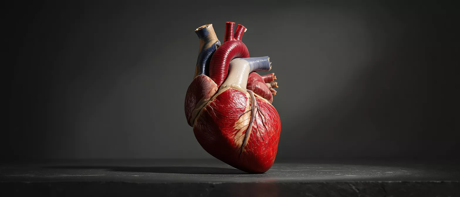
The interventricular septum is a sturdy, muscular wall that divides the lower two chambers of the heart, known as the left and right ventricles. Think of it as the internal partition or firewall that keeps the oxygen-rich blood on the left side completely separate from the oxygen-depleted blood on the right. This separation is essential for the circulatory system to function efficiently, ensuring the body receives fully oxygenated blood with every heartbeat. When patients hear this term during a consultation, it is simply the medical way of describing the heart's central dividing wall.
Understanding the interventricular septum is a vital part of grasping overall heart anatomy. Because it sits deep within the cardiac structure, it plays a dual role: acting as both a physical barrier and an active participant in the pumping mechanism. If this wall is compromised, the heart's ability to maintain proper blood flow and pressure is significantly affected. Recognizing its role helps patients understand why certain symptoms, like shortness of breath or fatigue, occur when structural issues are present.
From a structural standpoint, this dividing wall is foundational to normal cardiac performance. It is engineered to withstand immense pressure differences without faltering. When surgeons evaluate a heart for potential intervention, the integrity of this wall is always a primary focus, as it directly influences how well the organ can sustain life.
How does the interventricular septum function in the heart?
Ventricular septum function in cardiology: The primary function of this wall is to maintain strict separation between the two ventricles. The left ventricle pumps blood to the entire body, requiring immense pressure, while the right ventricle pumps blood to the lungs at a much lower pressure. By keeping these chambers isolated, the septum ensures the highly pressurized left side does not overwhelm the delicate right side.
Beyond simple separation, the interventricular septum actively contracts every time the heart beats. Composed largely of thick cardiac muscle, it works in perfect synchronization with the rest of the ventricular walls to squeeze blood out of the chambers. This coordinated muscular contraction is what generates the force needed to propel blood through the arteries. If the septum does not contract properly, the overall pumping efficiency of the heart drops, which can lead to a condition where the heart fails to circulate blood effectively.
The electrical pathways of the heart also rely heavily on this structure. Specialized fibers that carry the electrical signals telling the heart to beat run directly through the septum. This ensures the lower chambers contract from the bottom up, pushing blood out smoothly. Damage or thickening in this area can sometimes disrupt these electrical signals, leading to arrhythmias or irregular heartbeats that require careful medical monitoring and sometimes the use of cardiac monitoring devices to track rhythm changes over time.
What is the anatomy of the cardiac septum structure?
The cardiac septum structure is not uniform; it is composed of two distinct anatomical regions. The upper portion, known as the membranous septum, is thin and made mostly of fibrous tissue. This delicate area is located just beneath the heart's valves and is the most common site for congenital defects. Below this thin region is the muscular septum, which makes up the vast majority of the wall and provides the bulk of the structural integrity and contractile force.
Interventricular septum thickness variations: The thickness of this wall varies depending on the demands placed upon each side of the heart. The left ventricle, responsible for systemic circulation, is naturally thicker and more muscular than the right. Consequently, the septum is thickest on the left side, matching the powerful muscular build of the left ventricular wall. This asymmetry is entirely normal and is a crucial aspect of how the heart manages different pressure loads.
Because of its complex anatomical position, the septum interacts directly with the heart's valves. The aortic and mitral valves on the left, and the pulmonary and tricuspid valves on the right, anchor themselves near the top of this dividing wall. Any structural changes in the septum, such as thickening or thinning, can subtly alter how these valves open and close. Surgeons must account for these intricate relationships when planning procedures that involve the septal wall, ensuring that valve function is preserved alongside structural repairs.
What is a ventricular septal defect VSD heart condition?
A ventricular septal defect, commonly referred to as a VSD heart condition, occurs when there is a hole in the interventricular septum. This is one of the most common congenital heart defects, meaning it is present at birth. The hole allows oxygen-rich blood from the left ventricle to mix with oxygen-poor blood in the right ventricle. Depending on the size of the opening, this extra blood volume can overwork the lungs and the right side of the heart.
What are the symptoms of a VSD? Small defects often cause no noticeable symptoms and may even close on their own as a child grows. However, larger holes can cause significant symptoms in infants, including rapid breathing, poor feeding, and failure to gain weight. In adults, an undetected VSD can lead to progressive shortness of breath, fatigue, and an increased risk of lung infections. Recognizing these signs early is crucial for effective management and preventing long-term damage.
While many ventricular septal defects are congenital, they can occasionally develop later in life. Trauma, severe heart attacks, or complications from prior heart surgeries can create a break in the septal wall. These acquired defects are rare but require urgent medical attention. Understanding whether a defect is congenital or acquired is a critical step in determining the most appropriate medical or surgical intervention to restore normal heart anatomy.
How is septal wall motion assessed by cardiologists?
Cardiologists evaluate septal wall motion to determine how well the heart is pumping. Using non-invasive imaging techniques, they watch the septum contract and relax in real-time. Normal motion indicates the cardiac muscle is healthy and receiving adequate blood supply. If the wall moves abnormally—either too weakly or in a paradoxical, opposite direction—it can be an early indicator of underlying disease. Following an operation, patients may be advised to wear post-surgery compression garments to support overall circulation while the heart's pumping function normalizes.
How does a cardiologist see the septum? The primary tool for visualizing this wall is an echocardiogram. This ultrasound test provides clear, moving pictures of the heart's chambers, valves, and the interventricular septum itself. Doctors can measure the thickness, check for holes, and observe the wall's motion under the ultrasound. It is painless, quick, and provides a wealth of diagnostic information essential for clinical decision-making.
In some cases, more advanced imaging like a cardiac MRI or CT scan may be used to provide a highly detailed map of the septum. This is particularly helpful for complex congenital conditions or when planning surgical interventions. The detailed images allow surgeons to see exactly where a defect is located and how large it is before they ever enter the operating room. This level of precision is essential for successful surgical planning and minimizing operative risks.
What causes septal hypertrophy or interventricular septum thickness?
What is septal hypertrophy? Septal hypertrophy refers to the abnormal thickening of the interventricular septum. This usually occurs in response to chronic high blood pressure, which forces the left ventricle to work harder, causing the muscle wall to grow thicker over time. Another cause is hypertrophic cardiomyopathy, a genetic condition where the heart muscle grows excessively thick without a clear cause, obstructing blood flow out of the heart.
When the septum becomes too thick, it becomes stiff and less able to relax and fill with blood between beats. This can lead to diastolic dysfunction, a condition where the heart pumps fine but cannot fill properly. Patients may experience chest pain, dizziness, or fainting, especially during physical exertion. The thickened tissue can also disrupt the heart's electrical system, leading to dangerous arrhythmias that require specialized cardiac care.
Managing septal hypertrophy usually begins with medications to relax the heart muscle and lower blood pressure. In severe cases of hypertrophic cardiomyopathy where the thickened septum blocks blood flow, a surgeon may need to perform a septal myectomy. This procedure involves removing a small portion of the thickened muscle to widen the pathway for blood to exit the heart, relieving pressure and improving circulation dramatically.
How is septal wall heart disease treated?
The treatment for septal wall heart disease depends entirely on the specific condition affecting the wall. Small ventricular septal defects might simply be monitored by a cardiologist over time, as they often close naturally in childhood. Larger defects or those causing significant symptoms may require surgical closure to prevent long-term lung damage. The surgical approach is highly effective and involves placing a patch over the hole to permanently seal it.
For conditions involving thickened muscle, such as septal hypertrophy, treatment focuses on symptom management and preventing complications. Medications like beta-blockers can help the heart beat more slowly and with less force, reducing the strain on the thickened wall. If medications are insufficient, surgical removal of the excess tissue is the next step. This layered approach ensures patients receive the right level of intervention at the right time.
Post-treatment care is highly collaborative. Patients work closely with their cardiology team to monitor heart function, adjust medications, and make lifestyle modifications. Adhering to prescribed therapies and attending regular follow-up appointments are critical components of long-term success. This comprehensive approach helps ensure the septum remains healthy and the heart continues to function optimally.
What to expect during cardiac surgery for the septum?
When cardiac surgery is required to repair the interventricular septum, the surgical team will provide a detailed plan. Most septal repairs are open-heart procedures, meaning the surgeon will access the heart through an incision in the chest. The patient is placed on a heart-lung bypass machine, which temporarily takes over the job of pumping blood and oxygenating it so the surgeon can safely stop the heart and operate on the motionless septum.
For a VSD closure, the surgeon carefully opens the right atrium to access the septum. The hole is located, and a synthetic patch is stitched securely over the defect. The heart is then closed, the bypass machine is gradually withdrawn, and the heart resumes beating on its own. The entire process requires immense precision and is performed by highly specialized cardiovascular surgical teams in a hospital setting.
Knowing these steps beforehand often alleviates anxiety for both the patient and their family. The surgical team is highly experienced in protecting the heart during these procedures, ensuring the organ is safely stopped and restarted. Modern surgical techniques have refined this process, making septal repairs routine and highly successful. Patients are encouraged to ask their surgeon specific questions about their surgical approach to fully understand their individual care plan.
Recovery and prognosis after septal surgery
Recovery from septal surgery is a gradual process that typically begins in the intensive care unit for close monitoring. Patients usually spend a few days in the hospital before being discharged home. Full recovery takes several weeks to a few months, during which physical activity is slowly increased under the guidance of a cardiac rehabilitation team. The prognosis for septal surgery is generally excellent, with the vast majority of patients experiencing complete resolution of their symptoms and a return to normal heart function.
After repairing a structural defect, the heart's workload immediately normalizes. The right ventricle is no longer overwhelmed by extra blood volume, and lung pressures drop to healthy levels. Long-term follow-up with a cardiologist is essential to ensure the repair remains intact and the heart continues to function optimally. Patients are encouraged to maintain a heart-healthy lifestyle, managing blood pressure and cholesterol to protect their repaired heart.
Cardiac rehabilitation plays a pivotal role in this recovery journey. Through supervised exercise, education, and counseling, patients regain their strength and confidence. This structured program ensures that the transition back to daily activities is safe and progressive. The dedication to rehabilitation significantly impacts long-term outcomes, helping patients return to their favorite routines with renewed energy and a stronger heart.
Key takeaways about the heart septum anatomy
Understanding the interventricular septum provides a window into how the heart maintains its efficiency and power. As the central dividing wall, it is responsible for keeping blood flow separated and participating actively in the heart's pumping action. Whether dealing with a congenital defect, thickened muscle, or surgical repair, knowing the basics of this structure helps demystify the treatment process. The heart is a resilient organ, and with modern surgical techniques, conditions of the septum are highly treatable.
To summarize the essential points about the septal wall, consider the following:
- Barrier function: The septum prevents oxygen-rich and oxygen-poor blood from mixing.
- Contractility: It is a muscular wall that contracts to help pump blood out of the ventricles.
- Common issues: Ventricular septal defects and septal hypertrophy are primary conditions affecting this wall.
- Surgical success: Repairs to the septum, such as patching a VSD or thinning hypertrophied muscle, are highly effective and safe.
Ultimately, conditions affecting the septum are well-understood in modern cardiology. Through careful monitoring, medical management, and surgical intervention when necessary, patients can look forward to a healthy, active life after diagnosis and treatment.






