

Intramural Haematoma
Learn about intramural haematoma causes, symptoms, diagnosis and treatment.
What is intramural haematoma
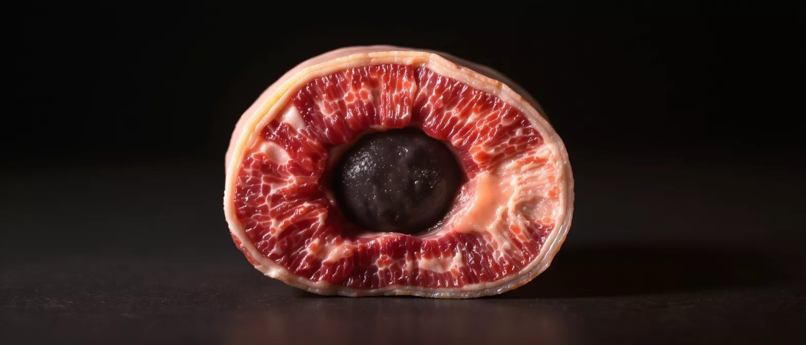
An intramural haematoma is a collection of blood that builds up within the wall of the aorta, the largest artery in the human body. Unlike a ruptured blood vessel where blood spills freely, this bleeding is contained within the layers of the aortic wall itself, creating a bruise-like area that can compromise the vessel's structure and function.
Why the aortic wall matters: The aorta is made of three distinct layers that provide both strength and elasticity. When blood leaks into the middle layer, it forces the inner and outer layers apart, which can reduce blood flow to vital organs or weaken the wall to the point of rupture. Understanding this anatomy helps patients and families grasp why prompt evaluation is essential.
Although the term may sound alarming, modern cardiology and cardiac surgery have refined the way an intramural haematoma is detected and treated. The condition is closely related to aortic dissection, but it has unique features that influence how specialists approach care. When diagnosed early and managed by an experienced team, many patients achieve good outcomes and return to a stable, active life.
How intramural haematoma forms in the aortic wall
The formation of an intramural haematoma typically begins when small blood vessels that supply the aortic wall rupture, causing blood to pool within the middle layer. This process can also occur when a tiny tear develops in the innermost lining, allowing blood to seep outward without creating a full flap that characterises a classic dissection.
Pressure dynamics in the aorta: Because the aorta handles high-pressure blood flow straight from the heart, any weakness in the wall layers can allow blood to dissect or pool. Over time, the accumulating blood expands, pushing against the outer wall and narrowing the central channel through which blood travels. This pressure can cause chest or back pain and may impair circulation to organs such as the kidneys or brain.
Cardiologists classify this condition as a variant of acute aortic syndrome, a group of sudden, life-threatening aortic events. Recognising the formation mechanism helps specialists distinguish an intramural haematoma from other emergencies and choose the right moment to intervene. The goal is always to stabilise the wall and prevent progression before a more dangerous tear or rupture occurs.
Intramural haematoma symptoms and warning signs
The symptoms of an intramural haematoma often mimic those of a heart attack, making immediate medical evaluation critical. The most common warning sign is sudden, severe chest or upper back pain, frequently described as a tearing or ripping sensation that may shift as the condition progresses.
Pain patterns to watch for: Some patients feel the pain migrate from the chest to the back, while others experience abdominal discomfort if the haematoma develops in the lower aorta. Additional warning signs can include shortness of breath, weakness or numbness in the limbs, and a noticeable difference in blood pressure readings between the arms. These symptoms reflect reduced blood flow and demand urgent attention.
It is important to note that not every patient experiences the classic tearing pain. Some may have milder, persistent discomfort that is easily mistaken for a muscle strain. If you or a loved one have risk factors for aortic disease and develop unexplained chest or back pain, seek emergency care without delay. A rapid, thorough cardiac evaluation can distinguish an intramural haematoma from other causes and ensure timely treatment.
Intramural haematoma vs dissection key differences
Distinguishing between an intramural haematoma and a classic aortic dissection is a core step in planning treatment. While both conditions involve bleeding within the aortic wall, a dissection features a visible tear in the inner lining that creates a true second channel for blood flow, whereas a haematoma is a contained pool of blood without that continuous tear.
Why imaging findings differ: On advanced imaging studies, a dissection typically shows a moving flap of tissue that separates the true and false channels. A haematoma appears as a crescent-shaped thickening of the wall with no flap and no direct communication with the central blood flow. These differences influence whether a patient is managed with emergency surgery or intensive medical monitoring.
Despite these distinctions, both conditions are treated as medical emergencies. A haematoma can evolve into a full dissection or rupture, so specialists monitor the wall closely. Understanding the difference helps patients and families participate meaningfully in discussions about surgical timing, follow-up imaging, and blood pressure targets. If you are seeking diagnostic clarity and specialist oversight, a comprehensive aortic aneurysm monitoring program provides structured surveillance to catch any progression early.
Types of intramural haematoma
Specialists classify an intramural haematoma by its location along the aorta, which determines the level of risk and the most appropriate treatment path. A Type A haematoma involves the ascending aorta, the first segment leaving the heart, and is generally considered more dangerous due to its proximity to the heart's valves and coronary arteries.
Type B and distal presentations: A Type B intramural haematoma occurs in the descending aorta, further from the heart. While still serious, it is often managed with blood pressure control and close imaging rather than immediate surgery, provided the haematoma remains stable and does not cause complications such as organ ischaemia.
- Type A: Involves the ascending aorta, often requires prompt surgical repair to protect heart valves and prevent rupture.
- Type B: Located in the descending aorta, typically managed first with intensive medical therapy and serial imaging.
- Wall thickness: Greater wall thickening on imaging may signal higher risk and influence the decision to intervene surgically.
Classification guides the entire clinical pathway, from the choice of imaging intervals to the target blood pressure range. Patients should ask their cardiologist or surgeon which type they have and how that classification affects both short-term management and long-term follow-up.
Intramural haematoma causes and risk factors
Uncontrolled high blood pressure is the single most significant risk factor for developing an intramural haematoma. Chronic hypertension places constant stress on the aortic wall, gradually weakening its inner layers and making them susceptible to microscopic tears or spontaneous bleeding from the small vessels that nourish the wall itself.
Underlying conditions: Certain connective tissue disorders, such as Marfan syndrome or Loeys-Dietz syndrome, weaken the structural fibres of the aorta from an early age. Other contributors include advancing age, atherosclerosis, traumatic chest injury, and previous aortic procedures. Recognising these risk factors allows clinicians to implement early screening and aggressive blood pressure management.
- Chronic hypertension: Long-standing high blood pressure remains the leading cause of wall stress and subsequent tearing.
- Genetic syndromes: Connective tissue disorders compromise aortic strength and raise the risk across all age groups.
- Age and atherosclerosis: Older patients with plaque buildup face increased susceptibility to wall degeneration.
- Sudden trauma: High-impact chest injuries can acutely damage the aortic wall.
Because many of these factors are modifiable, prevention plays a powerful role. Strict blood pressure control, smoking cessation, and routine imaging for patients with genetic syndromes form the foundation of risk reduction.
Intramural haematoma diagnosis and imaging
Diagnosing an intramural haematoma requires high-resolution imaging that can clearly visualise the layers of the aortic wall. A computed tomography angiogram (CTA) is the most commonly used first-line test in the emergency setting because it quickly captures detailed cross-sectional images of the entire aorta and highlights wall thickening or contrast leakage.
Transoesophageal echocardiography: In some cases, especially when a patient is too unstable to move to a CT scanner, a transoesophageal echocardiogram (TOE) provides immediate, highly detailed views of the ascending aorta and heart valves. Magnetic resonance imaging (MRI) offers excellent tissue detail without radiation exposure, making it a valuable tool for stable patients or for long-term follow-up imaging.
The diagnosis hinges on distinguishing a haematoma from a classic dissection or a ruptured aneurysm. Because the clinical signs overlap heavily with other acute cardiac events, specialists rely on imaging to confirm the precise location, extent, and stability of the haematoma. For patients navigating this diagnostic process, having access to a advanced cardiac diagnostic imaging service can ensure accurate, rapid results that directly guide the surgical or medical plan.
Intramural haematoma treatment and management
The treatment of an intramural haematoma depends on its type, size, and the patient's overall health. For Type A haematomas, emergency cardiac surgery is usually required to replace the damaged segment of the aorta and prevent rupture or extension into the pericardium, the sac surrounding the heart.
Medical management for Type B: When the haematoma is located in the descending aorta, specialists typically begin with intensive medical therapy focused on lowering blood pressure and reducing the force of the heartbeat. This approach reduces the stress on the aortic wall, giving the body a chance to reabsorb the pooled blood naturally. Patients are monitored closely in an intensive care setting, with repeat imaging performed within the first few days to ensure the haematoma is stabilising rather than expanding.
Surgery becomes necessary if a Type B haematoma grows, threatens to rupture, or blocks blood flow to vital organs. Endovascular techniques, which use minimally invasive stent grafts, are increasingly used to reinforce the weakened aortic wall from the inside. In complex cases that require open repair, choosing a specialised aortic root replacement device ensures the surgeon can precisely reconstruct the damaged segment while preserving heart function.
Intramural haematoma complications and prognosis
The prognosis for an intramural haematoma has improved dramatically with modern imaging and surgical techniques, but complications remain serious if the condition is left untreated. The most severe risks include progression to a full aortic dissection, complete rupture of the aortic wall, or compromise of blood flow to organs such as the kidneys, intestines, or brain.
Long-term outlook: Patients who receive prompt treatment and maintain strict blood pressure control often achieve favourable long-term survival. The risk of recurrence or progression, however, requires lifelong surveillance. Regular imaging ensures that any subtle changes in the aortic wall are caught early before they develop into emergencies.
Factors that influence prognosis include the patient's age, the presence of underlying connective tissue disease, and how quickly the diagnosis is confirmed. A multidisciplinary team of cardiologists, imaging specialists, and cardiac surgeons works together to tailor follow-up schedules and adjust medications as needed. Open communication with this team empowers patients to manage their condition confidently.
Intramural haematoma recovery and follow-up care
Recovery from an intramural haematoma involves both physical healing and significant lifestyle adjustments. In the immediate postoperative or post-diagnosis period, patients focus on pain management, gentle mobility, and monitoring in a specialised cardiac unit to ensure blood pressure remains tightly controlled and the aorta stays stable.
Cardiac rehabilitation: Structured rehabilitation programmes help patients regain strength safely while supervised by cardiac nurses and exercise physiologists. These sessions also provide education on heart-healthy nutrition, stress reduction, and safe activity levels. Beyond physical recovery, patients benefit from emotional support, as coping with an acute aortic event can be deeply unsettling for both the individual and their family.
Long-term follow-up revolves around serial imaging, usually with CT or MRI scans performed at scheduled intervals to monitor the aortic wall. Patients must adhere strictly to blood pressure medications and avoid high-intensity weightlifting or sudden strain. Building a strong partnership with a specialist is essential for a secure recovery; connecting with a dedicated cardiovascular rehabilitation specialist can bridge the gap between hospital discharge and confident, independent living.






