

Knowing your heart: the mitral valve
Learn how the mitral valve works, common diseases, symptoms, and repair vs replacement options.
What is the mitral valve?
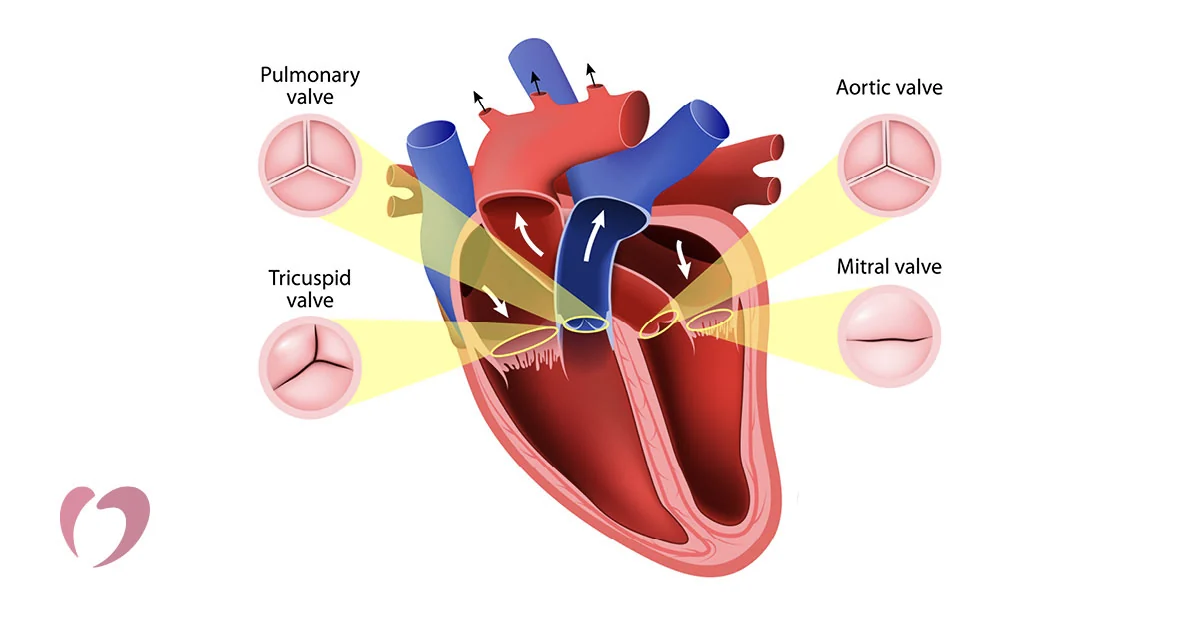
The mitral valve is one of the four critical doors inside your heart, tasked with keeping blood moving in exactly the right direction. For patients facing cardiac surgery, understanding this specific anatomy removes much of the fear of the unknown. It sits between the upper left chamber, known as the left atrium, and the lower left chamber, called the left ventricle. When functioning healthily, it acts as a one-way gate that opens to let blood pass downward into the main pumping chamber, then snaps shut to prevent any backward leakage.
Where is the mitral valve located? It sits squarely on the left side of your heart. Blood returns from the lungs full of oxygen, collecting in the left atrium before passing through the mitral valve into the left ventricle, which then pumps it out to the rest of the body. Its location is crucial because it handles the highest-pressure blood flow of any valve in the heart.
What structures make up the mitral valve? It is not just a simple flap, but a complex apparatus. It consists of two leaflets that open and close, a ring called the annulus that anchors them, and tough string-like cords that tether the leaflets to the heart muscle. If any of these supporting structures fail, overall valve function can deteriorate. In cases of severe structural damage, surgical intervention might require a tissue heart valve replacement to fully restore unidirectional blood flow.
What does the mitral valve do in the heart?
The primary function of the mitral valve is to facilitate forward blood flow while preventing any backflow into the left atrium. Every single heartbeat relies on this precise mechanical timing. When the heart relaxes, the valve opens fully to let the ventricle fill with freshly oxygenated blood. When the heart contracts, the valve closes tightly so that the blood is forced out through the aortic valve and into the body, rather than retreating upward.
How does mitral valve function affect blood flow? When the valve opens fully, the heart fills efficiently without any resistance. When it closes securely, it ensures that the left ventricle generates maximum force to circulate blood to your organs and muscles. This perfectly coordinated sequence is what maintains healthy circulation and proper blood pressure throughout your entire vascular system.
Any disruption to this sequence places extra strain on the heart muscle. If the valve leaks, the heart must work significantly harder to pump the same amount of blood forward. Over time, this extra workload can lead to the heart muscle stretching out and weakening, which is why addressing valve disease before permanent muscle damage occurs is a central goal of cardiac surgery.
What are the symptoms of mitral valve disease?
Patients often wonder what signs indicate a problem with their heart valves. In the earliest stages, you might not notice any symptoms at all because the heart is incredibly skilled at compensating for minor mechanical imperfections. However, as a valve becomes more diseased, the heart struggles to keep up, and distinct physical signs begin to appear in your daily life.
Early signs of mitral valve stenosis and regurgitation usually involve a noticeable drop in physical stamina. You might find yourself getting unusually short of breath during activities that used to be easy, such as climbing a flight of stairs or carrying heavy bags. Some patients experience a rapid, fluttering heartbeat, persistent fatigue, or swelling in the ankles and feet, which doctors call edema. These symptoms happen because blood pressure builds up in the lungs or the body as the valve fails to route blood efficiently.
It is vital to report these changes to your doctor promptly. Identifying the disease early provides your surgical team with more options, including the possibility of repairing your native valve rather than replacing it entirely. Ignoring these subtle warning signs can lead to severe heart enlargement over time.
What causes mitral valve prolapse and regurgitation?
Different underlying conditions can damage the delicate structures of the heart. The most common issues are stenosis, where the valve leaflets become stiff and narrow, and regurgitation, where they fail to close tightly and allow blood to leak backward. Both conditions force the heart to work harder and can eventually compromise its pumping ability if left untreated by a specialist.
What causes mitral valve prolapse? Prolapse occurs when the leaflets become floppy and stretchy, billowing backward into the left atrium during the heart's contraction. This is often a degenerative condition related to connective tissue disorders or simple age-related wear and tear. When the leaflets prolapse, they fail to form a tight seal, which directly results in regurgitation.
Other causes include rheumatic fever, which creates scarring and thickening of the leaflets over many years. A heart attack can also damage the muscles holding the valve in place, and endocarditis, an infection of the heart valves, can physically destroy the valve tissue. Sometimes patients require mechanical heart valve prosthesis when the native tissue is too badly destroyed to salvage.
How is mitral valve disease diagnosed?
Diagnosing a problem with the mitral valve is a straightforward process today, thanks to advanced cardiac imaging. Your physician will usually begin with a detailed physical examination, listening carefully to your heart through a stethoscope. If they hear an abnormal swishing sound, known as a heart murmur, it strongly suggests that blood is flowing turbulently over a diseased or damaged valve.
What tests check for mitral valve disease? The most definitive diagnostic tool is an echocardiogram, which is a painless ultrasound of the heart. This test gives the doctor a real-time moving picture of the valve's structure and function, showing exactly how the leaflets open and close. A chest X-ray or electrocardiogram (ECG) can provide additional context about the heart's rhythm and overall size.
In some complex cases, the surgical team might order a cardiac MRI or a CT scan to get highly detailed three-dimensional images of the heart's anatomy. These advanced imaging techniques help the surgeon plan the operation meticulously down to the millimeter, ensuring they know exactly what structures need repairing before they ever enter the operating room.
When does mitral valve disease need surgery?
Not every minor valve problem requires immediate surgical intervention. If the valve disease is mild and causing no symptoms, your cardiologist may simply monitor your condition regularly. However, surgery becomes necessary when the valve disease begins to negatively impact the heart's function or causes significant, life-limiting symptoms for the patient.
When is mitral valve surgery necessary? Surgeons typically recommend an operation when the left ventricle begins to enlarge significantly or its pumping function drops below a safe threshold. If you experience severe shortness of breath, fluid retention, or high blood pressure in the lungs due to a severely leaking or narrowed valve, surgical correction is required to prevent permanent heart failure. The goal is to intervene right before the heart muscle sustains irreversible damage.
For severe cases, minimally invasive mitral repair can often be performed through a small incision, avoiding the need to split the breastbone entirely. This modern approach greatly reduces postoperative pain and speeds up the return to normal daily activities compared to traditional open surgery.
Mitral valve repair vs replacement: which is better?
When planning surgery for a diseased valve, the primary goal is almost always to repair the native valve if at all possible. Preserving your own body's tissue generally yields superior long-term outcomes, particularly regarding life expectancy and freedom from long-term complications like blood clots.
Why choose mitral valve repair over replacement? Repairing the valve means you keep your own natural leaflets, which function far more efficiently than any artificial device. Crucially, repairing the valve eliminates the need to take lifelong blood-thinning medications, which are mandatory if you receive a mechanical replacement. Patients who undergo repair generally enjoy a better quality of life and lower risks of stroke or bleeding events over the long term.
However, replacement is necessary when the valve is too severely calcified or damaged to salvage. In these situations, the surgeon removes the diseased valve entirely and sews in a prosthesis. The decision between repair and replacement depends heavily on the underlying cause of the disease and the exact anatomical condition of the leaflets at the time of surgery.
What are the types of mitral valve prosthesis?
If replacement is necessary, your surgeon will choose between two main types of artificial valves, each with distinct advantages. The choice depends largely on your age, your ability to take long-term blood thinners safely, and your specific lifestyle preferences.
Mechanical vs biological mitral valve choices are fundamentally about durability versus lifestyle management. Mechanical valves are made from strong materials like carbon and titanium, lasting a lifetime without wearing out, but they require lifelong anticoagulant therapy. Biological valves are made from animal tissue, offering excellent natural blood flow without the strict need for blood thinners, but they eventually wear out.
- Mechanical valves: Highly durable, designed to last a lifetime, but require strict daily anticoagulant medication to prevent blood clots from forming on the metal.
- Biological valves: Made from cow or pig tissue, they function naturally and usually do not require blood thinners, but they typically last 12 to 15 years before needing replacement.
For older patients or those who cannot safely tolerate blood thinners, biological valves are usually the preferred choice. Younger patients might opt for mechanical valves to avoid facing a second major open-heart surgery later in their lifespan.
How long do mitral valve prostheses last?
A common worry for patients facing valve replacement is the lifespan of the artificial valve. The durability of the prosthesis depends entirely on the material it is made from and the biological environment of the patient's body. Understanding these timeframes helps patients make informed decisions about their long-term health planning.
How long does a mitral valve replacement last? A mechanical valve is designed to last permanently, easily enduring for the rest of the patient's natural life without needing structural replacement. Bioprosthetic valves, being organic tissue, have a limited lifespan. On average, a biological mitral valve lasts between 12 and 15 years before the tissue begins to stiffen or degenerate.
It is also important to note that the younger you are when a biological valve is implanted, the faster it tends to calcify and wear out. In younger patients, a biological valve might only last five to seven years. If a bioprosthetic valve eventually fails years later, it can sometimes be replaced using a transcatheter procedure without needing to fully reopen the chest.
What is recovery like after mitral valve surgery?
Recovery is a major concern for anyone facing heart surgery, but modern techniques have made the healing process much smoother than it was decades ago. The first few days are spent in the intensive care unit, where specialized nurses closely monitor your heart rhythm and vital signs to ensure stability immediately following the operation.
What to expect after mitral valve surgery involves a structured rehabilitation process. Most patients stay in the hospital for about a week, starting with simple breathing exercises and gradually progressing to walking in the hallways. Once discharged, you will be given specific guidelines about lifting restrictions, wound care, and gradually resuming normal daily activities.
Full recovery typically takes six to eight weeks, though patients who undergo minimally invasive surgery often bounce back much faster. Cardiac rehabilitation, a supervised exercise program, is highly recommended to help you safely rebuild your cardiovascular strength and return to an active, fulfilling life with a fully functional heart valve.






