

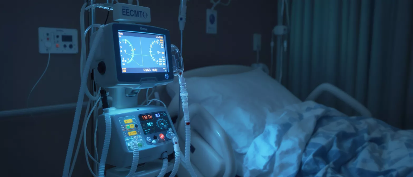
Veno-venous ECMO
Learn how veno-venous ECMO supports patients with respiratory failure, including indications, cannulation, and recovery at Cirugía de Corazón.
What is veno-venous ECMO
When the lungs are too sick or injured to add oxygen to the blood or remove carbon dioxide, doctors may use a heart-lung bypass machine to do that work. Veno-venous ECMO, also called VV ECMO or veno-venous extracorporeal membrane oxygenation, is a form of life support that bypasses the lungs while the heart keeps pumping. A doctor places large tubes (cannulas) in a patient's large veins to draw out dark, oxygen-poor blood. The blood goes through a special machine outside the body that adds oxygen and removes carbon dioxide. The bright red, oxygen-rich blood then returns to the patient through another vein. It is called veno-venous because blood leaves and returns through the veins only, so the machine does the work of the lungs but not the heart. For a frightened patient or family hearing this term for the first time, it helps to know that VV ECMO is a temporary bridge. It does not cure the lungs; it buys time for the lungs to heal.
Why veno-venous ECMO matters: It provides gas exchange so the body can survive when the lungs are failing. It is most often used for severe, reversible respiratory failure where a ventilator alone cannot keep up with the body's oxygen needs.
At specialized cardiac and respiratory care centers, a perfusionist or intensivist manages the circuit to ensure the blood gets the right amount of oxygen and clotting is prevented. VV ECMO is not a treatment for heart failure, so if the heart is also too weak, a different configuration called VA ECMO may be used. Understanding this difference is a key part of any veno venous ecmo protocol.
How vv ecmo lung bypass works
Understanding how vv ecmo lung bypass works can help families feel less anxious. The patient's own heart continues to beat and push blood through the body. The ECMO machine acts as an artificial lung. First, a pump gently draws blood from a large vein, usually in the neck or groin. The blood travels through plastic tubing to an artificial lung called an oxygenator. Inside this device, oxygen flows across a thin membrane and diffuses into the patient's blood while carbon dioxide diffuses out. The newly oxygenated blood is warmed and then returned to the patient through a second venous cannula.
What an artificial lung does: It performs the gas exchange that the native lungs cannot do. This allows doctors to lower the ventilator settings to very safe, gentle levels, which protects the injured lung tissue from further damage.
Blood thinners are used to prevent clots from forming inside the tubing and oxygenator. The entire circuit is closely monitored by specially trained staff. By constantly measuring pressures and oxygen levels, the team ensures the pump provides stable ecmo oxygenation support for the patient.
Vv ecmo indications and respiratory failure
Families often want to know exactly when a patient needs this support. Vv ecmo indications are usually severe, potentially reversible respiratory failure that has not improved with a conventional breathing machine. When a ventilator alone cannot provide enough oxygen or remove enough carbon dioxide, the care team may consider ecmo for respiratory failure. The goal is always to support the patient while the underlying problem, such as an infection or inflammation, is treated and resolves.
Common reasons to start ECMO: Pneumonia that causes severe lung injury, massive blood clots in the lungs, status asthmaticus, or trauma. In each case, the lung damage is severe but potentially recoverable within a few weeks.
- Severe oxygenation failure: When blood oxygen levels stay dangerously low even on maximum ventilator support.
- Carbon dioxide clearance: When the lungs cannot exhale carbon dioxide effectively, making the blood too acidic.
- Lung-protective ventilation: Using ECMO allows doctors to turn down the ventilator, preventing further injury to the lungs.
The decision to begin respiratory ecmo support is made by an intensive care team. They carefully weigh the risks and benefits, looking for signs that the lungs have a reasonable chance of healing if given enough time and rest.
Ecmo for ards and acute lung injury
One of the most common reasons a patient is placed on ECMO is a condition called ARDS (Acute Respiratory Distress Syndrome). Using ecmo for ards allows the team to rest the lungs while the underlying cause of ARDS, often a severe infection or sepsis, is treated. In ARDS, tiny air sacs fill with fluid, making the lungs stiff and difficult to oxygenate. High ventilator pressures are needed to push air in, but those high pressures can cause further inflammation and injury, a problem known as volutrauma.
How ECMO helps ARDS: By taking over the job of oxygenating blood, the machine allows the ventilator to be set to much lower, safer pressures. This is a key strategy for managing ecmo acute lung injury and preventing additional harm.
In cases of severe ecmo acute lung injury, the recovery path can be long, but the strategy of lung rest is highly effective. Providing advanced respiratory support equipment in the intensive care unit is essential for these patients to survive and eventually recover their lung function.
Vv ecmo cannulation and settings
Before starting ECMO, the care team must perform vv ecmo cannulation. This involves placing large tubes (cannulas) into the patient's veins. The most common method uses a two-site approach, with one cannula in a large vein in the neck and another in a vein in the groin. Alternatively, a single double-lumen cannula can be placed in the neck for certain patients. The cannulas are connected to the ECMO circuit, and blood flow is slowly started.
Adjusting the machine: Once the cannulas are in place, the team adjusts the vv ecmo settings. They set the pump speed to control how much blood flows through the circuit and adjust the oxygen flow (sweep gas) to control carbon dioxide removal.
These settings are fine-tuned continuously based on daily blood tests and the patient's clinical status. The goal is to provide enough support while minimizing stress on the patient's blood cells and ensuring the circuit runs smoothly and safely.
Vv ecmo flow rates and oxygenation support
The amount of blood passing through the ECMO machine is measured in liters per minute and is called the flow rate. Vv ecmo flow rates are adjusted to deliver the right amount of ecmo oxygenation support for the patient's body size and metabolic needs. Because blood leaving the ECMO machine mixes with the patient's own venous blood before entering the heart and lungs, the final oxygen level in the body depends on a mix of the patient's own lung function and the ECMO flow.
How flow is managed: If oxygen levels in the blood are low, the team may increase the pump flow or optimize the oxygenator's performance. If blood flow is not enough, they may need to evaluate the cannulas for positioning and patency.
In contrast to heart-lung bypass used in an operating room, which runs at very high flow to replace the heart entirely, VV ECMO uses lower, more gentle flow rates. This helps protect the blood cells from damage during their journey through the circuit and back into the body.
Vv ecmo vs va ecmo differences
A very common question from families is the difference between VV and VA ECMO. The key to vv ecmo vs va ecmo differences is where the blood is returned to the body. In VV ECMO (veno-venous), blood leaves a vein and returns to a vein. The heart is still responsible for pumping blood throughout the body. This is used for patients whose heart is strong but whose lungs are failing.
When VA ECMO is chosen instead: If the heart is failing and cannot pump blood effectively, doctors use VA ECMO (veno-arterial). In VA ECMO, blood leaves a vein but returns to an artery, effectively bypassing and supporting both the heart and the lungs.
For patients who need veno-venous extracorporeal membrane oxygenation, the heart is expected to work normally. If a patient on VV ECMO develops severe heart failure, the team may need to convert the configuration to VA ECMO to provide circulatory support as well.
Lung rest with ecmo support
The primary benefit of ECMO is not that it heals the lungs directly, but that it allows for lung rest. Lung rest with ecmo means doctors can safely turn down the ventilator. High-pressure breathing machines can damage fragile, inflamed air sacs. By letting the ECMO machine handle the work of breathing, the ventilator can be set to low, safe pressures and low breath sizes.
Why lung rest matters: Resting the lungs reduces the cycle of inflammation and mechanical injury. It allows the body's natural healing processes to repair damaged tissue without the constant stress of high-pressure ventilation.
Think of it like putting a broken bone in a cast. The cast does not heal the bone, but it holds everything still so the body can do the healing. Providing the lungs with a safe environment to heal, often supplemented by targeted nutritional and fluid management products, is the essence of why this therapy works.
Venovenous ecmo complications and risks
While ECMO is a life-saving technology, it is a major medical procedure with significant risks. Families should be aware of potential venovenous ecmo complications. Because blood is flowing through plastic tubes outside the body, patients require continuous blood thinners to prevent clots. This increases the risk of bleeding, which can happen anywhere in the body, including the brain or stomach.
Risks of being on the machine: Bleeding is the most common complication. Other risks include infection at the cannula sites, blood clots within the circuit, and the need for frequent blood transfusions.
- Bleeding and clotting: A delicate balance is required to prevent clots in the circuit while avoiding dangerous bleeding in the patient.
- Infection: Having large tubes entering the body creates a pathway for bacteria, requiring strict sterile technique.
- Transfusion needs: Because blood thinners and the circuit can affect blood cells, patients often require transfusions of red blood cells and other blood products.
Despite these risks, the intensive care team is highly trained to manage them. For a patient with severe, reversible lung failure, the potential benefits of survival often outweigh the serious risks of the procedure itself.
Recovery and conclusion
As the underlying lung condition improves, the medical team gradually reduces the ECMO flow. They monitor blood oxygen and carbon dioxide levels as the ventilator is slowly turned up to do more of the work. When the team is confident the lungs can support the patient independently, the cannulas are removed in a minor surgical procedure.
Final thoughts: Veno-venous ECMO is one of the most advanced therapies available for severe respiratory failure. It is a temporary support system that replaces lung function, allowing the body time to heal from infections or acute lung injury.
For patients and families navigating a frightening critical illness, understanding this technology brings empowerment. While the journey through critical illness is challenging, ECMO represents hope and the pinnacle of intensive care support, offering a lifeline when the lungs need it most.






