VV ECMO Versus VA ECMO: How the Two Types Differ
Learn how VV ECMO vs VA ECMO differ in cannulation, blood flow, and organ support.
What Is VV ECMO vs VA ECMO and How Do They Differ?
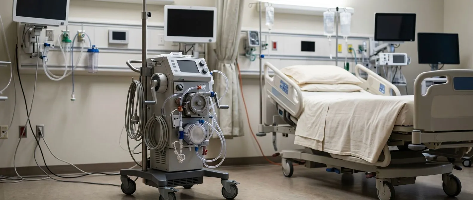
When a loved one is critically ill and their heart or lungs can no longer function on their own, doctors may mention a therapy called ECMO. The acronym stands for Extracorporeal Membrane Oxygenation, which simply means oxygenating blood outside the body. Understanding vv ecmo vs va ecmo begins with recognising that both approaches use the same machine but serve fundamentally different purposes depending on which organ needs support.
Two configurations for two different problems: VV ECMO (veno-venous) takes blood from a large vein, adds oxygen, removes carbon dioxide, and returns it to another vein — the heart then pumps that oxygen-rich blood to the body. VA ECMO (veno-arterial) takes blood from a vein, oxygenates it, and returns it directly into an artery, effectively bypassing both the heart and the lungs simultaneously. The choice between them depends on whether the patient's heart can still pump effectively on its own.
Both configurations represent the most advanced form of life support available in modern intensive care. They buy time for diseased organs to heal, for medications to work, or for a surgical intervention to take effect. Neither is a treatment itself — ECMO is a bridge that keeps someone alive while the underlying problem is addressed.
How Does VV ECMO Work for Lung Support?
VV ECMO is designed specifically for patients whose heart is still functioning but whose lungs have failed. The most common scenario is severe acute respiratory distress syndrome (ARDS), pneumonia, or pulmonary embolism where the lungs cannot exchange oxygen and carbon dioxide despite a ventilator. In these cases, the heart beats normally and circulates blood, but the blood it pumps carries too little oxygen.
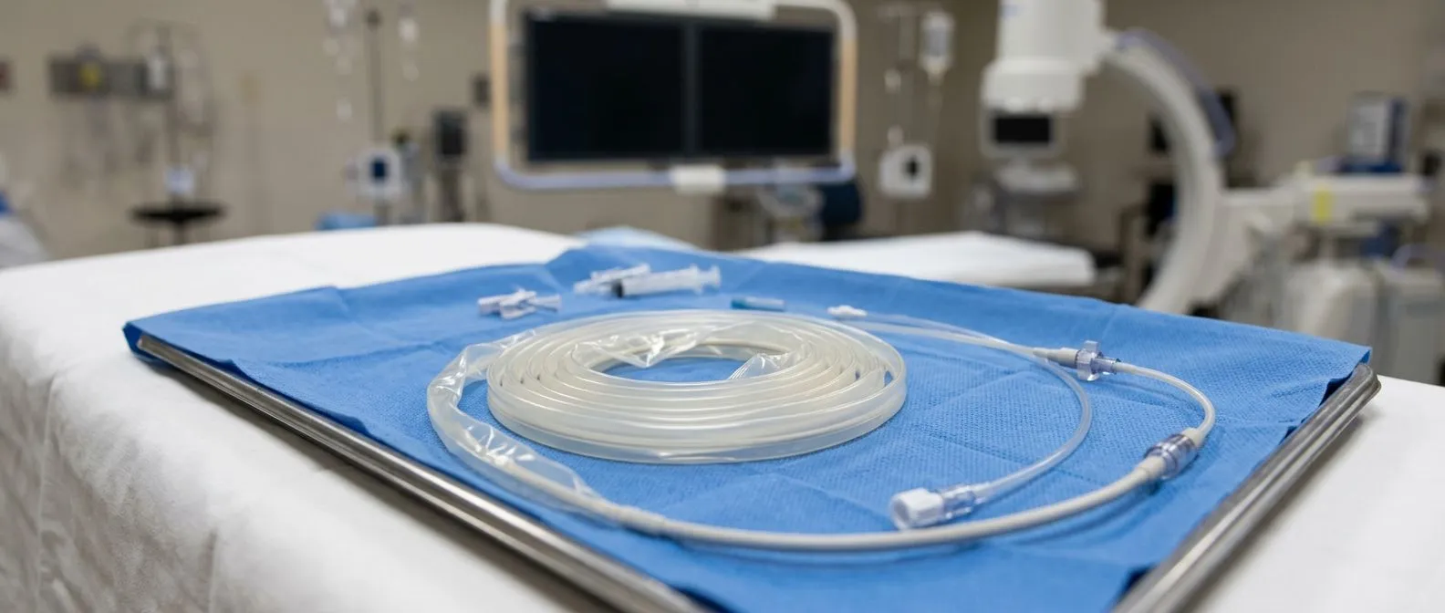
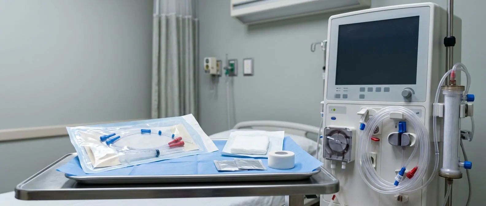
The circuit explained simply: A specialised tube called a cannula is placed into a large vein, typically in the neck or groin, and drains dark, deoxygenated blood out of the body. That blood passes through a membrane oxygenator — an artificial lung made of hollow fibres — where oxygen is added and carbon dioxide is removed. The now bright-red, oxygen-rich blood is returned through a second cannula into another large vein, and the patient's own heart pumps it through the body as normal.
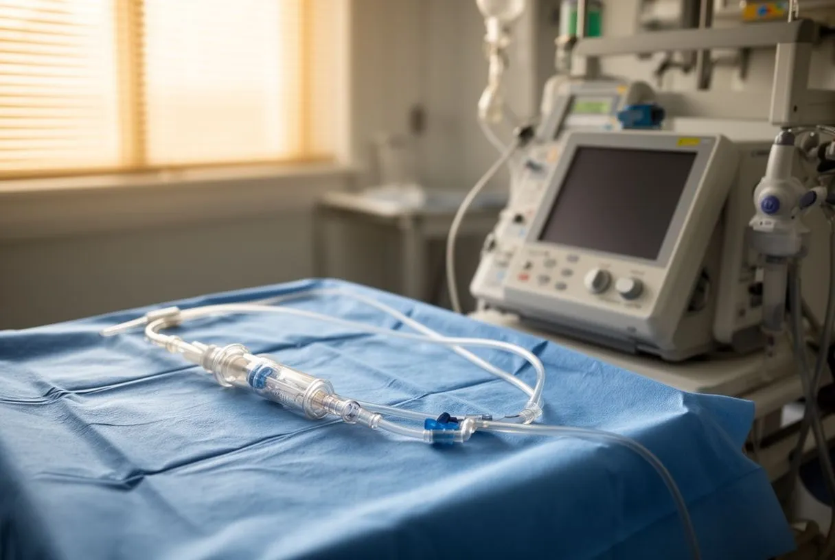
Why the heart still matters in VV ECMO: Because the blood is returned to the venous system, the patient's heart must be strong enough to pump that oxygenated blood through the lungs and out to the tissues. If the heart is also failing, VV ECMO alone will not be sufficient, and the team must consider switching to VA configuration or a hybrid approach. This is one of the most important distinctions in the vv ecmo vs va ecmo decision.
How Does VA ECMO Work for Heart and Lung Support?
VA ECMO takes over the work of both the heart and the lungs simultaneously. It is used when the heart cannot pump effectively — for example, after a massive heart attack, in severe cardiogenic shock, or during cardiac arrest where return of spontaneous circulation has not been achieved. It can also support patients after cardiac surgery when the heart needs time to recover, or serve as a bridge to a ventricular assist device or transplant.
Bypassing the heart entirely: Blood is drained from a large vein, passed through the oxygenator, and then returned directly into a large artery — usually the femoral artery in the groin or the ascending aorta in the chest. Because the blood enters the arterial system directly, the ECMO pump itself generates the blood flow to the body, meaning the patient's own heart does not need to function at all. This is the fundamental mechanical difference when comparing vv ecmo vs va ecmo.
VA ECMO provides full circulatory support, but it also introduces unique challenges. Because oxygenated blood from the machine and deoxygenated blood from the patient's own lungs can mix in the aorta, careful monitoring is essential to ensure the brain and upper body receive adequately oxygenated blood — a phenomenon known as differential hypoxemia or Harlequin syndrome.
When Is VV ECMO Used in Critical Care?
VV ECMO is most commonly deployed for severe, reversible respiratory failure. Typical scenarios include influenza or bacterial pneumonia that has progressed to ARDS, massive pulmonary embolism, or acute exacerbation of chronic lung disease. It may also be used as a bridge to lung transplantation in patients whose lungs are failing and who are already on a transplant waiting list. In each case, the heart must be strong enough to maintain circulation on its own.
Awake ECMO and the evolving landscape: In some centres, patients on VV ECMO remain awake and can even participate in light rehabilitation, a practice increasingly called awake ECMO. This is particularly relevant for patients bridging to lung transplant, where maintaining physical conditioning improves post-operative outcomes. The ability to communicate, eat, and perform gentle exercises while on support represents a meaningful quality-of-life advance compared to deep sedation and immobility.
Candidacy for VV ECMO depends on reversibility of the lung injury, absence of irreversible brain damage, and a reasonable prognosis if the lungs can recover. Teams also weigh whether the patient has conditions that make prolonged anticoagulation dangerous, as blood thinners are required to prevent clotting in the circuit.
When Is VA ECMO Chosen Over VV ECMO?
VA ECMO becomes necessary when the heart itself is the failing organ, or when both heart and lungs are compromised. The most common indications include cardiogenic shock from a large myocardial infarction, fulminant myocarditis, refractory cardiac arrest (known as ECPR), or post-cardiotomy shock where the heart cannot sustain adequate output after surgery. It may also support patients with life-threatening arrhythmias such as atrial flutter that compromise circulation.
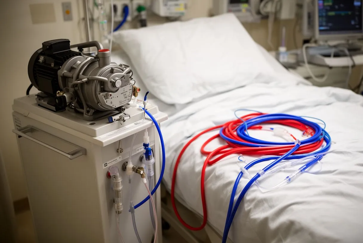
Centreal vs peripheral cannulation: VA ECMO can be configured peripherally, using vessels in the groin, or centrally, with cannulas placed directly into the heart and aorta during open chest surgery. Peripheral cannulation is quicker and less invasive, suited for emergency situations like cardiac arrest. Central cannulation is typically chosen in the operating room after cardiac surgery when the chest is already open and the ascending aorta is accessible for arterial return.
The decision to initiate VA ECMO carries significant weight because it involves cannulating an artery, which carries higher risk than purely venous access. Arterial cannulation can compromise blood flow to the leg, cause vascular injury, or create conditions for stroke if air or debris enters the arterial circuit. Teams must weigh these risks against the immediacy of the patient's condition.
What Are the Key Differences in Cannulation and Blood Flow?
The practical distinction between these two configurations comes down to where the blood is drawn from and where it is returned. VV ECMO uses only venous access — blood leaves a vein and returns to a vein. VA ECMO requires both venous drainage and arterial return, meaning a cannula must be secured in a major artery. This arterial line is the critical structural difference and the source of additional risk.
Flow dynamics and what they mean for the patient: In VV ECMO, the patient's heart determines how much oxygenated blood reaches the tissues — the ECMO circuit simply improves the oxygen content of the blood that the heart will pump. In VA ECMO, the pump itself controls systemic blood flow, and the native heart's contribution becomes secondary. If the heart begins to recover, native cardiac output and ECMO flow can interact in complex ways, requiring careful adjustment of flow rates to avoid competing pressures in the aorta.
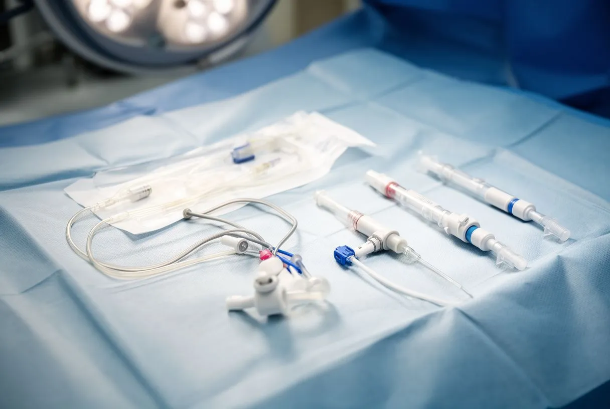
- VV ECMO access: Venous drainage and venous return — typically via the internal jugular and femoral veins, or a single dual-lumen cannula in the internal jugular vein.
- VA ECMO access: Venous drainage and arterial return — femoral vein to femoral artery is most common in emergencies; central cannulation uses the right atrium to aorta.
- Pump reliance: VV ECMO depends on native cardiac output for systemic perfusion; VA ECMO generates systemic flow independently of the heart.
One important consideration in VA ECMO is monitoring oxygen delivery to the upper body. When blood is returned to the femoral artery, it travels upward against the direction of normal aortic flow. If the patient's own lungs still function partially, blood leaving the left ventricle may carry less oxygen than the ECMO blood lower in the aorta, creating a gradient where the brain receives less oxygen than the lower body. Teams monitor this closely and may adjust cannulation strategy if differential hypoxemia develops.
What Are the Risks and Complications of ECMO Support?
ECMO is a high-stakes therapy, and both configurations carry substantial risks. Bleeding is the most common serious complication because the circuit requires continuous anticoagulation to prevent clots from forming as blood contacts artificial surfaces. Patients may develop bleeding at cannulation sites, in the brain, or in the gastrointestinal tract. The balance between too much anticoagulation (bleeding) and too little (clotting) is one of the most demanding aspects of daily ECMO management.
Infection, thrombosis, and circuit complications: Cannulas create large openings into the bloodstream, and patients on ECMO are critically ill with compromised immunity, making infection a constant concern. Blood clots can form within the circuit itself, occasionally requiring emergency circuit exchange. In VA ECMO specifically, leg ischaemia — inadequate blood flow to the limb — can occur if the arterial cannula obstructs the femoral artery. This risk may be mitigated by placing a small distal perfusion cannula to maintain blood flow to the lower leg. Evidence published in the BMJ and other major journals has helped refine protocols for these complications.
Additional risks include haemolysis (destruction of red blood cells by the pump), air embolism, and kidney injury from reduced perfusion or the inflammatory response triggered by blood contact with the circuit surface. Understanding these risks is essential when families weigh vv ecmo vs va ecmo, as the complications differ in frequency and severity between configurations.
How Long Do Patients Stay on ECMO Support?
There is no fixed duration for ECMO — it runs for as long as the underlying condition requires time to resolve. For VV ECMO in acute respiratory failure, a typical course might range from several days to two or three weeks. For VA ECMO in cardiogenic shock, support may be needed for days to weeks, depending on whether the heart is expected to recover, whether a durable ventricular assist device will be implanted, or whether transplantation is the plan.
The plateau question: If a patient shows no sign of organ recovery after a reasonable period, the team faces a difficult decision. Prolonged ECMO increases the cumulative risk of bleeding, infection, and thrombosis. Families should understand that ECMO is a bridge, not a destination — if the heart or lungs do not recover and no transplant or assist device is available, continuing support indefinitely is not a realistic option. This conversation is one of the hardest in critical care.
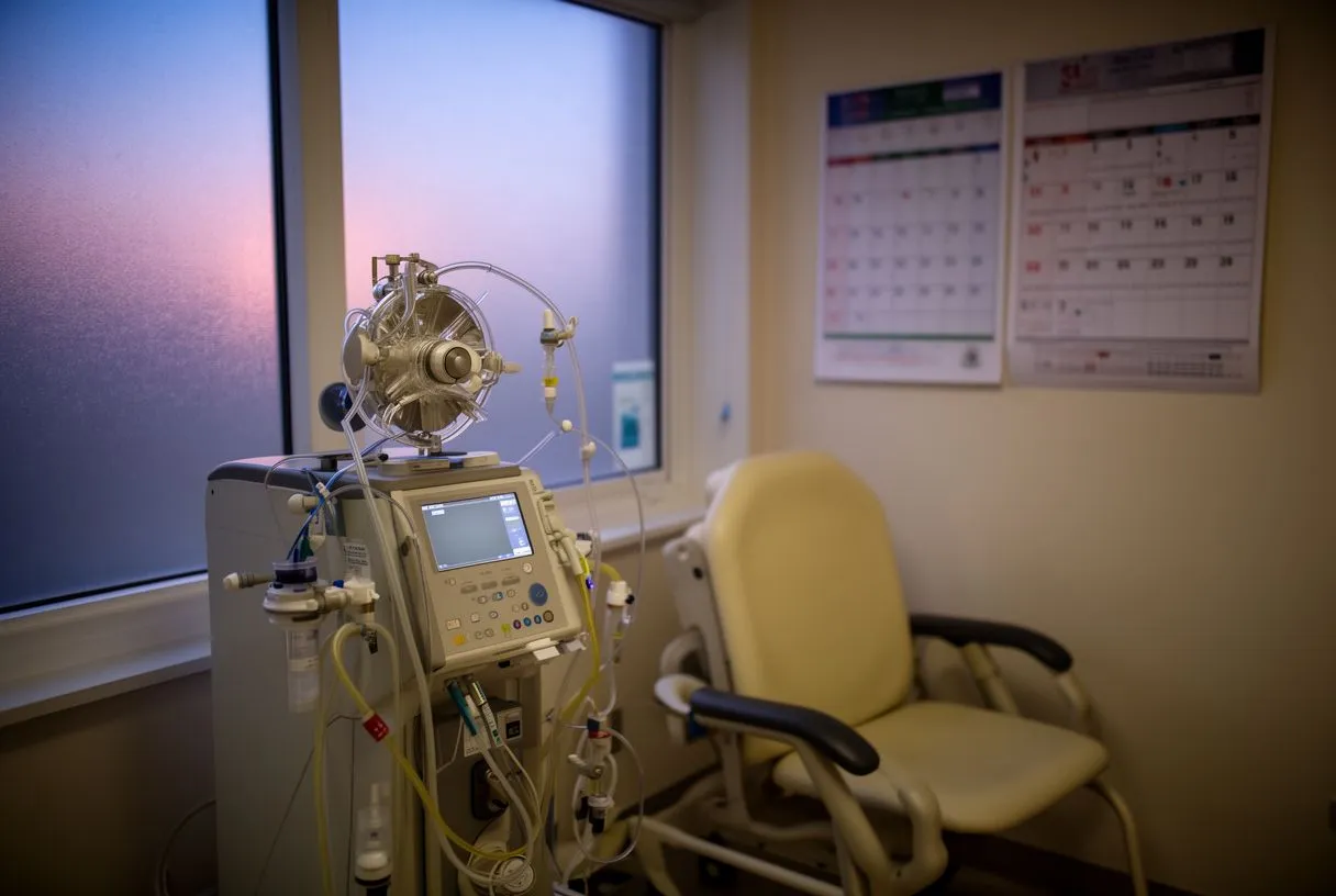
Decisions about duration are guided by daily assessments of native organ function, imaging, blood markers, and echocardiography. In VV ECMO, improving lung compliance and rising arterial oxygen levels signal that the lungs may be ready to take over. In VA ECMO, increasing native cardiac output and stable blood pressure with reduced pump flow suggest the heart is recovering. The CONSORT 2010 Explanation and Elaboration: updated guidelines for reporting parallel group randomised trials framework and similar structured reporting standards help ensure that data from ECMO studies are interpreted consistently across centres.
What Does Recovery Look Like After ECMO?
Recovery after ECMO is a marathon, not a sprint. The moment the circuit is removed — known as decannulation — is a significant milestone, but it marks the beginning of rehabilitation, not its end. Patients who have been on ECMO have typically spent days or weeks in intensive care, often deeply sedated, on a ventilator, and immobile. Muscle loss, weakness, and fatigue are nearly universal, and physical rehabilitation is a central part of the recovery phase.
Weaning and decannulation: Before removing cannulas, the team gradually reduces ECMO flow to test whether the heart or lungs can sustain function independently. For VV ECMO, this means increasing ventilator settings and watching oxygen levels. For VA ECMO, it means reducing pump flow while monitoring blood pressure and cardiac output. If the patient tolerates low flow for a defined period, decannulation is performed at the bedside, often with surgical removal of the cannulas and direct pressure or suturing of the vessel.
Post-decannulation, patients may still require a ventilator, dialysis, or circulatory medications as their organs continue recovering. Rehabilitation can take weeks to months, and some patients experience lasting cognitive effects from critical illness. Quality-of-life studies suggest that many survivors return to meaningful function, but the trajectory varies widely depending on age, underlying diagnosis, and duration of support.
How Do Doctors Decide Between VV ECMO vs VA ECMO?
The decision of vv ecmo vs va ecmo rests on a clear clinical question: is the problem primarily in the lungs, or does the heart also need support? If the heart is pumping effectively with adequate blood pressure and no signs of cardiogenic shock, but the lungs cannot oxygenate despite maximal ventilator support, VV ECMO is the appropriate choice. If the heart is failing — defined by low cardiac output, poor perfusion, or severe arrhythmia — VA ECMO becomes necessary. In some cases where both organs are compromised, a hybrid approach may be used.
- Lung failure with preserved heart function: Severe ARDS, pneumonia, or aspiration — VV ECMO is indicated.
- Heart failure with or without lung compromise: Cardiogenic shock, massive MI, myocarditis, or cardiac arrest — VA ECMO is indicated.
- Both heart and lungs failing: A hybrid configuration (VAV ECMO) may provide both circulatory support and adequate upper-body oxygenation.
Patients and families facing this decision should understand that ECMO is not offered unless conventional therapies have been exhausted. The International consensus statement on an update of the classification criteria for definite antiphospholipid syndrome (APS) illustrates how structured criteria guide high-stakes clinical decisions — similarly, ECMO candidacy is governed by established protocols that weigh reversibility, prognosis, and risk. If you or a loved one may need ECMO, the most important step is to have a direct conversation with your cardiac or intensive care team about the specific indications, expected duration, and realistic outcomes for your situation. We encourage you to book a consultation with our team at Cirugía de Corazón to discuss your individual case in detail.