

Antegrade Cerebral Perfusion: Brain Protection in Aortic Surgery
Antegrade cerebral perfusion aortic surgery protects your brain during complex aortic procedures.
If you or a family member has been told you need surgery on the aorta — the body's largest artery — you may have heard the phrase antegrade cerebral perfusion in aortic surgery and wondered what it means. In plain words, it is a technique that keeps blood flowing to your brain while your heart is temporarily stopped and your body is cooled during a complex operation. This guide explains how it works, when surgeons use it, what the risks and recovery look like, and how to think about choosing a team for this kind of procedure.
What Is Antegrade Cerebral Perfusion?
The basic idea. During aortic arch surgery — operations on the curved section of the aorta at the top of the chest, where the arteries supplying the brain branch off — the surgeon must often stop the circulation to work on blood vessels that carry blood to the brain. Antegrade cerebral perfusion means the surgical team deliberately sends oxygen-rich blood forward ("ante" = forward, "grade" = direction) into the brain's own arteries while the rest of the body has no circulation. The brain continues to receive freshly oxygenated blood the entire time, which is why this technique became a cornerstone of modern aortic surgery.
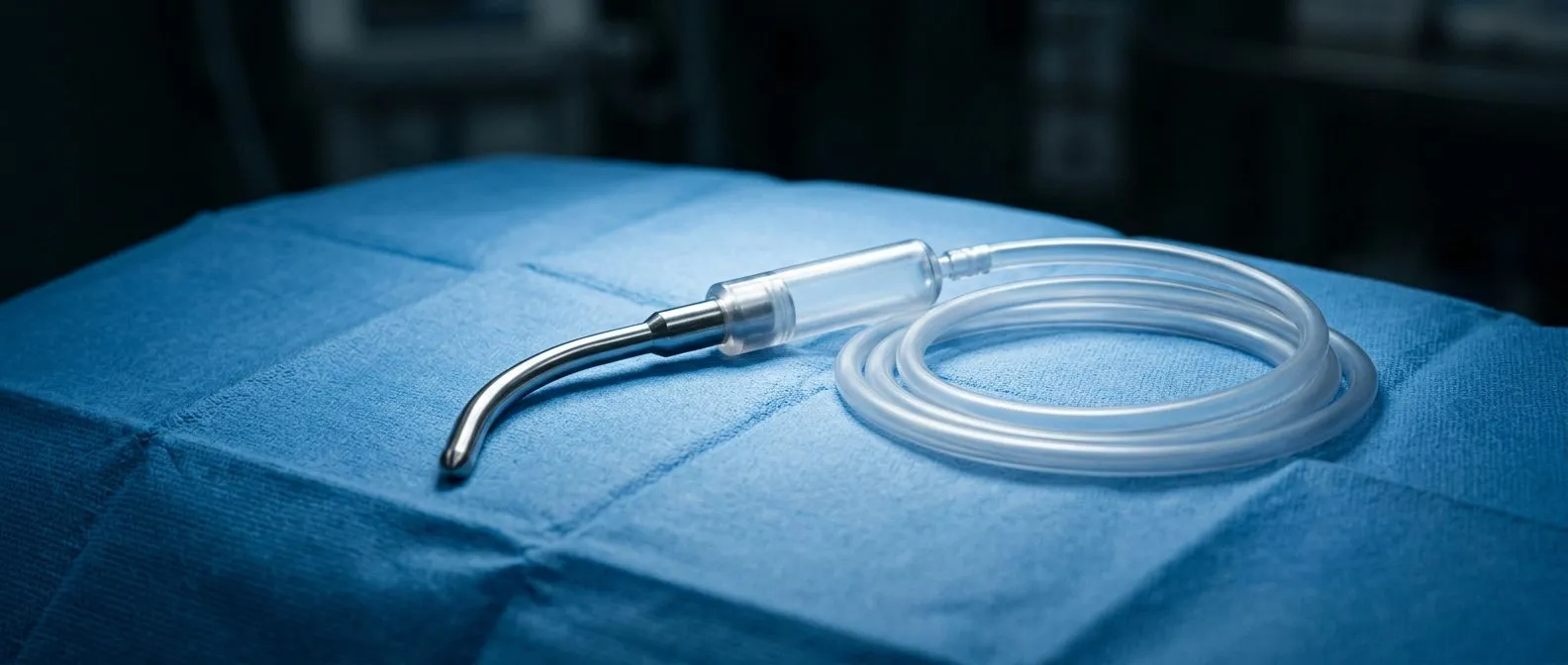
Why "selective" matters. You will often see the term selective antegrade cerebral perfusion. "Selective" means the perfusion is directed specifically into the arteries that feed the brain — usually the brachiocephalic (innominate), left common carotid, and left subclavian arteries — rather than flooding the whole body. The team may deliver blood through a cannula (a soft tube) placed in the right arm artery and through small tubes placed directly into the neck vessels. This selective delivery is what allows the brain to be protected while the surgeon works in a calm, bloodless field.
How it differs from older methods. Earlier generations of aortic surgery relied on deep cooling alone to protect the brain, a technique called deep hypothermic circulatory arrest. Cooling the body to very low temperatures slows the brain's metabolism so it can tolerate a period without blood. Cooling still plays a role today, but antegrade cerebral perfusion in aortic surgery added active blood delivery on top of it, extending the safe operating time and giving surgeons more room to do complex repairs. Our team performs conventional, minimally invasive, robotic and transcatheter procedures, and we recommend the less invasive route only where the evidence supports it for that patient — and for arch work, this perfusion strategy is part of that evidence-based planning.
Why Does Brain Protection Matter During Aortic Surgery?
The anatomy of the problem. The aortic arch gives rise to the vessels that supply the brain. Any operation on the arch — whether for an aneurysm (a ballooned, weakened section of the artery), a Type A Aortic Dissection (a tear in the artery's inner lining that is a true cardiovascular emergency), or a complex combined valve and arch repair — means interrupting or manipulating those very vessels. Without a protection strategy, the brain would be starved of oxygen within minutes. Brain protection during aortic surgery is therefore not an optional extra; it is the central technical challenge of the operation.
Stroke risk and how it is reduced. Historically, operations on the arch carried a meaningful risk of stroke, because clots, debris, or air could reach the brain during the procedure, and because the brain tolerated interrupted circulation poorly. Modern strategies — combining moderate cooling, selective antegrade cerebral perfusion, careful handling of the diseased aorta, and continuous monitoring of brain function — have substantially reduced that risk in experienced centers. MedlinePlus provides general patient information on stroke and its causes for readers who want a broader medical reference (MedlinePlus).
What the team monitors. Brain protection is not just about the plumbing. It also involves real-time monitoring. Our team reviews every case against international practice guidelines before a recommendation is given, and in the operating room this planning continues with monitoring of brain activity, blood flow, and temperature, so the perfusion strategy can be adjusted moment to moment. Patients are often surprised to learn how much technology is quietly working in the background during these operations.
How Does Antegrade Cerebral Perfusion Work, Step by Step?
Step 1 — Cooling. After the patient is connected to the heart-lung machine (the pump that takes over the heart's and lungs' work), the body is cooled in a controlled way. Cooling slows every cell's metabolism, including brain cells, so they need much less oxygen. This is the foundation on which everything else rests.
Step 2 — Lowering the circulation. Once the target temperature is reached, the circulation to the lower body is stopped or reduced. This is the circulatory arrest cerebral perfusion technique in action: the surgeon can now open the aorta and work inside vessels that would otherwise bleed heavily. The operating field becomes still and clear.
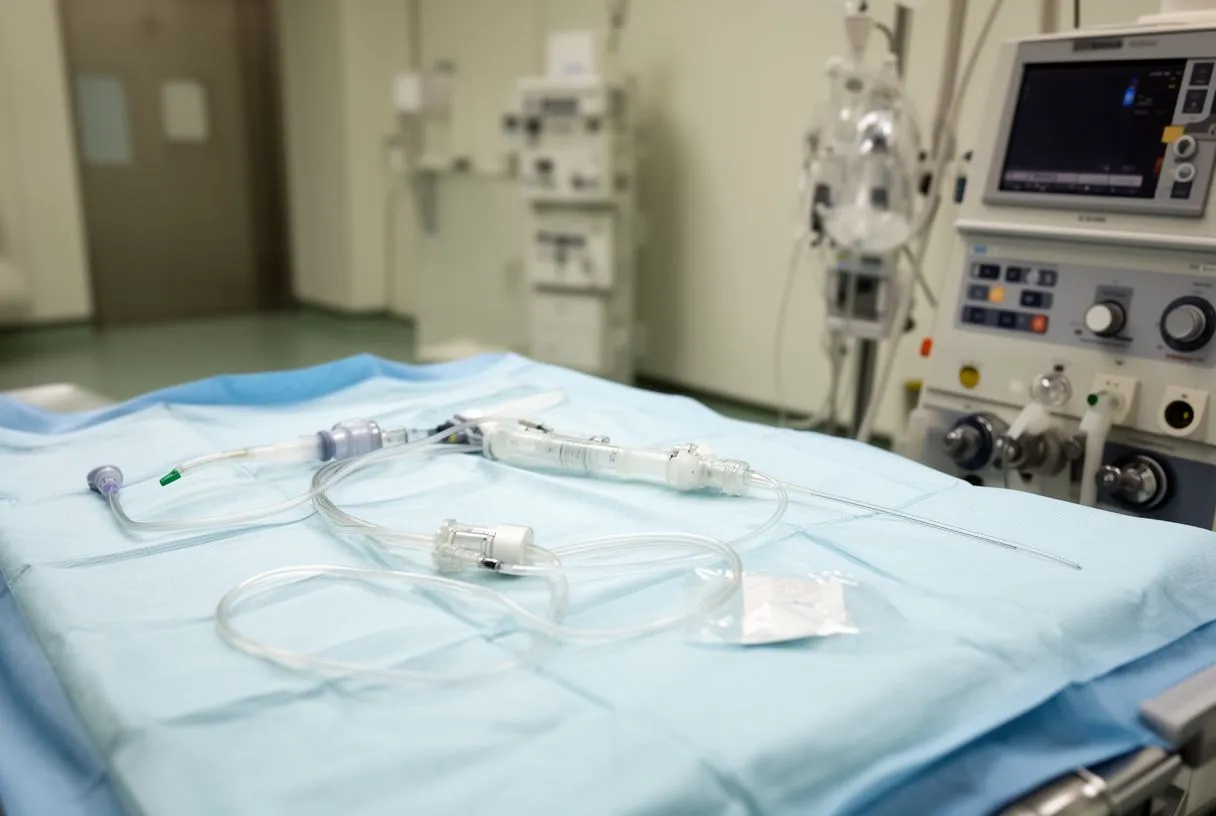
Step 3 — Delivering blood forward to the brain. While the rest of the body has no flow, oxygenated blood from the heart-lung machine is routed forward into the brain's arteries. Usually this is done through a cannula in the right arm artery, which connects to the circle of Willis — the ring of vessels at the base of the brain that distributes blood to both hemispheres. Sometimes additional small cannulas are placed in the left-sided neck vessels. The perfusion pressure and flow are watched continuously and kept within a narrow, deliberate range.
Step 4 — Rewarming. When the repair is complete, the body is gradually rewarmed, the circulation is restored to the whole body, and the patient is weaned off the heart-lung machine. The brain has never been left without oxygenated blood during the entire procedure — that is the entire point of the technique.
What Are the Types of Cerebral Protection Strategies?
An overview for families. There is no single "right" way to protect the brain during arch surgery; teams choose among several strategies based on the operation, the patient's anatomy, and the expected duration of repair. Understanding the main options helps you ask informed questions.
- Deep hypothermic circulatory arrest alone: the body is cooled deeply and the circulation is fully stopped for a defined period. Simple and gives a clear field, but limits the time the surgeon can work.
- Selective antegrade cerebral perfusion: oxygenated blood is pumped forward into the brain's arteries during the arrest. This is the most widely used modern strategy and extends the safe operating window considerably.
- Retrograde cerebral perfusion: blood is delivered backward through the veins that drain the brain. It is used by some teams, often as a supplement, and remains a topic of debate in the surgical literature.
- Moderate hypothermia with antegrade perfusion: newer protocols use less extreme cooling combined with continuous forward brain perfusion, shortening rewarming time and reducing complications related to very low temperatures.
How a strategy is chosen. The choice depends on the extent of the repair — an isolated ascending aorta replacement needs less protection time than a total aortic arch replacement with reimplantation of the neck vessels. It also depends on patient factors such as age, prior strokes, and the presence of a Thoracic Aortic Aneurysm extending into the arch. Every case is reviewed by the multidisciplinary team against international practice guidelines before a recommendation is given; the patient receives a clear proposal with the alternatives, and the final decision is always theirs.
What Is the Optimal Flow Rate for Antegrade Cerebral Perfusion?
Why flow rate matters. One of the most studied questions in this field is how much blood the brain actually needs during selective antegrade cerebral perfusion. Too little flow risks under-perfusion; too much flow can raise the pressure inside the skull and cause swelling. Research on minimal safe arterial blood flow during selective antegrade cerebral perfusion suggests that the brain does not need the full normal cardiac output — a modest, well-monitored flow is enough when combined with cooling, and excessive flow may actually be harmful. This is one of those counterintuitive findings that has shaped how perfusionists (the specialists who run the heart-lung machine) manage these cases.
Pressure versus flow. Surgeons and perfusionists track both the volume of blood delivered and the pressure in the brain's arteries. The target is to maintain adequate perfusion pressure — enough to push blood through the brain's vessels — without pushing so hard that the brain swells. Individual anatomy matters: some patients have narrowing or variations in their neck vessels that change how blood distributes. This is why imaging of the neck arteries before surgery is standard, and why the perfusion plan is individualized rather than taken from a formula.
What this means for you as a patient. You do not need to memorize numbers. What matters is knowing that your surgical team has a deliberate, monitored plan and that this question — the optimal flow for your anatomy — is one they should be able to answer clearly if you ask. In our consultation we take the time to explain what the studies actually show, in plain words, and to answer every question before any decision.
What Does Inadequate Cerebral Perfusion Mean?
The definition in everyday language. Cerebral perfusion simply means blood delivery to the brain. "Inadequate cerebral perfusion" means the brain is not receiving enough blood — or the blood is not reaching it at enough pressure — to meet its oxygen needs. This can happen inside the operating room if the perfusion strategy falls short, but it also happens outside surgery, for example when blood pressure drops severely, when a carotid artery becomes blocked, or during a cardiac arrest.
How teams detect it during surgery. During aortic operations, inadequate perfusion is watched for with several tools: near-infrared spectroscopy pads on the forehead measure the oxygen level in the brain tissue; pressure lines in the neck arteries confirm that the perfused blood is arriving; and in some centers EEG monitors brain electrical activity. If any of these signals drift outside the expected range, the team adjusts the flow, pressure, or temperature before a problem develops. Prevention is the whole design of the strategy.
How long does a brain perfusion scan take? This question usually refers to a Nuclear Perfusion Scan or a Ventilation-perfusion Scan of the lungs — diagnostic imaging studies done before surgery, not the intraoperative technique. These scans typically take a few hours including tracer injection and imaging, and they help the team understand blood flow to the brain or lungs before planning an operation. They are different from the perfusion delivered in the operating room, though both share the same underlying concern: making sure vital organs receive blood.
What Are the Symptoms of Poor Cerebral Perfusion?
Warning signs outside the operating room. For patients with aortic disease, recognizing poor brain perfusion matters long before any surgery. Symptoms of the brain not getting enough blood include sudden weakness or numbness on one side of the body, difficulty speaking or understanding speech, vision changes, severe dizziness, fainting, or a sudden severe headache. These can signal a stroke or a transient ischemic attack (a temporary, reversible lack of blood flow), and they warrant emergency evaluation — especially in someone known to have an aortic aneurysm or a dissection.
Symptoms related to aortic conditions specifically. An Acute Aortic Syndrome — the umbrella term for dissection, intramural hematoma, and Penetrating Aortic Ulcer — can present with tearing chest or back pain that radiates as the tear progresses. If the tear compromises the arteries supplying the brain, neurological symptoms appear alongside the pain. A Type A Aortic Dissection that involves the arch vessels is precisely the scenario where emergency surgery with antegrade cerebral perfusion is used: the technique allows the surgeon to repair the torn artery while the brain is perfused by the machine. Sudden tearing pain with any neurological symptom is an emergency — call emergency services, do not wait.
After surgery. In the intensive care unit, the team watches for subtle signs of neurological trouble: confusion that does not resolve as sedation wears off, weakness, or speech changes. Early detection allows early intervention, which is why patients are observed closely for the first days. Most patients wake up neurologically intact, and knowing the monitoring plan beforehand helps families feel prepared rather than frightened.
Which Conditions Require Antegrade Cerebral Perfusion?
Emergency scenarios. The clearest indication is a Type A Aortic Dissection — a tear in the ascending aorta or arch that requires immediate surgery to prevent rupture, stroke, or death. During this operation the arch vessels must be reconstructed, and antegrade cerebral perfusion is the standard way to protect the brain while that work is done. Time is critical in these cases, and the surgical team's familiarity with the technique directly influences outcomes.
Planned (elective) scenarios. The technique is also used in planned operations: repair of a Thoracic Aortic Aneurysm involving the arch, Aortic Arch Replacement with reimplantation of the neck vessels, combined valve and arch procedures, and complex reoperations where scar tissue makes the repair longer. Patients with a bicuspid aortic valve often develop ascending aortic dilation, and some of these operations extend into the arch. When an aortic valve replacement is planned and the aorta is also dilated, the surgical team evaluates whether the arch needs addressing in the same sitting; for patients at high surgical risk, options such as Transcatheter Aortic Valve Implantation (TAVR) or an Aortic Root Replacement may be discussed instead, each with its own trade-offs.
When the brain itself is at particular risk. Some patients have diseased or narrowed neck arteries, prior strokes, or anatomy that complicates perfusion delivery. In these cases the plan may include extra cannulation sites, shunts, or a modified strategy. The point is that the technique is adapted to the patient, not the other way around. In our team's experience, reviewing each patient's anatomy carefully before surgery — with the whole multidisciplinary group — is what prevents surprises in the operating room.
What Is Recovery Like After Aortic Arch Surgery?
The first days. After an arch operation you will wake up in an intensive care unit with a breathing tube that is usually removed within the first day, monitoring lines, and close neurological checks. Most patients are surprised by how quickly the tubes come out — often within 24 to 48 hours. Pain is managed with a planned regimen, and early mobilization begins as soon as it is safe, sometimes on day one or two.
The weeks that follow. Hospital stays for arch surgery commonly run from about a week to two, depending on complexity and how the recovery progresses. Once home, most people need several weeks to a few months to return to normal activities, with a graded return: walking first, then light activity, then driving and work. Cardiac rehabilitation — a supervised program of progressive exercise — is strongly recommended and measurably improves recovery. Some patients need blood-pressure medication long term, and everyone needs regular imaging of the remaining aorta, because aortic disease can progress in other segments over the years.
Quality of life after recovery. Patients who recover from arch surgery with intact neurological function generally return to full, active lives. The brain-protection strategy is a major reason for that. Families should also know what to watch for during recovery: new weakness, speech changes, fever, wound problems, or leg swelling all warrant a call to the surgical team. Families may find it helpful to keep a simple symptom diary during the first weeks at home.
How Do You Choose a Surgical Team for Complex Aortic Surgery?
Volume and training matter. Aortic arch surgery is among the most technically demanding operations in cardiac surgery, and outcomes are consistently better at centers and with teams that perform these procedures regularly. When you evaluate a team, ask directly: How many arch operations do you perform per year? What cerebral protection strategy do you use, and why for my anatomy? What is your monitoring protocol in the operating room? A confident team will welcome these questions.
Training and technology. Ask about the surgeon's training and the technologies available — minimally invasive approaches, perfusion capabilities, monitoring equipment. Dr Molina trained in cardiac, minimally invasive, robotic and structural surgery in Montreal and in structural cardiology and TAVR in Bordeaux, and the team also trained in Jena, Bad Berka and Buffalo. We are a multidisciplinary team of cardiac surgeons led by Dr Alejandro Molina, with consulting rooms in Guadalajara, Querétaro and Hermosillo.
Second opinions are normal. For an operation of this magnitude, a second opinion is not a sign of distrust — it is a standard part of making a good decision. We encourage second opinions and treat seeking one as a normal part of deciding about heart surgery. A reputable team will provide your imaging and records freely and will explain how their proposed plan compares with alternatives, including less invasive options where the evidence supports them.
Practical questions for international and traveling patients. If you are traveling to Mexico for surgery, ask about the preoperative workup timeline, how long you should plan to stay near the hospital, follow-up arrangements once you return home, and how coordination with your local doctors will work. For elective aortic surgery, the workup typically includes imaging of the whole aorta, evaluation of the neck arteries, heart function studies, and risk scoring. A transparent team will tell you which studies are genuinely needed and which are not.
Taking the next step. Antegrade cerebral perfusion is the quiet technology that makes modern aortic arch surgery possible: it keeps the brain supplied with oxygenated blood while surgeons repair the body's largest artery. If you have been diagnosed with an aortic aneurysm, a dissection, or complex valve-and-aorta disease, the most valuable thing you can do is sit down with a qualified cardiac team, understand your specific anatomy, and hear the options with their trade-offs. Our first contact is a free video consultation: we get to know the patient, talk through the condition, and say which studies are still needed so no unnecessary ones are ordered. Do not self-diagnose and do not delay — a conversation with a team that performs these operations regularly is the safest next move you can make.






