
Apex of the Heart: Where Heartbeats Are Felt and Measured
Find the apex of the heart location and learn why your heartbeat is felt there.
What is the apex of the heart location?
Understanding the apex of the heart location is the first step in making sense of how your cardiologist listens to and measures your heartbeats. The apex is the pointed, inferior end of the heart, directed downward, forward, and to the left. It is formed mainly by the left ventricle, the powerful muscular chamber responsible for pumping oxygenated blood to your entire body. When you feel your heartbeat through your chest wall, you are usually feeling the apex tapping against your ribs as it contracts.
Anatomically, the heart sits in the central compartment of the chest, resting just above the diaphragm. The relationship of the heart to the ascending aorta is crucial here, as blood exits the left ventricle through this main artery to reach the brain and body. The right side of the heart, specifically the right atrium, receives oxygen-poor blood returning from the veins. These landmarks explain why the apex is found where it is, typically in the fifth intercostal space on the left side of the chest. According to academic.oup.com, these anatomical coordinates are essential for clinical assessments.
Your cardiac team uses the apex as a physical reference point to gauge how hard the heart is working. Because the left ventricle has the thickest muscular wall, its contraction generates the most force. This is why the beat you feel on your chest is predominantly from the left side of the heart, even though the heart itself sits centrally. Recognising this physical landmark allows doctors to interpret symptoms accurately, often before any imaging is required.
How to find the apex of the heart location in the chest
Finding the apex of the heart location requires knowing the basic surface anatomy of the chest. For most adults, the apex is located in the fifth intercostal space, which is the area between the fifth and sixth ribs. If you draw an imaginary line straight down from the middle of your left collarbone (the midclavicular line), the apex is typically found just medial to this line. Your doctor will use these coordinates to place a stethoscope accurately during an exam.
Palpating the apex beat: Before listening, a physician will often try to feel the heartbeat through the chest wall. This tactile sensation, known as the apex beat, confirms the physical location and strength of the left ventricle's contraction. If the apex is difficult to feel, it may be due to a thick chest wall, obesity, or simply a heart that sits slightly further back in the chest. A shifted or absent apex beat can be an early indicator that further investigation is needed.
Sometimes, a patient may present with arm pain alongside chest discomfort, which can complicate the physical exam. This referred pain is a well-documented symptom of heart issues, particularly when the heart muscle is starved for oxygen. In such cases, finding the exact location of the apex helps the medical team determine if the pain is truly cardiac in origin. The physical exam remains a vital first step before any decisions are made regarding advanced monitoring.
What is the Point of Maximal Impulse (PMI)?
The Point of Maximal Impulse, commonly abbreviated as PMI, is a clinical term that directly corresponds to the apex of the heart location. It represents the specific spot on the chest where the heartbeat is felt most intensely. The PMI is not just a random strong pulse; it is the literal thrust of the left ventricle against the chest wall during each contraction. Identifying this point is a standard part of a routine cardiovascular physical examination.

PMI and heart size: The location of the PMI gives doctors an immediate clue about the size and function of the left ventricle. A normal PMI is a quick, localized tap. If the PMI is displaced further to the left or downward, it often suggests that the heart is enlarged or dilated. This non-invasive observation can prompt a cardiologist to order an echocardiogram to check for conditions like fluid overload or heart failure.
In emergency settings, the PMI is a rapid diagnostic tool. If a patient collapses and the PMI is absent, the medical team may immediately consider using an Automated External Defibrillator to restore a viable heart rhythm. The presence or absence of this maximal impulse, alongside other vital signs, dictates the urgency of the medical response. It serves as a bridge between basic physical assessment and life-saving intervention when every second counts.
Why the apex of the heart location matters for auscultation
Auscultation is the act of listening to the internal sounds of the body, usually with a stethoscope. The apex of the heart location is one of the primary listening posts for this practice. When a doctor places the stethoscope over the apex, they are listening specifically to the mitral valve, the valve that controls blood flow from the left atrium into the left ventricle. The sounds here are distinct from those heard at the base of the heart, where the aortic and pulmonary valves are best heard.
Listening for murmurs: The apex is the optimal location to detect murmurs associated with mitral regurgitation or mitral stenosis. A murmur is a swooshing sound that indicates turbulent blood flow. If the mitral valve does not close properly, blood leaks backward into the atrium during ventricular contraction, creating a distinct sound at the apex. Identifying these acoustic anomalies at the exact location helps diagnose structural heart disease early.
Advanced monitoring often complements auscultation. For example, a patient in intensive care might have an arterial line placed to provide real-time blood pressure readings. While the arterial line offers precise hemodynamic data, the sounds heard at the apex provide information about the mechanical function of the valves. Both diagnostic methods work together, giving a comprehensive picture of the patient's cardiovascular status without relying on a single metric.
Apex of the heart location and left ventricular function
The apex of the heart location is a direct window into the health of the left ventricle, which is the main pumping engine of the heart. The force and character of the apex beat reflect the ventricle's ability to contract and push blood out to the body. A strong, quick tap generally indicates a healthy, well-functioning muscle. Conversely, a diffuse or weak impulse can suggest that the ventricle is struggling to generate adequate force.

Pressure overload and volume overload: Different conditions affect the apex beat in distinct ways. If the heart is pumping against high resistance, such as severe untreated hypertension, the muscle thickens, creating a forceful but sustained apex beat. If the heart is stretched by too much blood volume, the apex beat becomes diffuse and shifting. Recognising these mechanical differences at the exact location helps doctors differentiate between types of heart stress before moving to imaging.
When the left ventricle fails, the body struggles to receive oxygenated blood, which can have systemic effects. A comprehensive review by Dementia prevention, intervention, and care: 2024 report of the Lancet standing Commission highlights how poor cardiovascular health, including heart failure, is linked to cognitive decline. The inadequate pumping function that alters the apex beat can ultimately reduce blood flow to the brain. This connection underscores why a simple physical exam at the chest wall is so vital for long-term overall health.
When apex of the heart location shifts: cardiomegaly
Cardiomegaly is the medical term for an enlarged heart. When the heart muscle grows or the chambers dilate, the physical shape of the organ changes, causing the apex of the heart location to shift. A displaced apex beat is often the first physical sign of cardiomegaly. If the apex is found further down the chest or further to the left than expected, it strongly suggests the left ventricle has expanded beyond its normal anatomical boundaries.
Causes of displacement: An enlarged heart is not a disease itself, but a response to an underlying stressor. Common causes include untreated high blood pressure, damaged heart valves, or coronary artery disease. When a portion of the heart muscle dies from an atypical infarction—a heart attack with non-standard symptoms—the surviving muscle stretches to compensate, shifting the apex. Understanding the cause of the shift is critical for planning the right medical or surgical intervention.
Treating cardiomegaly usually involves managing the underlying condition to reduce the strain on the heart muscle. Medications are often prescribed to lower blood pressure or reduce fluid retention. In cases where severe coronary blockages are the root cause, a patient might require balloon angioplasty to open the narrowed vessels and restore blood flow. Relieving this mechanical stress can sometimes allow the heart to return to a more normal size and shift the apex back toward its original location.
How doctors image the apex of the heart location
While the physical exam locates the apex through touch and sound, imaging provides a detailed look at its structure and movement. An echocardiogram, or heart ultrasound, is the most common tool used to image the apex of the heart location. The technician places a probe on the chest wall directly over the PMI to get a clear view of the left ventricle. This allows the cardiologist to watch the apex contract in real-time and measure the thickness of the muscle walls.
Advanced imaging technologies: Echocardiography has limitations in patients with dense chest walls or lung disease, so other modalities are sometimes required. Cardiac MRI offers a high-resolution, three-dimensional view of the apex, revealing scarring or subtle structural defects that ultrasound might miss. According to The Internet of Things for Health Care: A Comprehensive Survey, modern remote monitoring and imaging integration are becoming essential for tracking these structural changes over time, especially for international patients managing chronic conditions.
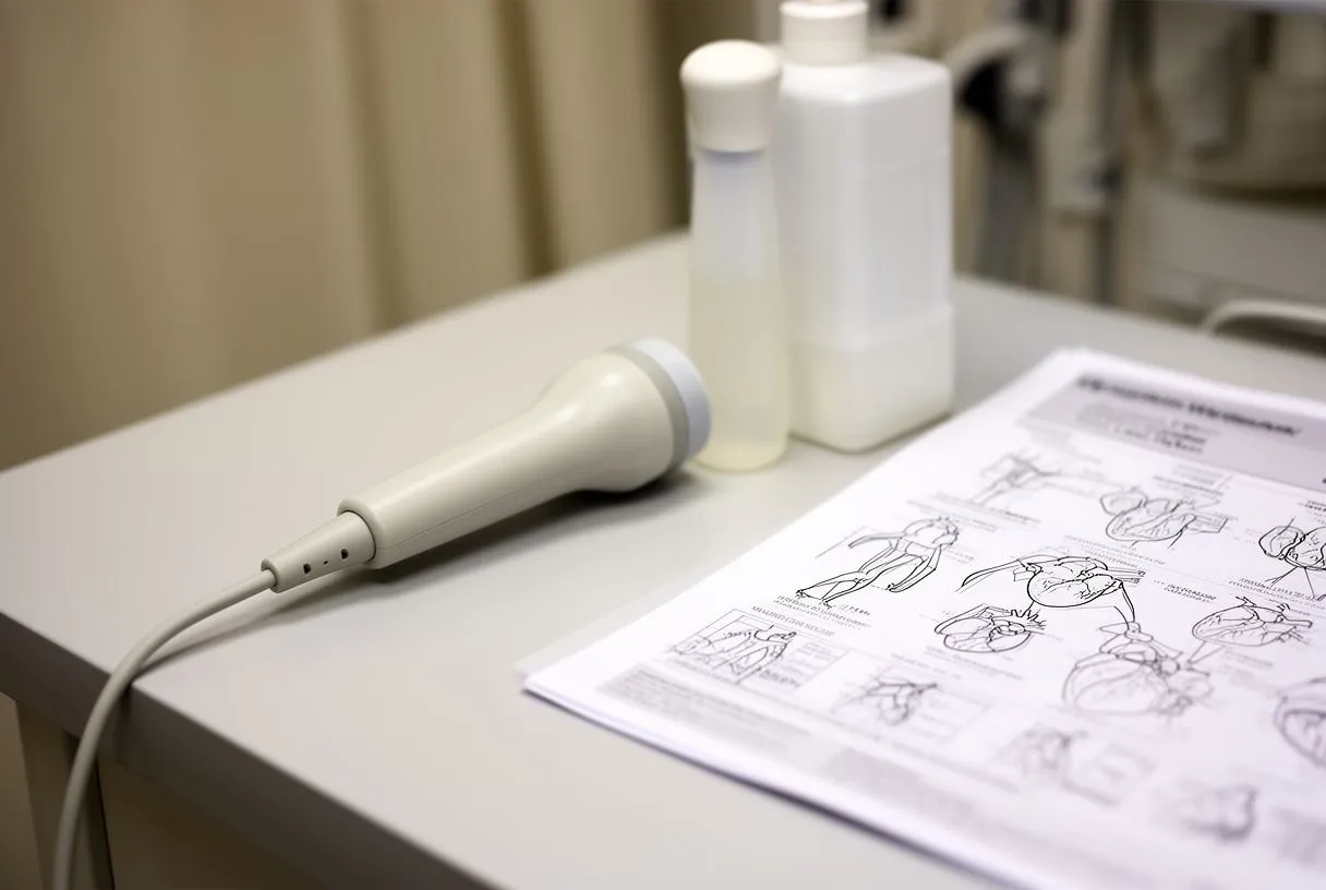
Imaging the apex is also crucial when planning complex surgical interventions. If a patient requires a coronary bypass, the surgeon must understand the exact anatomy and viability of the left ventricle. The images confirm whether the muscle at the apex is healthy enough to benefit from restored blood flow, or if the damage is permanent. This diagnostic step ensures that surgical risks are minimized and outcomes are optimized for the patient's specific situation.
What apex beats reveal about heart failure
Heart failure occurs when the heart cannot pump enough blood to meet the body's needs. The characteristics of the apex beat change significantly as heart failure progresses. Instead of a quick, localized tap, the apex beat in a failing heart often becomes a sustained, heaving impulse that lasts longer than a normal heartbeat. This altered physical sensation at the apex of the heart location indicates that the ventricle is struggling to empty completely with each contraction.
Diastolic versus systolic dysfunction: The feel of the apex can also hint at the type of heart failure. If the muscle is thick and stiff, preventing it from relaxing to fill with blood, the apex beat may be strong but localized. If the muscle is weak and stretched, the apex beat will be diffuse and displaced. Distinguishing between these types is critical because the medication strategies for each are vastly different, directly impacting the patient's quality of life.
Patients with advanced heart failure sometimes require specialised support. In extreme cases where the heart cannot sustain circulation, a team might consider awake ECMO (Extracorporeal Membrane Oxygenation), a temporary life support system that oxygenates blood outside the body. While the physical apex beat is obscured by these massive interventions, the initial assessment of the impulse remains the trigger that escalates care to this level. Monitoring the basic physical signs guides the transition from standard care to life support.
Apex of the heart location during physical examination
A thorough physical examination is a cornerstone of cardiac care, and finding the apex of the heart location is a fundamental component. The doctor will usually have the patient roll slightly onto their left side, which brings the heart closer to the chest wall and makes the apex beat easier to feel and hear. This specific positioning, known as the left lateral decubitus position, enhances the sensitivity of the exam for subtle abnormalities that might be missed when lying flat on the back.

Integrating the full exam: Examining the apex is not done in isolation. A cardiologist will correlate the findings at the apex with pulses in the neck, blood pressure readings, and breathing sounds in the lungs. For instance, if the apex is displaced and the patient has irregular lung sounds, it might indicate fluid backing up due to left ventricular failure. The following list highlights what a doctor evaluates during this crucial physical step:
- Location: Is the apex beat in the normal fifth intercostal space, or has it shifted due to enlargement?
- Intensity: Is the tap strong and localized, or weak and diffuse, indicating muscle weakness?
- Duration: Does the beat last a fraction of a second, or is it a sustained heave suggesting high pressure?
- Accompanying sounds: Are there murmurs or extra sounds like gallops when listening over the apex?
During surgery, continuous assessment is vital. Patients receiving a bypass might have an arterial graft placed to bypass a blockage. Monitoring the apex and overall cardiac function ensures the new graft is supplying adequate blood to the heart muscle. The physical and intraoperative exams work together to ensure the mechanical structure of the heart is responding well to the surgical repair.
Living with conditions affecting the apex of the heart
Living with a condition that alters the apex of the heart requires active management and a strong partnership with your medical team. While a displaced or altered apex beat may sound alarming, it is often a manageable sign of a chronic condition rather than an immediate emergency. The goal of treatment is to address the underlying cause—whether that is lowering blood pressure, repairing a valve, or improving blood flow—so the heart can function as efficiently as possible. Regular check-ups allow your cardiologist to monitor the apex location over time.
Medication and lifestyle management: Many conditions affecting the apex are managed with a combination of medications and lifestyle adjustments. Patients may be prescribed Aspirin to prevent blood clots in narrowed arteries, or statins to manage atheromatous plaque build-up in the vessels. Adhering to these medical routines is essential to prevent further stress on the heart muscle. The following list outlines the pillars of managing these conditions long-term:
- Medication adherence: Taking prescribed drugs exactly as directed to reduce cardiac workload and prevent clots.
- Regular monitoring: Attending follow-up appointments to track the apex beat and overall heart size.
- Activity modification: Engaging in supervised, heart-safe exercise to maintain muscle tone without overstraining the ventricle.
- Dietary balance: Maintaining a low-sodium, heart-healthy diet to prevent fluid overload and additional heart stress.
Ultimately, understanding your own heart anatomy empowers you to participate actively in your care. If you have concerns about your heart, palpitations, or have been told your heart is enlarged, the most important step is to seek professional evaluation. We encourage you to discuss your specific situation with a qualified cardiac team. Booking a consultation allows specialists to perform a thorough physical exam, locate your apex, and provide a tailored diagnosis rather than relying on self-assessment. Your heart health is too important to leave to guesswork.