

Cardiac Tamponade: Fluid Around the Heart in an Emergency
Cardiac tamponade treatment saves lives when fluid builds around the heart.
What Is Cardiac Tamponade and Why Is It an Emergency?
Understanding the pericardium and its function: Your heart sits inside a thin, double-layered sac called the pericardium. Between the two layers, a small amount of natural lubricating fluid lets the heart beat and shift without friction. When too much fluid, blood, or pus accumulates in that narrow space, pressure rises rapidly. The fluid squeezes the heart chambers from the outside, preventing them from filling properly with blood between each beat. This compression is what doctors call cardiac tamponade, and it is a life-threatening emergency that requires immediate cardiac tamponade treatment to relieve the pressure and restore normal circulation.
Why the pressure builds so quickly: The pericardial sac is relatively stiff and cannot stretch far. If fluid enters it fast — for example, from a bleeding vessel near the heart — even a small volume (sometimes as little as 150 to 200 millilitres) can overwhelm the sac's ability to expand. The heart's pumping chambers collapse inward with each attempted fill, cardiac output drops sharply, and the body's organs receive less oxygenated blood. Without intervention, blood pressure can fall to dangerous levels within minutes or hours, depending on how rapidly the fluid accumulates.
Why it qualifies as a true emergency: Cardiac tamponade does not improve on its own. The mechanism is mechanical — no medication can remove the fluid or release the external compression. The only effective solution is to drain the fluid or create an opening in the pericardium so the heart can fill and pump normally again. If you or a family member develops sudden chest pressure, breathlessness, fainting, or a feeling of fullness in the chest after surgery, trauma, or a known illness, this combination warrants calling emergency services without delay.
What Causes Fluid to Build Up Around the Heart?
Common medical causes of pericardial effusion: Fluid around the heart, called a pericardial effusion, can develop for many reasons. Viral infections are among the most frequent triggers, often producing inflammation of the pericardium (pericarditis) that weeps fluid into the sac. Autoimmune conditions such as lupus or rheumatoid arthritis can also inflame the pericardium. Kidney failure, hypothyroidism, and certain cancers — particularly lung cancer, breast cancer, and lymphoma — may cause slow, progressive fluid accumulation that eventually tips into tamponade if left unmonitored.
Post-surgical and trauma-related causes: Patients who have recently undergone cardiac surgery are at particular risk. Bleeding from the ascending aorta suture line, a coronary artery, or a small vessel near an arterial graft can send blood directly into the pericardial space in the hours or days after an operation. This is why cardiac surgical teams monitor drainage tubes closely and watch for signs that the heart is being compressed. Blunt chest trauma from a car accident, a fall, or a sports injury can also tear a vessel and cause rapid blood pooling around the heart. For a broad clinical overview of these mechanisms, you can refer to information published by Healthline.
Medication-related and less common triggers: Certain drugs, including some that prevent blood clots, can increase the risk of bleeding into the pericardium. Patients on long-term anticoagulation — for example, those taking aspirin or warfarin for atrial fibrillation — may be more vulnerable if pericarditis develops at the same time. Radiation therapy to the chest for cancer treatment can also irritate the pericardium months or years later. Rarely, a tear in the heart muscle itself from a heart attack can cause blood to leak outward, a complication that demands immediate surgical attention.
Recognizing the Symptoms of Cardiac Tamponade
Early warning signs that are easy to miss: Cardiac tamponade can begin subtly, especially when fluid accumulates slowly over days or weeks. Patients often describe a feeling of fullness or pressure in the chest that does not quite feel like typical angina. Shortness of breath, particularly when lying flat (orthopnoea), is one of the most reliable early symptoms. Some people notice increasing fatigue, dizziness when standing, or a dry cough that seems to come from deep inside the chest rather than the airways.
Classic signs that demand immediate attention: When tamponade progresses, the body shows clearer distress signals. Blood pressure may drop noticeably, while the pulse becomes weak and rapid. Veins in the neck — the jugular veins — may appear swollen and pulsing because blood cannot return efficiently to the compressed heart. A doctor performing auscultation (listening with a stethoscope) may notice that heart sounds are muffled or distant, as if heard through a cushion. Together with low blood pressure and distended neck veins, this trio is known as Beck's triad, a well-known clinical sign of tamponade. Some patients also report radiating discomfort similar to arm pain, which can be confused with a heart attack.
Symptoms that mean the situation is critical: If the heart cannot fill adequately, the brain and other organs receive insufficient blood. This can lead to confusion, extreme drowsiness, cold and clammy skin, and eventually collapse. At this stage, the window for safe intervention is narrowing rapidly. Family members who see someone with known heart disease suddenly become pale, breathless, and unable to respond normally should treat it as a medical emergency and call for an ambulance rather than waiting for a scheduled appointment.
How Doctors Diagnose Cardiac Tamponade
The role of echocardiography as the first-line test: When a physician suspects tamponade, an echocardiogram — an ultrasound of the heart — is almost always the first and most important diagnostic tool. It shows the fluid collection directly, reveals how much space exists between the pericardium and the heart muscle, and, critically, demonstrates whether the heart chambers are collapsing inward during the filling phase. This "chamber collapse" on ultrasound is a hallmark sign that the compression is haemodynamically significant and that drainage is needed.
Other imaging and monitoring tools: A chest X-ray may show an enlarged, globular heart shadow, sometimes called a "water-bottle heart," though this sign appears only when a large volume of fluid has already collected. An electrocardiogram (ECG) can reveal low-voltage electrical signals because the fluid dampens the heart's electrical activity, and occasionally shows a pattern called electrical alternans, where the QRS complex changes size beat to beat as the heart swings within the fluid pool. If an arterial line is already in place — a thin catheter placed in an artery to monitor blood pressure continuously — it may reveal a concerning drop in blood pressure during inspiration, a phenomenon known as pulsus paradoxus, which strongly supports the diagnosis.
How quickly the diagnosis must happen: In a genuine emergency, doctors do not wait for every test to come back. If the clinical picture and a rapid bedside echo point to tamponade, the team moves straight to drainage. In stable patients where fluid has been building gradually, the team may have time for additional tests such as a CT scan or MRI, which can show fluid density and help identify whether it is simple fluid, blood, or pus. The guiding principle is always the patient's haemodynamic stability: if the heart is struggling, diagnostic perfection gives way to prompt action.
Cardiac Tamponade Treatment Options in an Emergency
The immediate goal of any intervention: Cardiac tamponade treatment always begins with the same principle: relieve the pressure on the heart as quickly and safely as possible. The approach chosen depends on the patient's stability, the suspected cause of the fluid, what equipment is available, and whether the situation allows for a planned procedure or demands an emergency manoeuvre at the bedside. In every case, the aim is to restore the heart's ability to fill so that cardiac output and blood pressure recover.
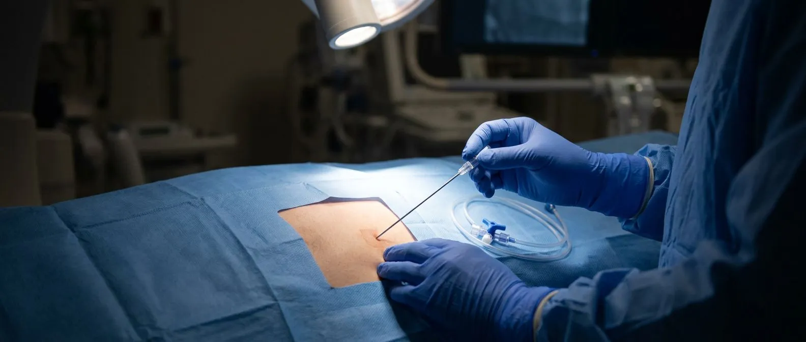
Pericardiocentesis as the primary emergency procedure: For most patients, the fastest available option is pericardiocentesis — a procedure in which a needle is guided into the pericardial space to withdraw fluid. When performed under echocardiographic guidance, this technique is generally the safest and least invasive first step. The doctor watches the needle tip on ultrasound in real time, aspirates fluid, and often leaves a temporary drain in place for 24 to 48 hours to prevent re-accumulation. This is particularly effective for larger effusions caused by inflammation, kidney failure, or cancer.
When surgery becomes necessary: Not every case can be managed with a needle alone. If the fluid is thick blood that will not pass through a needle, if it has clotted, or if the source is active bleeding from a cardiac structure, surgical drainage is required. Surgical options include creating a pericardial window — a small opening that lets fluid drain continuously into the chest cavity where it can be reabsorbed — or a formal pericardiectomy in rare, recurrent cases. The choice between needle drainage and surgery is made by the treating cardiac team based on imaging findings, the patient's overall condition, and the suspected underlying cause.
What Happens During Pericardiocentesis
Preparing the patient and positioning: Pericardiocentesis is usually performed at the bedside or in a cardiac catheterisation room. The patient lies with the head of the bed raised to about 30 to 45 degrees so that fluid pools at the bottom of the pericardial sac, closest to the skin. The skin below the breastbone is cleaned and numbed with local anaesthetic. If the patient is awake but anxious, a mild sedative may be given through an intravenous line to maintain comfort without suppressing breathing.
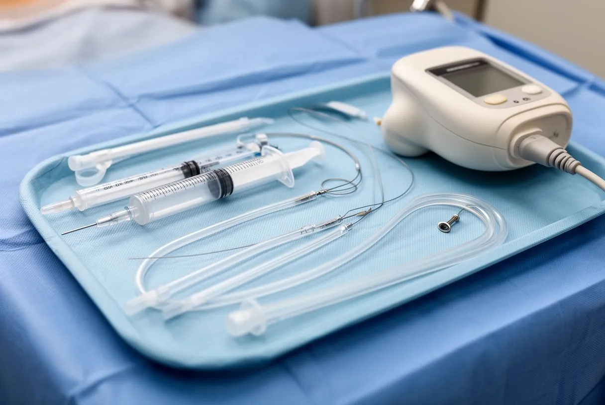
Guiding the needle with ultrasound: The cardiologist identifies the clearest path to the fluid using an echocardiogram probe, then advances a thin needle through the skin and into the pericardial space. When fluid appears in the syringe, its colour and consistency provide immediate clues — straw-coloured fluid suggests inflammation, while dark blood may indicate a haemorrhagic source. Research into the inflammatory cascades that worsen cardiac injury, including the role of innate immune signalling in myocardial damage, is an active area of investigation; one such line of study is described in the work IRF3 and type I interferons fuel a fatal response to myocardial infarction. Once fluid is confirmed, a guidewire is passed through the needle, and a soft drainage catheter is threaded over it for ongoing drainage.
What to expect after the procedure: Most patients feel rapid relief as the pressure on the heart is released — blood pressure typically rises, the pulse slows, and breathing becomes easier within minutes. The drainage catheter usually stays in place for one to two days, and the collected fluid is sent to the laboratory for analysis to help identify the underlying cause. Nurses monitor the drain output closely, check vital signs frequently, and watch for any sign of re-accumulation. If the drain produces little fluid for 24 hours and the echocardiogram confirms no significant residual effusion, it can be safely removed.
Surgical Approaches for Draining the Pericardial Sac
Pericardial window (subxiphoid or thoracoscopic): A pericardial window is a surgical opening created in the pericardium to allow continuous drainage. The subxiphoid approach makes a small incision just below the breastbone and is often performed under local or regional anaesthesia, making it suitable for patients who are too unstable for general anaesthesia. The thoracoscopic (VATS) approach uses keyhole instruments inserted between the ribs to create a larger opening and place a drain under direct visualisation, which is particularly useful when fluid is loculated or when a biopsy of the pericardium is needed.
When a full pericardiectomy is indicated: Pericardiectomy — the surgical removal of most or all of the pericardium — is reserved for cases of constrictive pericarditis, where the pericardium has become thick, scarred, and calcified, restricting the heart chronically. This is a major open-heart operation performed through a sternotomy (splitting the breastbone) and carries a longer recovery. It is not a first-line treatment for acute tamponade but becomes relevant when repeated effusions or chronic constriction develop despite prior drainage procedures.
Surgical drainage after recent cardiac operations: Patients who develop tamponade days after heart surgery present a unique challenge. The fluid is often clotted blood that cannot be aspirated through a needle, making a return to the operating room necessary. Surgeons reopen the chest, evacuate the clot, identify and control the bleeding source, and confirm that all chambers — including the atrium — are filling properly before closing. This scenario is one reason why cardiac surgery programmes maintain close postoperative monitoring protocols and rapid-response teams for any sign of haemodynamic deterioration.
Recovery After Cardiac Tamponade Treatment
The first 24 to 48 hours: Immediately after drainage, the priority is confirming that the heart is functioning normally and that fluid is not returning. Patients typically stay in a high-dependency or intensive care unit for continuous cardiac monitoring. An echocardiogram is repeated within 12 to 24 hours to verify that chambers are filling well and that no significant residual effusion remains. Intravenous fluids and, occasionally, medications to support blood pressure are administered while the body re-establishes stable circulation.
Managing the underlying cause: Successful drainage addresses the immediate emergency but not the reason the fluid appeared. If a viral infection caused the pericarditis, anti-inflammatory medications may be prescribed for several weeks. If kidney failure is the culprit, dialysis may need to be adjusted. If cancer is identified in the fluid, the patient is referred to an oncology team for staging and treatment planning. Addressing the root cause is essential to prevent recurrence, which can happen within days if the underlying condition is not treated. The importance of tissue engineering and regenerative approaches in cardiac care is growing, as illustrated by platforms such as the one described in Cardiopatch platform enables maturation and scale-up of human pluripotent stem cell-derived engineered heart tissues.
What patients and families should watch for at home: After discharge, the most important warning sign is any return of the original symptoms — chest pressure, breathlessness, dizziness, or swelling of the neck veins. Patients are usually advised to avoid strenuous activity for two to four weeks, attend a follow-up echocardiogram within a week or two, and report fevers, increasing fatigue, or wound problems promptly. A calm, structured recovery plan reduces anxiety for the whole family and ensures that any re-accumulation is caught early, while it is still manageable.
Long-Term Outlook and Follow-Up Care
Recurrence risk and how it is managed: The likelihood of cardiac tamponade recurring depends almost entirely on the underlying cause. Viral or idiopathic pericarditis has a moderate recurrence rate, and some patients benefit from a course of colchicine or corticosteroids to reduce inflammation. Malignant effusions, by contrast, tend to recur more frequently and may require a planned pericardial window to provide permanent drainage. Patients with chronic kidney disease on dialysis are also at ongoing risk and may need repeated drainage if fluid management is challenging.
Quality of life after successful treatment: Many patients return to their normal activities within a few weeks of a straightforward pericardiocentesis, provided the underlying condition is controlled. The heart muscle itself is rarely damaged by tamponade unless the compression was prolonged or a concurrent heart attack occurred. This means that once the pressure is relieved, cardiac function often returns to its previous baseline, and long-term heart failure is uncommon. Patients with rhythm disturbances such as atrial flutter may need additional cardiology follow-up, but the tamponade episode itself usually does not cause permanent electrical problems.
Building a long-term surveillance plan: A practical follow-up plan typically includes a clinic visit within two weeks of discharge, a repeat echocardiogram at that visit or shortly after, and an ongoing schedule determined by the underlying diagnosis. The goal is to give each patient a clear roadmap: what to monitor, when to call, and when a routine check is sufficient versus when urgent re-evaluation is needed.
When to Seek Immediate Help for Heart Symptoms
Red-flag symptoms that should never wait: Certain combinations of symptoms are too dangerous to manage at home. Sudden chest pressure or fullness accompanied by breathlessness, fainting, or a rapid weak pulse — especially in someone with recent heart surgery, cancer, kidney disease, or a known pericardial effusion — warrants calling emergency services immediately. Waiting to see whether symptoms resolve on their own can allow tamponade to progress to a point where even emergency drainage becomes more difficult and risky.
Lower-risk symptoms that still need prompt evaluation: Not every chest sensation is an emergency, but new or changing symptoms deserve timely assessment. A gradual increase in shortness of breath over several days, trouble lying flat without feeling breathless, or unexplained swelling in the legs with neck vein distension should prompt a same-day medical review. In these situations, an echocardiogram can quickly determine whether fluid is accumulating and whether intervention is needed before the situation becomes critical.
- Call emergency services if: there is sudden severe chest pressure with fainting, extreme breathlessness, or a rapid weak pulse — this may indicate active tamponade or another cardiac emergency.
- Seek same-day evaluation if: breathlessness is worsening over days, you cannot lie flat comfortably, or you notice swollen neck veins — these may signal a growing pericardial effusion.
- Attend scheduled follow-up if: you have a known effusion or recent drainage and feel well — repeat echocardiography confirms the heart remains free of dangerous fluid re-accumulation.
Why early action changes outcomes: The single most important factor in surviving cardiac tamponade is the speed of intervention. When drainage occurs before blood pressure collapses, outcomes are generally excellent, and the heart recovers quickly. Delay allows the cycle of falling cardiac output, falling coronary blood flow, and worsening heart function to compound, making recovery harder. If you are caring for someone who has had a pericardial effusion or recent cardiac surgery, knowing these warning signs and acting on them promptly is the most valuable contribution you can make to their safety.
If you or a family member has been told there is fluid around the heart, or you are recovering from a pericardial drainage procedure, the most reassuring step you can take is to speak directly with a qualified cardiac team. Every patient's anatomy, underlying condition, and recovery trajectory is different, and no online guide can replace a personal consultation. We encourage you to book a consultation with Dr Alejandro Molina and the team at Cirugía de Corazón to discuss your specific situation, review your imaging, and build a clear plan for monitoring and care.






