

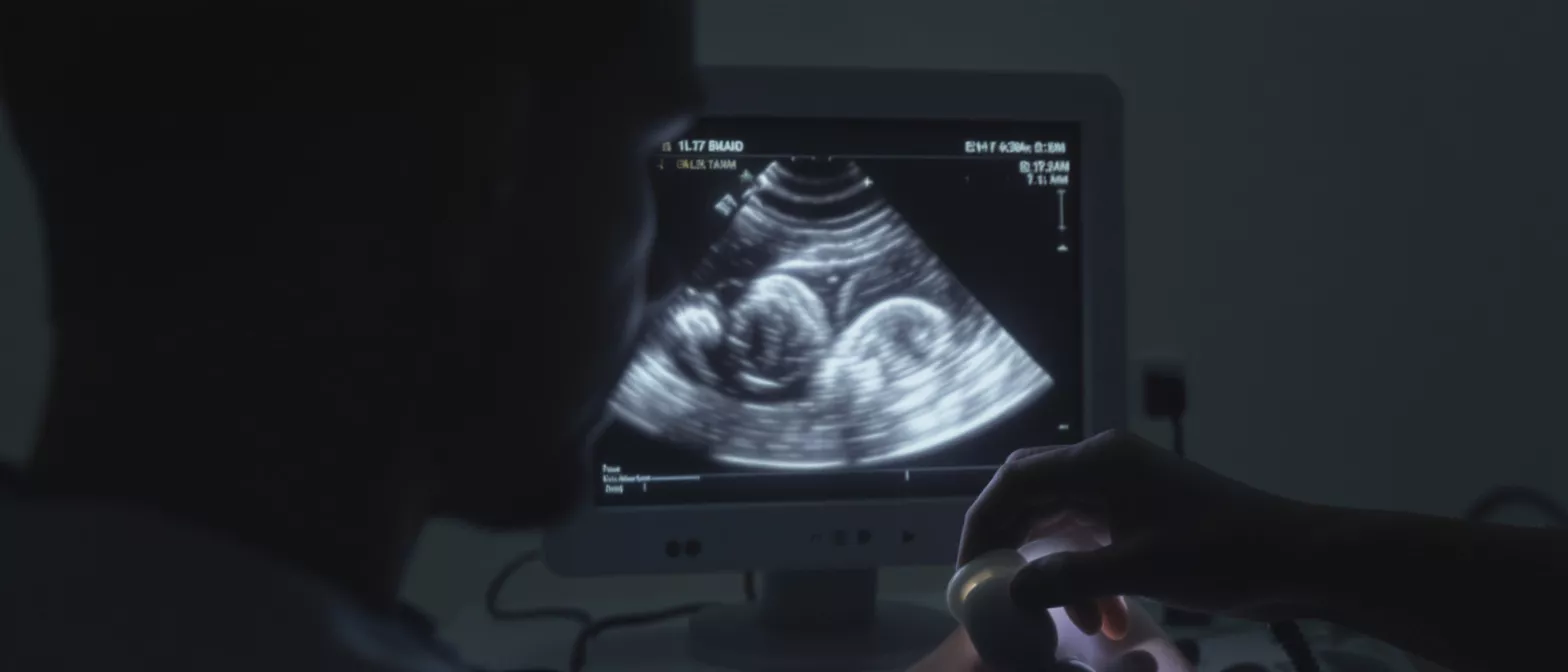
Annual Echocardiogram
Learn how an annual echocardiogram works and why heart monitoring matters after surgery.
What is an annual echocardiogram
Defining the diagnostic test: An annual echocardiogram is a non-invasive medical test that uses high-frequency sound waves to create detailed moving pictures of your heart. By bouncing sound waves off the structures inside your chest, this technology allows cardiologists to see your heart muscle, valves, and chambers in real time. It provides a completely safe and painless way to look at how well your heart is functioning without using any radiation.
Why heart surgery patients need yearly monitoring: If you have previously undergone heart surgery, your cardiovascular system requires ongoing observation to ensure that the surgical repairs remain effective. A yearly heart screening acts as a preventive measure, allowing your medical team to catch subtle changes in your heart's structure or pumping ability before you ever feel any symptoms. This proactive approach is crucial for long-term cardiac health.
Regular monitoring builds a baseline of your heart's unique anatomy and function. When you visit a specialized cardiac care center for your routine imaging, doctors compare your current results to previous years. This ongoing record helps them identify even the smallest alterations in your heart's performance, ensuring that any post-surgical complications are addressed early.
How does an echocardiogram work
The science behind sound wave imaging: During a heart ultrasound screening, a trained technician applies a small amount of warm gel to your chest and moves a handheld device called a transducer over your skin. This device emits sound waves that travel through your tissues and bounce back when they hit solid structures like heart valves and muscle walls. The machine translates these returning echoes into precise visual images on a monitor.
What the echo heart test shows your doctor: This diagnostic tool reveals critical information about your cardiovascular system. It shows the size and shape of your heart, the thickness of your muscle walls, and how well your valves open and close. Most importantly, it measures the pumping strength of your left ventricle, which tells doctors exactly how effectively your heart circulates blood to your body.
The entire procedure is completely painless and requires no needles or incisions. The gel used during the test simply helps eliminate air between the transducer and your skin, providing a clear path for the sound waves to travel. Understanding this simple mechanism often helps anxious patients feel much more comfortable during their appointment.
Why do I need a yearly heart screening
Monitoring heart valve function post-surgery: Over time, surgically repaired or replaced heart valves can experience wear or develop new issues. A heart valve ultrasound performed annually ensures that your valves continue to open fully and close tightly. Detecting a slow decline in valve performance early allows your surgeon to intervene before serious symptoms develop.
Tracking heart muscle strength and pumping capacity: After a major cardiac operation, the heart muscle needs time to heal and adapt. Annual heart imaging tests measure your ejection fraction, which is the percentage of blood your heart pushes out with each beat. Tracking this number yearly confirms that your heart muscle remains strong and capable of meeting your body's demands.
Consistent monitoring provides peace of mind for both you and your family. Knowing that your heart is healing properly and functioning as expected relieves the anxiety that often accompanies recovery. It transforms your follow-up care from a reactive process into a structured, confident health strategy.
How to prepare for an echocardiogram procedure
Simple steps before your appointment: Preparing for an echocardiogram procedure requires very little effort. You can eat and drink normally before the test, and you should continue taking your prescribed medications unless your doctor specifically tells you otherwise. The only physical requirement is removing clothing from the waist up and wearing a medical gown for the exam.
What to wear and bring to the clinic: Wear comfortable, loose-fitting clothing that is easy to remove from the upper body. Leave valuable jewelry at home, as you will need to take off necklaces and remove clothing from the waist up. Bring your identification, a list of your current medications, and any previous imaging records if you are visiting a new specialized heart clinic for the first time.
Patients often feel nervous about medical appointments, but echocardiogram preparation is incredibly straightforward. Because the test is non-invasive and does not require anesthesia, there is no need to arrange for transportation home afterward. You will be able to drive yourself and immediately resume your normal daily activities.
What to expect during a cardiac ultrasound exam
The actual test experience: During a cardiac ultrasound exam, you will lie comfortably on an examination table on your left side. The technician will attach small sticky patches called electrodes to your chest to monitor your heart rhythm simultaneously. They will then apply the warm gel and move the transducer wand gently across your skin to capture different views of your heart.
Duration and physical sensations during the test: A standard echo heart test typically takes about thirty to forty-five minutes to complete. You will feel slight pressure from the transducer as the technician presses firmly to get clear images, especially if they need to view your heart from beneath your ribs. You may hear a swooshing sound during the procedure, which is simply the audio representation of your blood flowing through your heart valves.
- Comfortable positioning: Lying on your left side brings your heart closer to the chest wall for clearer imaging.
- Electrode placement: Sticky patches connect to an ECG machine to time the ultrasound images with your heartbeat.
- Warm gel application: The gel improves sound wave transmission and does not harm your skin or clothing.
- Doppler sound: Hearing your blood flow is normal and helps the technician assess valve health.
Understanding your echo test results
How cardiologists interpret the findings: When you review your echo test results, your cardiologist looks at several key metrics to evaluate your heart health. They assess the size of your heart chambers, the thickness of your muscle walls, and the movement of your valves. These numbers tell a comprehensive story about how well your heart handles its daily workload.
Key measurements in your diagnostic report: Your report will contain specific medical terms that describe your heart's function. Understanding these terms helps you actively participate in your cardiac care. A comprehensive cardiovascular diagnostic report will clearly outline these measurements for your referring physician.
- Ejection Fraction (EF): Measures the percentage of blood pumped out with each beat, indicating muscle strength.
- Valve Function: Evaluates whether valves open fully and close tightly to prevent backward blood flow.
- Chamber Size: Checks for enlargement or thickening of the heart walls, which can indicate high blood pressure or valve disease.
- Diastolic Function: Assesses how well the heart muscle relaxes and fills with blood between beats.
How much does an echocardiogram cost
Factors influencing the price of a heart imaging test: The echocardiogram cost varies depending on several factors, including the facility type, your geographic location, and whether you have health insurance. Hospital-based imaging centers typically charge more than private cardiology clinics. The specific type of echo performed, such as a standard transthoracic echo versus a stress echo, also impacts the final price.
Navigating insurance and payment options: Most major health insurance plans cover a yearly heart screening when deemed medically necessary by your physician. If you are paying out of pocket, many clinics offer transparent pricing or payment plans. Always ask the billing department for a complete cost breakdown before scheduling your procedure to avoid unexpected charges.
For international patients or those traveling for specialized care, understanding the cost structure is especially important. Facilities often provide dedicated international patient coordinators who can quote exact prices for your cardiac diagnostic test. This allows you to budget accurately for your ongoing heart health management.
Is an annual heart check up safe
Safety profile of ultrasound technology: An annual heart check up using echocardiography is completely safe for patients of all ages. Unlike X-rays or CT scans, this imaging method uses no ionizing radiation. It relies entirely on harmless sound waves to create its images, meaning you can safely repeat this test every year without accumulating radiation exposure over your lifetime.
Risks versus benefits of yearly monitoring: There are no known risks or side effects associated with a standard transthoracic echocardiogram. The benefits of annual monitoring far outweigh the minimal effort required to attend the appointment. Early detection of structural deterioration or valve dysfunction prevents severe emergencies, making this simple test a powerful tool for protecting your long-term health.
Because the procedure requires no incisions, contrast dye, or anesthesia, the risk of infection or allergic reaction is zero. You will not need recovery time, and you can safely drive yourself home immediately after the technician removes the electrodes and wipes away the gel.
Finding an echocardiogram near me
Choosing the right facility for your heart care: When searching for an echocardiogram near me, it is vital to select a facility with modern equipment and experienced cardiac technicians. Look for clinics affiliated with reputable cardiac surgeons and specialized cardiovascular centers. The quality of the images depends heavily on both the ultrasound machine and the skill of the person operating it.
Why international patients choose Guadalajara for cardiac care: Many patients travel to Mexico for high-quality, accessible cardiac evaluations.
When evaluating facilities, consider the accessibility of the clinic, the availability of follow-up consultations, and the communication style of the medical team. A good facility will guide you through the entire process, ensuring your images are promptly reviewed by a board-certified cardiologist who can explain your results clearly.
Next steps after your annual cardiac screening
Reviewing results with your cardiologist: After your annual cardiac screening, a cardiologist will review your images and compile a detailed report. You should schedule a follow-up appointment to discuss these results in person. This consultation is your opportunity to ask questions about any changes since your last visit and to understand how your heart is adapting post-surgery.
Creating a long-term heart health strategy: Your echo results help your medical team adjust your treatment plan. If the test shows perfect heart function, you can continue your current medications and lifestyle habits with confidence. If the screening reveals minor changes, your doctor might adjust your medications or recommend specific lifestyle modifications to support your cardiovascular system.
Your yearly heart imaging test is a critical checkpoint in your lifelong cardiac care journey. By committing to regular screenings, you take an active role in preserving your heart health and ensuring the longevity of your previous surgical repairs. Consistent monitoring, combined with expert medical guidance, empowers you to live a full and healthy life.






