

Knowing your heart: the aortic valve
Learn how aortic valve disease affects your heart and explore symptoms, diagnosis, and treatment options from experts in Guadalajara.
What is the aortic valve and what does it do?
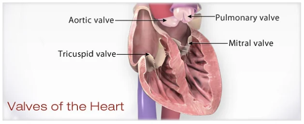
The aortic valve is one of the four essential gates inside your heart, acting as a critical passageway between the left ventricle and the aorta, the largest artery in your body. Every time your heart beats, this valve opens to allow oxygen-rich blood to flow out to your brain and body, and then closes tightly to prevent any blood from slipping backward. When this valve is healthy, it operates seamlessly, making a quiet but vital contribution to your circulatory system.
Understanding this structure is the first step in grasping heart valve disease. The valve itself is made of three thin leaflets, or cusps, that open and close in perfect synchrony with each heartbeat. Because the left ventricle is the strongest chamber of the heart, pumping blood to the entire body, the aortic valve must withstand immense pressure every single second of your life.
When a patient hears they have a heart murmur, it often means the physician has detected a slightly altered sound caused by blood moving through a valve that is not opening or closing perfectly. For those exploring heart valve replacement options, understanding the specific mechanics of this valve helps clarify why certain interventions are necessary when it fails to function properly.
What are the common aortic valve disease types?
Aortic valve disease primarily manifests in two distinct forms: stenosis and regurgitation. Stenosis occurs when the valve leaflets become stiff, calcified, or fused together, narrowing the valve opening and making it significantly harder for the heart to push blood through. Over time, this narrowing forces the heart muscle to work much harder, leading to thickening of the ventricle wall.
Differentiating stenosis from regurgitation: While stenosis is a problem of restricted forward flow, aortic valve regurgitation is a problem of backward leakage. In a leaky aortic valve, the leaflets do not close completely, allowing blood that has already been pumped into the aorta to fall back into the left ventricle. This creates a cycle of inefficiency, forcing the heart to pump the same blood twice.
Both conditions place an extraordinary burden on the heart, though they do so through entirely different mechanical failures. Regardless of whether the valve is narrowed or leaking, the ultimate result is a heart that is overworked, which can eventually lead to heart failure if left untreated. Recognising the specific type of disease is crucial for determining the correct surgical or medical approach.
How does aortic valve stenosis develop over time?
Aortic valve stenosis is typically a slow, progressive condition that takes many years to become severe. The most common cause in older adults is calcification, where calcium deposits build up on the valve leaflets over decades. This is not the same calcium build-up associated with dietary calcium or a poor diet, but rather a natural inflammatory process that occurs as we age, similar to the way pipes might gather rust over time.
Rheumatic fever and congenital factors: In younger patients, aortic stenosis might be the result of rheumatic fever, a complication of untreated strep throat that causes inflammation and long-term scarring of the valve. Alternatively, some people are born with a bicuspid aortic valve, meaning they have only two leaflets instead of three, which predisposes them to early calcification and narrowing.
Because the narrowing happens so gradually, the heart initially compensates for the restricted blood flow without producing any noticeable symptoms. This long, silent phase is what makes regular cardiac check-ups essential, particularly as we age, so that the progression can be monitored before the heart muscle suffers irreversible damage.
What causes a leaky aortic valve?
Aortic valve regurgitation, commonly referred to as a leaky aortic valve, can be caused by several factors that stretch, damage, or perforate the valve leaflets. One major cause is an enlargement of the aortic root, the part of the aorta closest to the heart. When the aorta dilates, it pulls the valve leaflets apart, preventing them from meeting cleanly in the middle to seal the chamber.
Endocarditis, a serious infection of the heart valves, can also destroy valve tissue and create holes in the leaflets, leading to severe, rapid leakage. Additionally, structural issues such as a congenital bicuspid valve can lead to regurgitation over time, as the abnormal leaflets endure higher stress and wear down or prolapse.
Exploring minimally invasive cardiac surgery techniques is often the next step when a patient discovers their leaky valve is causing the heart to enlarge or pump inefficiently. Your surgical team will assess the underlying cause of the leak to determine whether the valve can be safely repaired or if it requires a full replacement.
What are the silent aortic valve symptoms?
One of the most challenging aspects of heart valve disease is that it often remains completely asymptomatic for years, a period sometimes called the latent or silent phase. During this time, the heart is working harder than normal to compensate for the diseased valve, but the patient feels entirely normal. This is why the disease is so frequently discovered only after a doctor hears a murmur through a stethoscope during a routine exam.
When the heart finally loses its ability to compensate, patients typically begin to experience a recognizable set of warning signs. These aortic valve symptoms often include progressive shortness of breath, initially only with heavy exertion but eventually during routine activities. Other common complaints are unusual fatigue, a feeling of tightness or pressure in the chest, and dizziness or fainting spells, particularly when standing up or exercising.
Recognising aortic stenosis symptoms early:If you begin to notice chest pain, breathlessness, or dizzy spells that were not there before, it is a sign that the compensatory phase has ended. The heart is now struggling to maintain adequate blood flow to the body, and these symptoms indicate that the valve disease has reached a critical stage that requires immediate medical evaluation.
How is aortic valve diagnosis performed?
Diagnosing aortic valve disease requires a combination of a thorough physical examination and advanced imaging technologies. The process usually begins with a doctor listening to the heart with a stethoscope; the sound of turbulent blood flow, known as a heart murmur, is often the first indicator of a valve problem. To confirm the diagnosis, an echocardiogram is ordered, which uses ultrasound waves to create detailed moving pictures of the heart's structures.
The echocardiogram allows cardiologists to see exactly how the valve leaflets are moving, measure the size of the valve opening, and estimate the severity of any leakage. Further diagnostic tools may include an electrocardiogram (ECG) to check the heart's electrical activity, a chest X-ray to see if the heart is enlarged, and sometimes a cardiac CT scan or MRI for highly detailed cross-sectional images of the heart and aorta.
Patients often benefit greatly from expert cardiac catheterisation insights when non-invasive tests leave questions about the coronary arteries or the exact pressure gradients across the valve. This procedure involves threading a thin tube into the heart to take direct pressure measurements, providing definitive data that helps surgeons plan the safest, most effective intervention.
What are the best aortic valve treatment options?
Aortic valve treatment options range from careful, watchful waiting to complex surgical interventions, entirely depending on the severity of the disease and the patient's overall health. In the early, asymptomatic stages, a doctor may simply recommend regular monitoring with echocardiograms to track the progression of the stenosis or regurgitation. Medical therapy can also help manage mild symptoms, though it cannot fix the underlying structural valve problem.
Weighing surgical vs. transcatheter approaches: When the valve disease becomes severe and symptoms appear, surgery is usually unavoidable. Open-heart surgery allows the surgeon to directly repair or replace the damaged valve, and it remains the gold standard for long-term durability and versatility. However, transcatheter procedures like TAVR offer a viable alternative for patients who are at high risk for traditional open surgery.
- Active surveillance: Monitoring mild valve disease with regular ultrasounds to catch progression early.
- Open repair/replacement: The traditional method allowing full visual access for complex valve corrections.
- Transcatheter (TAVR): A less invasive option threaded through a blood vessel, ideal for high-risk surgical candidates.
How does aortic valve replacement surgery work?
When repair is not possible, aortic valve replacement involves removing the diseased valve and sewing in a prosthetic substitute. There are two main types of prostheses: mechanical valves and biological valves. Mechanical valves are crafted from highly durable materials like titanium and carbon, designed to last a lifetime, but they require the patient to take blood-thinning anticoagulant medications permanently to prevent clots from forming on the metal.
Biological valves, on the other hand, are made from animal tissue, typically from a pig or a cow, or occasionally from human donor tissue. These valves function very similarly to natural human valves and generally do not require long-term blood thinners. The main trade-off is durability; biological valves gradually wear out and typically need to be replaced after twelve to fifteen years.
Choosing the right prosthesis is a highly personal decision that depends on the patient's age, lifestyle, and tolerance for lifelong medication. Younger patients often receive mechanical valves to avoid multiple surgeries over their lifespan, while older patients frequently opt for biological valves to avoid the risks associated with anticoagulation therapy.
Are minimally invasive aortic valve surgeries safe?
Minimally invasive aortic valve surgeries have been developed to achieve the same surgical goals as open-heart surgery but with smaller incisions and less trauma to the body. Instead of a full sternotomy, where the entire breastbone is split, surgeons use a small incision, often around five centimeters, to access the heart. Specialized 3D cameras and fine instruments allow the surgeon to perform the repair or replacement with exceptional precision.
Recovery and surgical safety profiles: Because the chest is not fully opened, patients generally experience less postoperative pain, require fewer blood transfusions, and enjoy a noticeably faster return to normal daily activities. The safety profile of these techniques is excellent when performed by experienced surgical teams, though not every patient is a candidate based on their individual anatomy.
Transcatheter aortic valve replacement, or TAVR, pushes the minimally invasive concept even further by avoiding surgery altogether. A replacement valve is compressed into a catheter, threaded through an artery in the groin, and expanded directly inside the old, diseased valve. This approach is a major breakthrough for elderly or frail patients who simply cannot withstand the physiological stress of traditional surgery.
What to expect during aortic valve recovery?
Recovery after aortic valve surgery is a gradual process that requires patience, dedication, and a structured cardiac rehabilitation program. Immediately after the procedure, patients typically spend a day or two in the intensive care unit for close monitoring, followed by a few days in a regular hospital room to regain strength. Most patients are discharged from the hospital within a week, though the full recovery at home takes several weeks to a few months.
During the first few weeks at home, patients are encouraged to walk daily and gradually increase their physical activity, while strictly avoiding heavy lifting or strenuous activity that could strain the healing breastbone. Cardiac rehabilitation, a medically supervised exercise and education program, is crucial for safely rebuilding cardiovascular fitness and confidence. Follow-up appointments and echocardiograms are scheduled to ensure the new valve is functioning perfectly and the heart is healing well.
Ultimately, most patients experience a profound improvement in their quality of life and life expectancy once they have fully recovered. The fatigue and breathlessness that plagued them before the surgery vanish as the heart can finally pump efficiently again, allowing them to return to the activities and lifestyle they love.






