

Autograft
Learn how an autograft uses your own tissue for heart valve repair, reducing rejection risk.
What is an autograft in cardiac surgery?
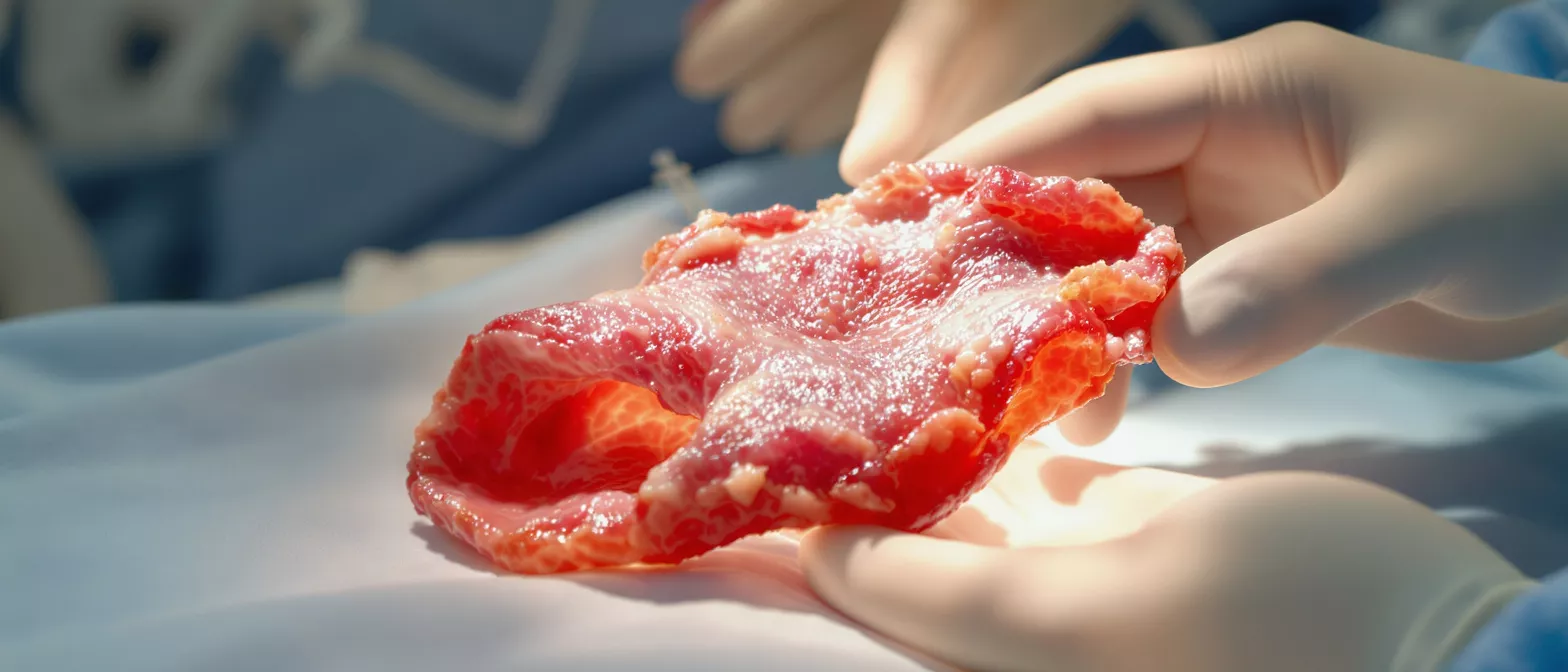
When you first hear the term autograft in the context of heart surgery, it can sound highly technical and intimidating. However, the concept is straightforward and remarkably natural: an autograft is tissue that your surgeon takes from one part of your own body and transplants to another area that needs repair. In cardiac procedures, this usually means moving a healthy valve or blood vessel to replace a damaged one. Because the tissue comes from you, it is genetically identical to the rest of your body, which eliminates the risk of biological rejection.
Why this matters for a cardiac-surgery patient: Your heart has four valves that keep blood flowing in the correct direction, and sometimes these valves become diseased or leak. Instead of using an artificial mechanical valve or tissue from an animal or human donor, your surgeon might use your own tissue to fix the problem. This approach uses your body's natural materials to restore normal heart function, offering a biological solution that integrates seamlessly with your living system.
Understanding this autograft definition in cardiology helps reduce the fear of the unknown. Knowing that your surgical team can use your own healthy tissue to rebuild your heart provides reassurance that the repair is both natural and durable. It is a well-established technique that cardiovascular specialists have refined over decades, offering a safe and effective option for many patients facing valve or vascular repair.
How does an autograft heart procedure work?
The autograft procedure steps are carefully planned and executed with extreme precision. The most famous cardiac autograft technique is the Ross procedure, primarily performed on younger patients or children with a diseased aortic valve. During this operation, the surgeon removes your healthy pulmonary valve—the one controlling blood flow from your heart to your lungs—and uses it to replace your damaged aortic valve, which controls blood flow to the rest of your body.
How the surgical transfer happens: Once the surgeon relocates your healthy pulmonary valve to the aortic position, they must replace the pulmonary valve they just removed. This is where a homograft, or a donated human tissue valve, is placed in the pulmonary position. It is a two-part repair: your strongest living tissue goes to the most demanding, high-pressure area of your heart, while the donor tissue comfortably handles the lower-pressure pulmonary circuit.
This complex but highly effective autograft cardiac procedure requires an experienced surgical team. The benefit of this approach is that your relocated valve continues to live and grow within your heart. It functions exactly as it did in its original location, responding dynamically to your body's changing demands for blood flow without requiring the permanent use of blood thinners.
What are the main autograft benefits for heart repair?
The autograft benefits are significant, particularly for patients looking for a long-lasting, natural solution. Because the transplanted tissue is your own, your immune system recognizes it as self. This means you do not need to take powerful immunosuppressant medications, which are typically required when receiving organs from donors to prevent rejection. Your body accepts the relocated tissue immediately and begins integrating it into the surrounding heart structure.
Living tissue that grows and adapts: One of the most remarkable autograft benefits is that the transplanted valve or vessel remains alive. For younger patients, this means the autograft can grow alongside the rest of their body as they mature. Even in adults, the living tissue adapts to physiological changes, maintaining excellent flexibility and blood flow dynamics that mechanical valves simply cannot replicate.
- No rejection risk: Because the tissue is your own, your immune system leaves it alone.
- Growth potential: The living valve can grow with the patient, making it ideal for younger recipients.
- Excellent blood flow: The natural valve opens and closes smoothly, mimicking native heart function perfectly.
When is an autograft valve replacement necessary?
An autograft valve replacement becomes a primary consideration when a patient has a severely damaged aortic valve but possesses a perfectly healthy pulmonary valve. The aortic valve faces the highest pressure in the heart, and when it fails, symptoms like severe fatigue, chest pain, and shortness of breath occur. If a surgeon determines that an artificial mechanical valve is not the best fit—perhaps due to a patient's age or reluctance to take lifelong blood thinners—an autograft is a powerful alternative.
Matching the patient to the procedure: This specific autograft tissue transplant is most commonly recommended for younger adults, teenagers, and children. The reasoning is twofold: younger patients have a longer life expectancy, and a living autograft can last much longer than traditional animal tissue valves in this demographic. Furthermore, because younger patients are still physically active, the hemodynamic performance of a living valve provides a significant quality-of-life advantage.
Older patients might also be candidates, though the decision depends on overall health, anatomy, and surgical risk. Your cardiologist will evaluate the condition of your pulmonary valve through detailed echocardiograms before recommending you for this type of autograft cardiac procedure. Ensuring the donor valve within your own body is pristine is the most critical factor in surgical planning.
Autograft vs allograft vs xenograft in cardiology
Understanding the difference between an autograft vs allograft and a xenograft is essential for grasping why your surgeon chooses one method over another. An autograft, as discussed, comes from your own body. An allograft, sometimes called a homograft, is healthy tissue taken from a deceased human donor. A xenograft is tissue sourced from a different species entirely, such as a pig or cow, which has been chemically treated to make it safe for human use.
The clinical trade-offs: Each type of graft has a specific role in cardiac surgery. Autografts offer superior longevity and growth potential but require a more complex double-valve surgery. Allografts are excellent for replacing infected valves or reconstructing heart structures, as they resist infection well, but they do not last forever and do not grow with the patient. Xenografts are readily available and do not require immunosuppression, but they tend to wear out faster than living autografts.
The choice between an autograft vs xenograft often comes down to patient age and lifestyle. If you want to avoid blood thinners and prioritize long-term durability, and if your anatomy supports it, the autograft is usually the preferred biological option. Your surgical team will weigh these factors carefully to ensure you receive the best tissue type for your specific heart condition.
What is the autograft healing process and recovery time?
The autograft healing process begins the moment your surgery is complete. Because the relocated tissue is alive and native to your body, it immediately starts receiving nutrients from your coronary blood supply. Unlike synthetic materials that the body merely tolerates, an autograft integrates at the cellular level, forming new connections with surrounding heart tissue and maintaining its natural structural integrity over time.
What to expect during recovery: The standard autograft recovery time in the hospital is similar to other major open-heart procedures, typically requiring a stay of about a week. During this time, the medical team monitors your heart rhythm, blood pressure, and the function of your newly transplanted valve. Once home, the first few weeks focus on resting, managing incision pain, and slowly increasing physical activity under supervised cardiac rehabilitation.
Full recovery takes several months, but the transplanted tissue itself heals remarkably fast because it is living and vascularized. You will have regular follow-up echocardiograms so your cardiologist can watch the autograft adapt to its new high-pressure environment. Most patients find that once the surgical trauma subsides, their heart functions as if it had never been diseased, allowing them to return to normal activities without the restrictions associated with mechanical valves.
Understanding the autograft donor site and tissue engineering
When discussing an autograft, it is important to remember that you are both the recipient and the donor. The autograft donor site is the area of your body from which the surgeon harvests the healthy tissue. In the context of the Ross procedure, the donor site is the pulmonary valve position. Once the healthy valve is moved to the aortic position, the donor site must be reconstructed, usually with a human donor valve, to maintain proper blood flow to the lungs.
The role of future biological innovations: While current procedures move tissue from one anatomical spot to another, the field of autograft tissue engineering is rapidly evolving. Scientists are exploring ways to take a small sample of a patient's own cells and grow them in a laboratory to create entirely new heart valves. Although still largely in the experimental stages, this approach promises to eliminate the need for a secondary donor valve entirely.
For now, the traditional autograft remains a testament to the body's ability to heal itself with surgical assistance. The careful management of the donor site is just as crucial as the placement of the autograft itself. Surgeons must ensure that the replacement tissue at the donor site is perfectly sized and functional, creating a balanced system where both the aortic and pulmonary circuits operate harmoniously.
What are the potential autograft complications?
While using your own tissue offers profound advantages, it is vital to understand the potential autograft complications. The most significant risk is not rejection, since the tissue is your own, but rather the structural changes that can occur over time. The pulmonary valve, which is designed for the low-pressure environment of the lungs, is suddenly tasked with handling the high-pressure output of the aorta, which can sometimes lead to valve dilation or leakage years down the line.
Risks of the dual procedure: Because an autograft valve replacement usually involves operating on two valves instead of one, the surgery takes longer and carries the standard risks associated with extended time on a heart-lung bypass machine. These risks include bleeding, infection, and adverse reactions to anesthesia. However, in the hands of an experienced cardiac surgeon, these risks are meticulously managed and remain quite low.
Long-term autograft complications might also involve the homograft used to replace the pulmonary valve. Over decades, this donor tissue may wear out and require a percutaneous valve replacement via a catheter. Patients must remain vigilant with annual cardiac check-ups so their care team can catch and address any minor changes before they become significant issues.
What are the long-term autograft success rates?
The autograft success rates are generally excellent, particularly in specialized centers that frequently perform the Ross procedure. Many patients enjoy decades of normal heart function without needing additional surgeries. Because the living autograft tissue remains supple and grows with the patient, it provides a durability that far exceeds traditional biological valves, making it a highly effective long-term solution for younger demographics facing valve disease.
Factors that influence longevity: The success of an autograft cardiac procedure depends heavily on surgical technique and the patient's individual anatomy. Surgeons use various techniques to reinforce the new aortic valve to prevent the dilation that can sometimes occur under high pressure. When performed correctly, the autograft acts like a native valve, providing quiet, efficient blood flow that allows patients to live vigorous, unrestricted lives.
- High durability: Living tissue lasts significantly longer than animal valves in younger patients.
- No blood thinners: Patients avoid the bleeding risks and lifestyle restrictions of anticoagulation therapy.
- Active lifestyle: The valve tolerates vigorous exercise and pregnancy exceptionally well.
Autograft definition cardiology and future trends
To summarize the autograft definition in cardiology, it is the ultimate biological repair strategy, utilizing your body's own resources to heal your heart. By transferring a healthy valve or blood vessel to replace a failing one, surgeons provide a solution that lives, breathes, and grows with you. It is a procedure that demands high surgical expertise but rewards the patient with unparalleled quality of life and freedom from long-term medication.
The future of cardiac tissue repair: As cardiac surgery advances, the principles of the autograft continue to inspire new techniques. The push toward regenerative medicine and tissue engineering aims to harness the body's healing power even further. While the current autograft vs allograft debate will continue based on patient-specific needs, the foundational truth remains that your own tissue is the best material your heart can use to rebuild itself.
Ultimately, an autograft represents hope and resilience. If you or a loved one is facing a valve replacement, discussing the possibility of an autograft with your cardiologist could open the door to a highly natural, durable recovery. Understanding the procedure, the benefits, and the realistic recovery time empowers you to face surgery with confidence, knowing your heart has the remarkable ability to heal itself with the right surgical guidance.






