

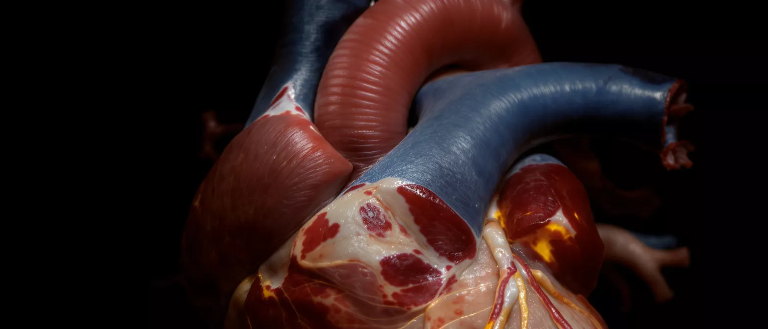
Circumflex Artery
Understand the circumflex artery, its anatomy, function, and role in heart health.
What Is the Circumflex Artery?
The circumflex artery is a major blood vessel that wraps around the back of the heart, playing a critical role in supplying oxygen-rich blood to the cardiac muscle. As a key branch of the left main coronary artery, it works alongside the left anterior descending artery to ensure the heart receives the nutrients it needs. Understanding this vessel is essential for patients preparing for cardiac surgery, as its health directly impacts overall heart function.
Where is the circumflex artery located? It originates at the bifurcation of the left main coronary artery, curving around the left side of the heart in the coronary sulcus. It travels posteriorly, eventually reaching the back of the organ where it gives off various terminal branches. Because of its pathway, it is often referred to as the left circumflex artery (LCX).
What is the circumflex artery medical definition? In clinical terms, it is defined as the branch of the left coronary artery that runs in the atrioventricular groove. It is responsible for providing the arterial supply to the left atrium and the posterolateral wall of the left ventricle. Recognizing this definition helps patients better understand their surgical plans and recovery needs.
How Circumflex Artery Anatomy Supports Heart Function
Circumflex artery anatomy is uniquely designed to support the continuous, heavy workload of the heart. By curving around the lateral and posterior aspects of the heart, the vessel ensures that the left ventricle—the main pumping chamber—receives a steady stream of oxygenated blood. Without this blood flow, the heart muscle cannot effectively pump blood to the rest of the body.
Why does the left circumflex artery anatomy vary between patients? Human hearts are not identical, and the anatomical dominance of the coronary circulation varies from person to person. In most individuals, the right coronary artery supplies the inferior wall, but in about 15% of the population, the circumflex artery is dominant. This means it supplies the posterior descending artery, a crucial detail for surgeons to map out before any procedure.
This anatomical variation is why detailed preoperative imaging is so important. Identifying whether a patient has a right-dominant or left-dominant system helps the surgical team anticipate the exact blood flow patterns and plan interventions that protect the heart muscle during operations. Knowing this helps patients feel more confident about their personalized care plan.
What Does the Left Circumflex Artery Supply?
The primary role of the left circumflex artery is to provide blood to specific regions of the heart that are vital for coordinated pumping. It primarily supplies the left atrium, which receives oxygenated blood returning from the lungs, and the lateral and posterior walls of the left ventricle. This circumflex artery supply ensures the heart muscle remains viable and functional.
What happens if circumflex artery blood flow is reduced? When the blood flow through this vessel is compromised, the affected heart muscle can become ischemic, meaning it is starved for oxygen. Patients might experience angina, or chest pain, particularly during physical exertion when the heart demands more oxygen than the narrowed artery can deliver. Over time, chronic reduction in flow can weaken the cardiac tissue.
Understanding the specific territory this artery covers helps explain why blockages in this area can be so serious. The lateral wall of the left ventricle is crucial for maintaining adequate blood pressure and cardiac output. Protecting cardiac health monitoring devices becomes part of a lifelong strategy for patients who have experienced compromised flow in this region.
Types of Circumflex Artery Branches
As the circumflex coronary artery wraps around the heart, it gives off several important branches that supply distinct regions of the cardiac muscle. These branches ensure that blood reaches all the necessary areas of the lateral and posterior heart walls. The most notable of these are the obtuse marginal arteries, which provide the primary blood supply to the left ventricle's lateral wall.
What is the obtuse marginal artery? The obtuse marginal artery is the largest branch of the LCX artery, running along the outer curvature of the heart. It is critical because it supplies the thick muscular wall of the left ventricle, which generates the force needed to pump blood throughout the body. Surgeons pay close attention to this branch during bypass operations to ensure complete revascularization.
Depending on the individual's anatomy, the circumflex artery may also give rise to the posterior descending artery if the patient has a left-dominant coronary system. This vast network of branches means that maintaining the health of the main trunk is vital for preserving the downstream blood supply to the heart.
- Obtuse Marginal Arteries: Supply the lateral wall of the left ventricle.
- Sinus Node Artery: Supplies the sinus node, the heart's natural pacemaker, in a small percentage of patients.
- Posterolateral Arteries: Branches that supply the back of the left ventricle and help coordinate pumping.
What Causes Circumflex Artery Stenosis?
Circumflex artery stenosis occurs when the artery narrows, restricting blood flow to the heart muscle. The primary cause is atherosclerosis, a condition where fatty plaques build up inside the arterial walls. Over time, these plaques harden and narrow the vessel, making it difficult for blood to pass through freely.
How does plaque affect circumflex artery disease? Plaque does not just physically narrow the vessel; it also creates turbulent blood flow that can lead to clot formation. If a plaque ruptures, it can trigger a sudden blockage, which is a common pathway for heart attacks. Recognizing and treating stenosis early is critical to preventing these acute events.
Risk factors for developing this narrowing include high cholesterol, high blood pressure, smoking, and diabetes. Patients preparing for heart surgery often have these risk factors managed aggressively to prevent the progression of stenosis. Adopting a heart-healthy lifestyle is a fundamental part of protecting the LCX artery and preventing future complications.
Circumflex Artery Occlusion and Heart Attack Risk
While stenosis refers to a narrowing, circumflex artery occlusion is a complete blockage of the vessel. A total occlusion prevents any blood from reaching the downstream heart muscle, leading to a myocardial infarction, commonly known as a heart attack. Because the circumflex artery supplies the lateral wall of the left ventricle, an occlusion here can significantly impair the heart's pumping ability.
Why is circumflex artery occlusion sometimes called a "silent" heart attack? Occlusions in the circumflex artery can sometimes present with atypical symptoms compared to blockages in the front of the heart. Patients may experience discomfort in the back, jaw, or neck rather than classic crushing chest pain. This atypical presentation makes diagnosis challenging and highlights the need for thorough medical evaluation.
When an occlusion occurs, rapid intervention is vital to restore circumflex artery blood flow. Cardiac surgeons and interventional cardiologists must work quickly to remove the blockage, often using advanced circulatory support equipment to stabilize the patient during the procedure. Time is muscle, and restoring flow quickly saves heart tissue.
Best Treatments for Circumflex Artery Disease
Treating circumflex artery disease involves a combination of lifestyle changes, medication, and surgical interventions. The best treatment depends on the severity of the blockage and the patient's overall health. For mild stenosis, doctors often prescribe medications to lower cholesterol, reduce blood pressure, and prevent clotting.
When is surgery needed for circumflex artery stenosis? When the vessel is severely narrowed or completely occluded, more invasive treatments are required. Angioplasty with stent placement is a common minimally invasive procedure where a small mesh tube is inserted to prop the artery open. This restores immediate blood flow and relieves symptoms of ischemia.
In cases where the blockage is complex or located in a difficult area, coronary artery bypass grafting (CABG) may be the preferred surgical route. During a bypass, surgeons use a healthy blood vessel from another part of the body to reroute blood around the blocked circumflex artery.
How Circumflex Artery Blood Flow Is Tested
Determining the health of the circumflex artery requires specialized diagnostic testing. The most direct method is a coronary angiogram, an imaging test where a special dye is injected into the bloodstream. X-rays are then used to track the dye as it moves through the heart's vessels, revealing any blockages or narrowing in the LCX artery.
What non-invasive tests show circumflex artery disease? Before moving to an angiogram, doctors often use non-invasive tests like a stress test or an echocardiogram. During a stress test, patients exercise while their heart is monitored to see if reduced blood flow causes any symptoms. An echocardiogram uses sound waves to create images of the heart, allowing doctors to see if the left ventricle wall is moving properly.
For patients preparing for cardiac surgery, these tests provide a complete map of the heart's plumbing. Understanding the exact location and severity of any circumflex artery blockage is crucial for the surgical team to plan their approach. Accurate testing ensures that every affected area of the heart is treated.
Are Circumflex Artery Blockages Dangerous?
Any blockage in the heart's blood supply is serious, and circumflex artery disease is no exception. Because the LCX artery supplies the left ventricle, a blockage here can lead to significant heart muscle damage if left untreated. The danger level depends largely on how quickly the blockage is addressed and whether the patient has developed collateral blood vessels to compensate.
Can you live with a partially blocked circumflex artery? Yes, many people live with mild to moderate stenosis without realizing it, especially if they have good collateral circulation. The body can sometimes grow tiny new blood vessels to bypass a slow-growing blockage. However, this does not cure the disease, and the underlying atherosclerosis must still be managed to prevent progression.
The risk arises when a partial blockage suddenly becomes a total occlusion. This is why regular check-ups with a cardiologist are essential for anyone diagnosed with heart artery blockages. Proactive management reduces the risk of a sudden cardiac event and protects the long-term health of the heart.
Recovering After Circumflex Artery Surgery
Recovery from surgery involving the circumflex artery is a gradual process that requires patience and commitment. Whether a patient has had a stent placed or a bypass graft performed, the immediate goal is to allow the heart to heal while preventing further blockages. Patients will typically spend time in the intensive care unit for close monitoring before moving to a regular hospital room.
What is the long-term outlook for circumflex artery disease? With proper treatment and lifestyle modifications, the long-term outlook is generally very positive. Patients are usually placed on a comprehensive cardiac rehabilitation program that includes supervised exercise, nutritional counseling, and medication management. Adhering to this plan helps keep the newly opened vessels clear and prevents future blockages.
Understanding the role of the circumflex artery demystifies much of the cardiac surgical process. By learning how this vessel supports the heart, what causes it to become diseased, and how it is repaired, patients can approach their recovery with confidence. A strong partnership between the patient and the medical team is the best defense against future heart problems.






