

Circumflex Artery Blockage: Symptoms and Treatment
Circumflex artery blockage symptoms and treatment explained by Guadalajara heart surgery specialists.
A circumflex artery blockage affects one of the most strategically important vessels supplying your heart muscle with oxygen-rich blood. The left circumflex artery wraps around the left side of your heart, nourishing the left Atrium and in many people the critical lateral wall of the left ventricle—the main pumping chamber. When plaque narrows or completely blocks this vessel, the consequences can range from subtle fatigue during ordinary activities to a life-threatening heart attack. Understanding circumflex artery blockage symptoms and treatment options empowers you and your family to act quickly and make informed decisions with your cardiac team.
The circumflex artery branches from the left main coronary artery, sharing its origin with the more famous left anterior descending artery. While public awareness often centers on blockages in the "widowmaker" artery, left circumflex artery blockage carries comparable risk and deserves equal attention. Anatomical variation means some individuals receive substantial blood supply to their heart's posterior wall from the circumflex rather than the right coronary artery, making this vessel even more vital in their particular heart architecture.
This article explains what happens when the circumflex becomes diseased, how doctors identify the problem, and which treatment paths—from medication to balloon Angioplasty to bypass surgery—fit different clinical situations. All information reflects established cardiovascular understanding; we encourage you to discuss your specific case with a qualified cardiac team rather than self-diagnose.
What Is a Circumflex Artery Blockage and Why Does It Matter?
The circumflex artery emerges from the left main coronary artery and travels within the Atrioventricular groove—the shallow groove separating the Atrium from the ventricles on the heart's left side. In its course, it gives off branches called obtuse marginals that penetrate the heart muscle itself, delivering oxygen and nutrients to the working myocardium. A circumflex artery blockage occurs when Atheromatous Plaque—fatty deposits mixed with inflammatory cells and calcium—accumulates within the vessel wall, progressively narrowing the channel through which blood must flow.
Acute versus chronic blockage defines your clinical experience. Chronic circumflex artery stenosis develops gradually over years, often allowing the heart to develop collateral circulation—tiny spontaneous bypass vessels that partially compensate for reduced flow. Acute circumflex artery occlusion, typically caused by plaque rupture and sudden clot formation, interrupts blood supply abruptly without warning. This distinction matters because chronic disease may cause predictable exertional symptoms, while acute occlusion frequently presents as a "silent" heart attack or with atypical symptoms that delay recognition and treatment.
Anatomical dominance determines how much heart muscle depends on the circumflex. Cardiologists classify coronary circulation as "left-dominant," "right-dominant," or "co-dominant" based on which artery gives rise to the posterior descending artery supplying the heart's inferior wall. In approximately 15% of people, the circumflex artery continues beyond its usual territory to become the posterior descending artery—making them left-dominant. For these individuals, circumflex artery blockage threatens not just the lateral wall but also the inferior wall and the electrical conduction system housed near the posterior septum. The pig: a model for human infectious diseases has informed researchers about anatomical similarities between human and porcine cardiovascular systems, though clinical relevance here centers on your individual coronary anatomy as visualized during cardiac catheterization.
The location of blockage along the circumflex also shapes prognosis. Proximal circumflex artery blockage—near its origin from the left main—affects all downstream branches and causes more extensive ischemia than distal disease limited to a single marginal branch. Your interventional cardiologist will map exact lesion location when planning treatment strategy.

What Are the Warning Signs of Circumflex Artery Blockage?
Circumflex artery blockage symptoms often prove frustratingly subtle compared to classic "Hollywood" heart attack presentations. Because the circumflex supplies the lateral and posterior heart walls rather than the anterior territory served by the left anterior descending artery, its ischemia frequently manifests atypically. Patients and families must recognize these less familiar patterns to avoid dangerous delays in seeking emergency care.
Exertional discomfort without classic crushing chest pain commonly signals circumflex ischemia. Many patients describe vague left-sided chest discomfort, unusual fatigue with ordinary activity, or pressure that radiates to the jaw, back, or arm—particularly the left Arm Pain pattern that cardiologists associate with lateral wall involvement. Some experience dyspnea (shortness of breath) as their predominant or sole symptom, representing "angina equivalent" where reduced cardiac output with exertion masquerades as a lung problem. This symptom spectrum explains why circumflex-related heart attacks carry higher misdiagnosis rates in emergency departments compared to anterior territory events.
Nausea, sweating, and unexplained weakness may constitute the entire presentation. The circumflex artery's territory includes portions of the sinoatrial node blood supply in many individuals, meaning its occlusion can trigger arrhythmias—irregular heart rhythms—that present as palpitations, dizziness, or syncope (fainting) rather than pain. Atrial Flutter and other supraventricular arrhythmias sometimes herald otherwise silent circumflex territory infarction. Women and individuals with diabetes especially report these non-pain predominant symptoms, though the pattern affects all demographics.
Key symptom patterns to monitor include:
- Effort intolerance: Previously manageable walks or stair climbing now cause disproportionate breathlessness or fatigue
- Postural symptoms: Discomfort worsening when lying flat and improving when sitting forward, suggesting posterior ischemia
- Episodic palpitations with weakness: Heart rhythm disturbances accompanying vague ill-defined malaise
Symptom stability provides crucial information. Stable symptoms predictably triggered by exertion and relieved by rest suggest chronic circumflex artery stenosis with preserved collateral reserve. Progressive symptoms occurring with decreasing activity levels, or symptoms at rest, indicate unstable disease requiring urgent evaluation. The transition from stable to unstable pattern may signal plaque rupture or thrombosis that mandates immediate medical attention.
How Does Circumflex Artery Blockage Differ from Other Coronary Blockages?
Not all coronary blockages behave identically, and understanding circumflex-specific characteristics helps patients grasp why their experience may diverge from friends or family members with other vessel disease. The circumflex artery's unique anatomical course, variable territory, and relationship to cardiac structures create distinct clinical signatures that influence both recognition and treatment.
The circumflex artery's location within the Atrioventricular groove places it near the mitral valve apparatus and the left Atrium. This proximity means severe circumflex disease can contribute to mitral regurgitation—leakage of the mitral valve—when ischemia weakens the papillary muscles anchoring the valve leaflets. Patients may notice worsening shortness of breath from this valve dysfunction even before recognizing classic angina symptoms. This mechanism differs from anterior territory disease, which more typically causes direct pump failure without primary valve involvement.
Electrical instability represents another circumflex-specific concern. The artery's branches supply the left Atrium and, in left-dominant systems, portions of the atrioventricular node. Circumflex artery occlusion therefore carries elevated risk of Atrial Flutter, atrial fibrillation, and heart block—dangerous rhythm disturbances requiring specific monitoring and intervention. While any myocardial infarction can trigger arrhythmias, circumflex events show particular predilection for these supraventricular and conduction abnormalities.
Diagnostic challenges compound these clinical distinctions. Standard electrocardiogram (ECG) findings of circumflex ischemia prove less reliable than for other territories. The characteristic ST-elevation pattern may appear subtly in posterior leads (V7-V9) rather than the standard 12-lead tracing, or may be entirely absent despite significant occlusion. This "STEMI equivalent" or NSTEMI presentation requires heightened clinical suspicion and urgent angiography rather than reassuring negative initial tests. Application of Collagen Scaffold in Tissue Engineering: Recent Advances and New Perspectives illustrates how biomedical research advances understanding of vascular healing, though clinical management of acute circumflex syndrome currently relies on established revascularization strategies rather than emerging scaffold technologies.
How Is Circumflex Artery Blockage Diagnosed?
Accurate diagnosis of circumflex artery blockage demands integration of clinical history, non-invasive testing, and ultimately invasive coronary angiography when indicated. The diagnostic pathway follows established cardiology protocols while accounting for the circumflex's propensity for atypical presentations that can obscure initial assessment.
Initial evaluation begins with detailed symptom characterization and risk factor assessment. Your cardiologist will inquire about diabetes, hypertension, smoking history, family premature coronary disease, and lipid profiles. Physical examination may reveal signs of heart failure in advanced cases—elevated jugular venous pressure, pulmonary congestion sounds on lung Auscultation, or peripheral edema—though many patients appear deceptively normal at rest despite significant circumflex artery stenosis.
Stress testing with imaging provides crucial functional assessment. Standard exercise treadmill ECG testing misses circumflex disease more frequently than anterior territory disease due to the lateral wall's electrical representation being less prominent on surface tracings. Stress echocardiography or nuclear perfusion imaging improves detection by visualizing wall motion abnormalities or blood flow heterogeneity under stress. These tests document ischemia's location, severity, and threshold—guiding urgency of invasive evaluation. A stress test showing extensive lateral or inferolateral ischemia typically prompts expedited cardiac catheterization.
Coriac catheterization—insertion of catheters through wrist or groin vessels to inject contrast dye directly into coronary arteries—remains the definitive diagnostic study. During this procedure, your interventional cardiologist will engage the left coronary ostium and obtain multiple angiographic views specifically profiling the circumflex artery's course and branches. Quantitative analysis measures percentage diameter stenosis, while assessment of lesion characteristics (calcification, thrombus, bifurcation involvement) determines intervention feasibility. Fractional flow reserve (FFR) measurement—assessing pressure drop across a narrowing during maximal vasodilation—helps confirm hemodynamic significance of angiographically intermediate lesions, preventing unnecessary stenting of flow-limiting but anatomically moderate circumflex stenosis.
What Are the Treatment Options for Circumflex Artery Blockage?
Treatment selection for circumflex artery blockage follows the same evidence-based hierarchy applied to coronary disease generally, with specific technical considerations for this vessel's anatomy. Optimal medical therapy forms the foundation for all patients; revascularization—restoring blood flow through percutaneous or surgical means—addresses anatomically suitable disease causing significant symptoms or high-risk features.
Medical management stabilizes plaque and reduces cardiac workload. Every patient with circumflex artery blockage receives anti-atherosclerotic therapy regardless of revascularization plans. High-intensity statin therapy lowers low-density lipoprotein cholesterol and stabilizes Atheromatous Plaque. Aspirin provides antiplatelet protection against thrombotic events. Beta-blockers reduce myocardial oxygen demand by slowing heart rate and decreasing contractility. Angiotensin-converting enzyme inhibitors or angiotensin receptor blockers offer additional protection, particularly in patients with diabetes or reduced ejection fraction. Nitrates and calcium channel blockers relieve angina symptoms by reducing preload and afterload or by direct coronary vasodilation.
Revascularization decisions integrate symptom burden, ischemia severity, anatomy, and patient comorbidities. The SYNTAX score—a measure of coronary anatomical complexity—helps guide choice between percutaneous coronary intervention (PCI) with stenting and coronary artery bypass grafting (CABG). For circumflex disease specifically, technical factors including vessel tortuosity, calcification, and lesion length influence PCI feasibility, while the location relative to other diseased vessels determines bypass grafting strategy.
Key factors influencing treatment selection include:
- Lesion complexity: Long, heavily calcified, or bifurcation circumflex lesions often favor surgical revascularization
- Left ventricular function: Reduced ejection fraction shifts balance toward complete revascularization achievable with multi-vessel CABG
- Diabetes mellitus: The FREEDOM trial established CABG superiority over multi-vessel PCI in diabetic patients with complex disease
Your cardiac team will discuss these factors without imposing a single "best" approach, instead framing options as lowest risk for your specific situation given your anatomy, comorbidities, and personal priorities.
Is Angioplasty the Right Choice for Circumflex Artery Blockage?
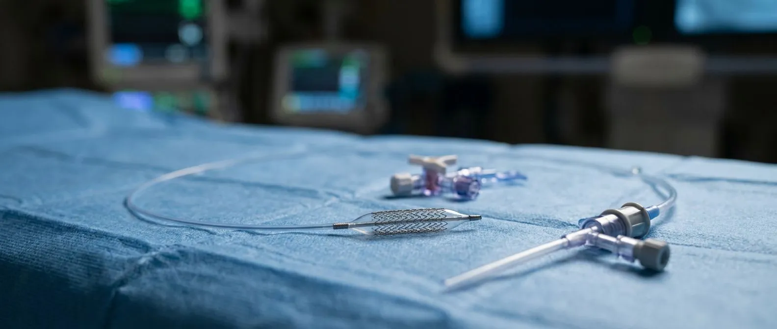
Balloon Angioplasty with intracoronary stent placement represents the most common intervention for focal circumflex artery blockage, offering effective symptom relief and prognosis improvement in appropriately selected patients. Modern techniques address many historical challenges that made circumflex intervention technically demanding.
The circumflex artery's variable anatomy—ranging from large dominant vessels to small non-dominant branches—requires careful procedural planning. Guide catheter selection must provide stable backup support without damaging the left main coronary artery. Wire navigation through tortuous segments demands specialized hydrophilic-coated guidewires. Lesion preparation with cutting Balloon Aortic Valvuloplasty-style scoring balloons or rotational atherectomy for heavy calcification improves stent expansion and apposition—the critical determinants of long-term patency.
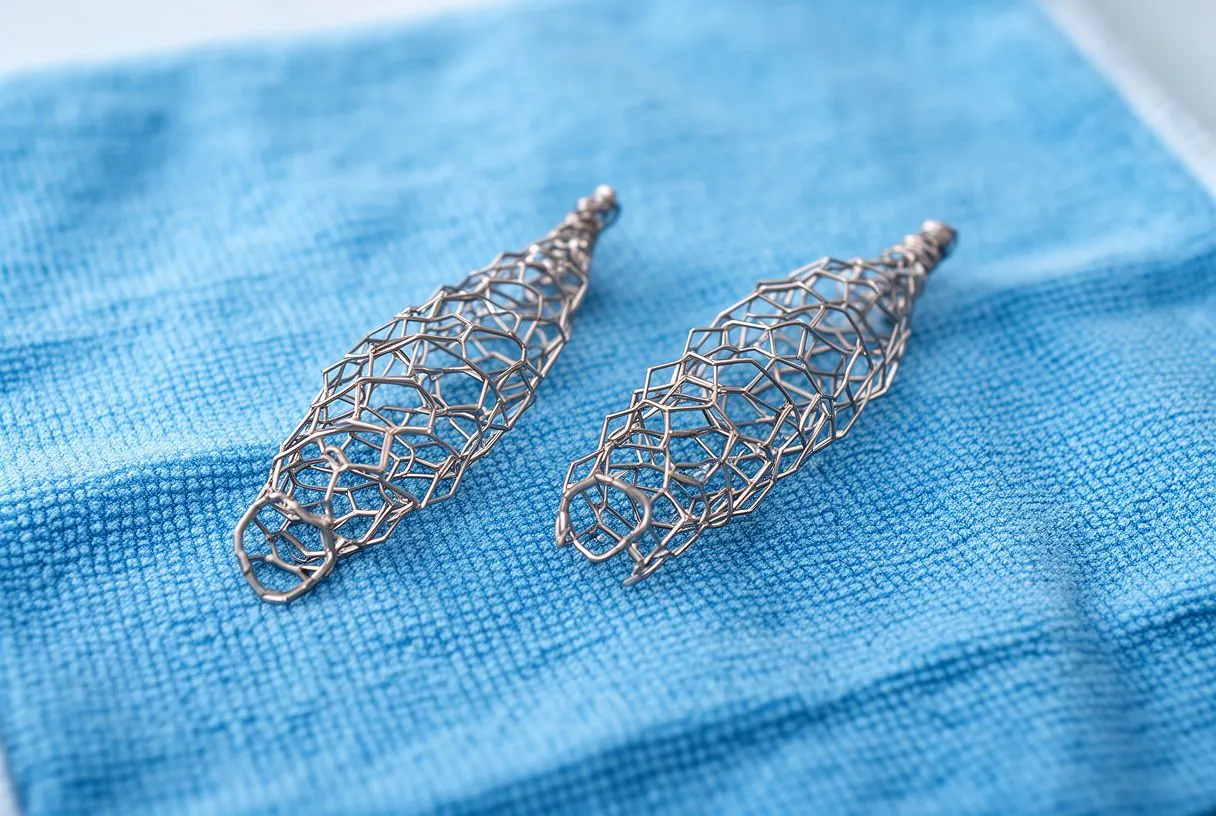
Drug-eluting stents have revolutionized circumflex PCI outcomes by dramatically reducing restenosis—re-narrowing at the treated site—compared to bare-metal predecessors. Second- and third-generation platforms with biocompatible or biodegradable polymers minimize late inflammatory reactions. Dual antiplatelet therapy with Aspirin plus a P2Y12 inhibitor (clopidogrel, ticagrelor, or prasugrel) for 6-12 months after drug-eluting stent placement prevents stent thrombosis, a catastrophic complication of early discontinuation. Your cardiologist will tailor duration based on bleeding risk, planned non-cardiac surgery, and specific stent type.
Technical success rates for circumflex PCI now approximate other territories in experienced centers. However, specific anatomic challenges persist: distal circumflex lesions near obtuse marginal bifurcations may require two-stent techniques with their attendant complexity; heavily calcified proximal segments may resist balloon expansion; and very distal vessels may be too small for stent placement, requiring balloon angioplasty alone. These technical realities sometimes prompt referral for surgical consultation when complete revascularization appears unachievable percutaneously.
When Is Bypass Surgery Needed for Circumflex Artery Blockage?
Coronary artery bypass surgery using an Arterial Graft or venous conduit becomes preferable to angioplasty in specific circumflex disease patterns, particularly when multi-vessel involvement, diabetes, or complex lesion anatomy favors complete surgical revascularization. Understanding these indications helps patients appreciate why their cardiologist might recommend surgery despite the appeal of less invasive alternatives.
The left internal mammary artery (LIMA)—the standard conduit to the left anterior descending artery—can sometimes be routed to circumflex territory when origin proximity permits, though this requires specific anatomic suitability. More commonly, the circumflex receives a saphenous vein graft from the Aorta or a radial artery Arterial Graft. The right gastroepiploic artery, harvested from the stomach, offers another Arterial Graft option for posterior circumflex branches in selected patients. Arterial conduits demonstrate superior long-term patency compared to vein grafts, with LIMA grafts maintaining >90% patency at 10 years—a statistic that influences graft selection when conduit availability and target vessel quality permit.
Multi-vessel disease patterns strongly influence surgical recommendation. Circumflex artery blockage rarely occurs in isolation; concurrent left anterior descending or right coronary disease creates the "three-vessel disease" pattern for which CABG demonstrates survival advantage over medical therapy alone and often over PCI in complex disease. The SYNTAX trial established that patients with high anatomical complexity scores—reflecting diffuse disease, bifurcations, and chronic total occlusions—experience better outcomes with CABG. Circumflex lesions contributing to high SYNTAX scores thus drive surgical referral.
Minimally invasive surgical approaches have expanded circumflex bypass options. Off-pump CABG avoids cardiopulmonary bypass, potentially reducing inflammatory response and coagulopathy. Robotic-assisted techniques enable left thoracotomy approaches with smaller incisions, though sternotomy remains standard for multi-vessel disease requiring circumflex grafting alongside other territories. Hybrid revascularization— combining LIMA to left anterior descending with PCI of circumflex lesions—represents another evolving strategy for selected patients.
What Does Recovery Look like After Circumflex Artery Treatment?
Recovery trajectory following circumflex artery blockage treatment varies substantially by intervention type, baseline cardiac function, and presence of myocardial damage prior to revascularization. Setting realistic expectations helps patients and families plan for the weeks and months ahead while engaging actively in rehabilitation.
After uncomplicated PCI with drug-eluting stent placement, most patients experience rapid functional recovery. Same-day or next-day discharge has become standard for stable elective procedures. Immediately post-procedure, patients must maintain strict compliance with dual antiplatelet therapy—discontinuation even briefly risks stent thrombosis with potentially fatal consequences. Activity generally resumes progressively: walking within 24 hours, light household activities within days, and return to desk work within one week for many. Radial artery access (wrist entry) permits faster mobilization than femoral access (groin entry), now the preferred approach in most centers.
Post-infarction recovery extends substantially longer. Patients whose circumflex artery blockage caused actual myocardial infarction—death of heart muscle tissue— face a different recovery calculus. The damaged lateral or posterior wall undergoes healing and remodeling over 6-8 weeks, during which strict activity restriction applies. Cardiac rehabilitation—a structured program of supervised exercise, education, and risk factor modification—typically begins 2-6 weeks post-event and continues for 3-6 months. This program demonstrably improves functional capacity, psychological well-being, and secondary prevention adherence. Stanford Health Care and other major centers emphasize cardiac rehabilitation as integral to recovery rather than optional adjunct.
Surgical recovery following CABG requires the longest convalescence. Sternotomy healing demands 6-12 weeks of lifting restrictions. Saphenous vein harvest site management—typically from the leg—adds wound care considerations. Return to driving, employment, and full activity follows individualized timelines based on wound healing, rhythm stability, and functional recovery. The Arterial Line and other monitors used in the intensive care unit are removed within 24-48 hours as stability permits, though some patients require temporary pacing wires for conduction disturbances more common after circumflex territory surgery given the vessel's proximity to the electrical conduction system.

What Is the Long-Term Outlook After Circumflex Artery Blockage?
Prognosis after circumflex artery blockage treatment depends critically on four modifiable factors: completeness of revascularization, preserved left ventricular function, rigorous risk factor control, and sustained lifestyle modification. Patients who optimize these domains can expect outcomes approaching population norms despite established coronary disease.
Left ventricular ejection fraction—the percentage of blood pumped with each contraction—emerges as the strongest prognostic indicator. Circumflex territory infarction affecting substantial lateral wall mass reduces ejection fraction and increases risk of subsequent heart failure, arrhythmic sudden death, and recurrent events. Conversely, prompt revascularization preserving viable myocardium maintains pump function and prognosis. Modern cardiology emphasizes "time is muscle": the shorter the interval from symptom onset to restored blood flow, the more myocardium salvaged. This principle underscores emergency department goals for door-to-balloon times under 90 minutes for STEMI presentations.
Secondary prevention targets aggressive risk factor modification. LDL cholesterol targets have progressively lowered: current guidelines advocate <70 mg/dL for very high-risk patients, often requiring statin-ezetimibe combinations or PCSK9 inhibitors. Blood pressure control to <130/80 mmHg reduces recurrent events. Smoking cessation—absolute rather than reduced consumption—provides the single largest mortality benefit of any intervention. Diabetes optimization with newer agents demonstrating cardiovascular benefit (SGLT2 inhibitors, GLP-1 receptor agonists) addresses the metabolic driver of disease progression.
Surveillance following treatment includes periodic functional assessment and symptom review. Stress testing may repeat at 1-3 year intervals to detect new disease or graft/stent failure. Echocardiography monitors left ventricular function and valvular status. Some patients with reduced ejection fraction qualify for implantable cardioverter-defibrillators (ICDs) or cardiac resynchronization therapy—devices preventing sudden death or improving pump efficiency through coordinated ventricular contraction.
When Should You Seek a Cardiologist for Circumflex Artery Symptoms?
Recognizing when symptoms warrant urgent versus routine cardiology evaluation can prevent catastrophic delays in treating circumflex artery blockage. The atypical symptom patterns described earlier complicate this decision, requiring heightened vigilance rather than dismissal of seemingly benign complaints.
Emergency presentation—call emergency services immediately—includes: chest discomfort persisting >15-20 minutes unrelieved by rest; new severe shortness of breath at rest; syncope or near-syncope; palpitations accompanied by hypotension or chest discomfort; and acute neurological symptoms suggesting embolic stroke from atrial arrhythmia. These patterns may indicate acute coronary syndrome, malignant arrhythmia, or hemodynamic compromise requiring immediate Automated External Defibrillator availability and advanced cardiac life support. Do not self-transport; emergency medical services provide monitoring, early defibrillation capability, and prehospital notification activating catheterization laboratory teams.
Routine but expedited cardiology evaluation—within days to weeks—applies to: new exertional symptoms even if mild; progressive reduction in exercise tolerance; unexplained fatigue disproportionate to activity level; and new or worsening symptoms in patients with known coronary disease. These presentations suggest chronic circumflex artery stenosis or progression of established disease that merits diagnostic workup and potential intervention before acute decompensation occurs.
For patients in Mexico seeking specialized cardiac evaluation, geographic access to experienced interventional cardiologists and cardiac surgeons varies. Guadalajara, Querétaro, and Hermosillo represent established centers with comprehensive cardiac catheterization facilities and surgical programs capable of addressing complex circumflex disease. International patients considering evaluation should verify center volumes and outcomes transparency, as procedural experience correlates with technical success for challenging circumflex interventions. Insurance coverage, including international policies and out-of-network benefits, requires pre-authorization verification before elective procedures.
The journey through circumflex artery blockage diagnosis and treatment demands partnership between informed patients and experienced cardiac teams. No article substitutes for personalized clinical assessment; if you or a family member recognize symptoms discussed here, we encourage scheduling consultation with a qualified cardiologist to discuss your specific situation, review your risk profile, and develop an individualized management plan.






