

De-airing
Learn how de-airing prevents air embolism in open heart surgery.
What is de-airing in cardiac surgery?
De-airing is a critical step in cardiac surgery where the surgical team actively removes trapped air bubbles from inside the heart chambers and major blood vessels before restoring normal blood flow. When the heart is opened during a procedure, room air can enter. If this air is not expelled, it can travel to the brain or other organs when the heart begins beating normally again. The de-airing process ensures that only blood, not air, is pumped through the body.
Understanding air removal in cardiac surgery: The concept relies on simple physics. Air is lighter than blood, so it naturally rises to the highest point of a chamber when the heart is positioned. Surgeons use this principle to their advantage, placing small vents or suction catheters at the highest points of the heart to draw the air out.
This practice is a routine and expected part of any open procedure on the heart. Rather than an emergency intervention, it is a planned, meticulously executed phase that bridges the gap between the repair of the heart and the restarting of its normal pumping function.
Why de-airing matters for heart surgery patients
The importance of thorough de-airing cannot be overstated. If air bubbles are allowed to enter the systemic circulation, they can cause an air embolism. An air embolism acts like a tiny roadblock in a blood vessel, cutting off the supply of oxygen-rich blood to vital tissues. For heart surgery patients, the primary concern is protecting the brain, as even a tiny bubble in a cerebral artery can lead to a stroke.
Protecting the brain and heart muscle: Beyond the brain, air can obstruct the coronary arteries that feed the heart muscle itself. If air enters these vessels, it can cause temporary oxygen deprivation, leading to irregular heartbeats or muscle damage during the critical moments when the heart is trying to recover from the operation.
Patients and families often worry about surgical complications. Knowing that the operating room team dedicates a specific, carefully timed phase solely to purging trapped air provides reassurance. It is a direct, proactive measure to prevent a known hazard.
How the de-airing technique is performed
The execution of the de-airing technique relies on a combination of positioning, venting, and perfusion management. Once the primary surgical repair is complete, the heart remains open while the surgical team prepares to close it. The patient is positioned slightly head-down, allowing air to migrate upward toward the base of the heart and the aorta.
Using specialized suction and vents: The team places a small tube, often called a vent, directly into the aortic root or through the left heart chambers. This tube is connected to gentle suction that continuously pulls air out as it collects. The lungs are also ventilated to push blood from the pulmonary vessels back into the heart, helping to push residual air upward.
This phase is coordinated between the surgeon, the anesthesiologist, and the perfusionist. Each member of the team watches the heart and monitors the heart-lung machine to ensure that air is effectively evacuated before normal circulation is fully restored.
De-airing maneuvers for the left ventricle and aortic root
Specific de-airing maneuvers target the left ventricle and the aortic root because these are the highest-pressure areas of the heart. When the heart begins to contract, any air in these locations will be ejected forcefully into the rest of the body. To prevent this, surgeons often gently agitate the heart or use manual massage to dislodge tiny bubbles trapped within the muscular walls of the ventricle.
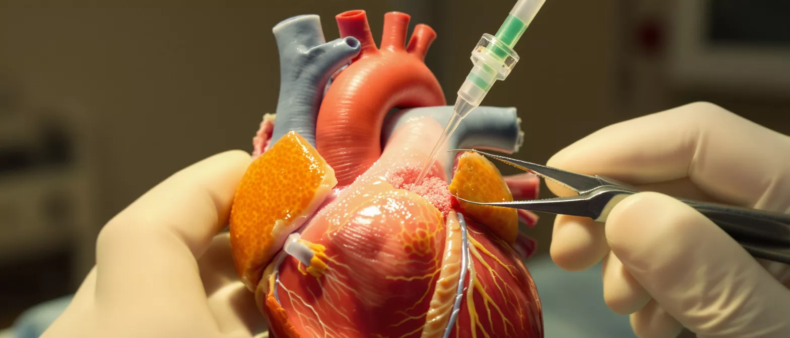
Aspirating air from the highest points: The aortic root, the very beginning of the body's largest artery, is a common final collection point for trapped air. A needle is often placed directly into this root to aspirate the last remnants of air just before the surgeon ties the final sutures. This ensures a completely clean exit for the blood.
Visual and ultrasonic confirmation: In many modern operating rooms, surgeons use transesophageal echocardiography, an ultrasound of the heart, to visually confirm that all air has been removed. This real-time imaging allows the team to see even microscopic bubbles and continue venting until the chambers are clear.
What are the risks of air embolism prevention surgery?
De-airing is itself the primary preventive measure against air embolism. The primary risk associated with incomplete de-airing is an arterial air embolism. If a significant amount of air reaches the brain, it can cause transient neurological symptoms or, in rare cases, permanent injury. In the heart, trapped air can trigger ventricular fibrillation, a dangerous irregular rhythm.
These risks are precisely why the de-airing process is so rigorous. The surgical team does not rush this step. They utilize multiple simultaneous methods—gravity, manual manipulation, and active suction—to help ensure that the risk is minimized before the heart takes over its normal workload.
- Cardiac monitoring: Continuous electrocardiogram tracking ensures no dangerous arrhythmias develop as air is evacuated.
- Neuroprotection: Adequate de-airing is the most direct line of defense against brain injury during valve or bypass operations.
Are de-airing procedures safe for open heart recovery?
Yes, the de-airing procedure is extremely safe and is an expected, standard part of open heart recovery. The techniques used today have been refined over decades of practice. Because the process occurs while the patient is safely supported by the heart-lung machine, the heart itself is resting and tolerates the necessary manipulation well.
Standardized safety protocols: Operating rooms follow strict protocols for air removal. Anesthesia teams use specialized monitoring equipment to track blood pressure and oxygenation continuously. The perfusion team manages the heart-lung machine to provide a smooth transition back to normal circulation only when de-airing is confirmed complete.
For the patient, there is no specific recovery time dedicated solely to the de-airing step. It is integrated seamlessly into the overall intra-operative timeline, meaning the recovery process is identical to the standard recovery for the primary procedure.
De-airing steps in heart surgery explained
Understanding the sequence helps families visualize the operation. After the main repair is finished, the heart is still open. The team begins ventilating the lungs to fill the left side of the heart with blood, displacing the air upward. The surgeon actively suctions air from the highest points of the chambers.
Final closure and root venting: The left side of the heart is filled completely with a clear fluid to push out any remaining air. The surgeon closes the incision in the heart, and a final venting needle is placed in the aortic root to remove the last traces of air before normal pumping resumes.
- Positioning: The operating table is tilted to let air rise naturally toward the aorta.
- Filling: The heart is flooded with blood and clear fluid to displace air.
- Aspiration: Final suction removes the remaining trapped bubbles.
How teams monitor cardiac surgery air evacuation
Monitoring is the cornerstone of safe air evacuation. The most vital tool is transesophageal echocardiography, often called TEE. This ultrasound probe sits in the esophagus behind the heart, providing a continuous, real-time view of all heart chambers. The anesthesiologist watches this screen closely, looking for the bright, reflective signatures of micro-bubbles.
Team communication during de-airing: The anesthesiologist communicates constantly with the surgeon. If the ultrasound shows residual air in the left atrium or left ventricle, the surgeon will perform additional manipulation or prolong the suctioning. This loop continues until the ultrasound confirms a clear field.
Beyond imaging, the perfusionist monitors the pressure within the heart-lung machine circuit. Sudden changes in pressure can indicate a change in volume or the presence of air, prompting an immediate pause and reassessment before proceeding.
De-airing complications and how surgeons prevent them
Complications directly related to de-airing are rare because so much effort is dedicated to preventing them. The primary concern is an undetected air embolism. To prevent this, surgeons rely on redundancy. They do not rely on a single method; instead, they combine gravity, manual manipulation, and mechanical suction simultaneously to ensure no single point of failure exists.
Preventing residual air pockets: Sometimes, tiny air pockets hide behind artificial valves or within the deep muscular folds of the left ventricle. Surgeons prevent this by flooding the chambers with special solutions and using specific instruments to reach into these deep spaces, ensuring every corner is washed clear of air before the heart is closed.
By adhering to these strict protocols, the risk of complications is kept exceptionally low. The operating team treats air evacuation with the same precision as the primary surgical repair, to protect patient safety.
Recovery and what to expect after the de-airing procedure
Because de-airing is an intra-operative step, it does not add specific complications or unique symptoms to the post-operative recovery period. Patients wake up in the intensive care unit having already benefitted from this safety measure. The focus of recovery shifts entirely to healing the surgical incisions and strengthening the heart muscle.
Post-operative monitoring: The immediate hours after surgery involve careful monitoring of neurological function. Nurses check reflexes, grip strength, and responsiveness. This monitoring serves as the final confirmation that the intra-operative air evacuation was successful and that blood flow was safely restored.
Families can take comfort in knowing that the operating room team has handled the invisible threat of air embolism before the patient ever leaves the operating table. The de-airing process is a quiet, essential guardian of surgical safety, allowing the patient to focus entirely on a smooth and healthy recovery.






