

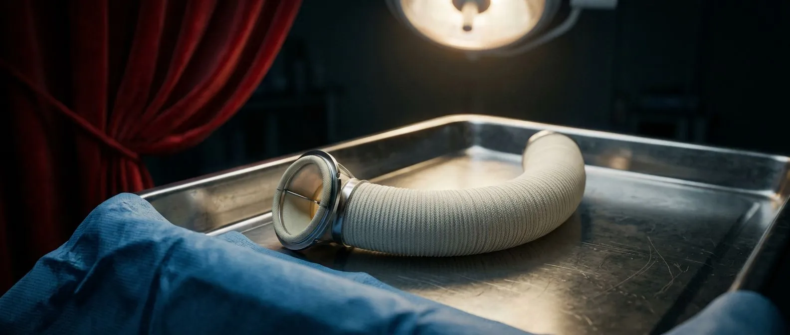
Bentall Procedure: Aortic Root Replacement Explained
Bentall procedure aortic root replacement explained by Guadalajara heart surgeons.
In the heart, quite literally, every millimetre counts. When the aortic root — the first part of the great artery that carries blood from the heart to the rest of the body — becomes dilated or diseased, life can be at risk. In those cases, one of the most effective and well-established operations is the Bentall procedure, which combines surgical precision, anatomical knowledge and advanced technology to repair or replace the aortic root and the aortic valve.
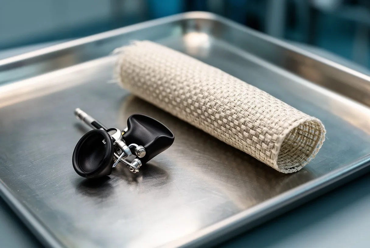
Anatomy of the aortic root
The aortic root is the first part of the aorta, arising directly from the left ventricle. It is made up of three main structures:
- Aortic annulus: the narrowest point, where the aortic valve sits.
- Sinuses of Valsalva: three natural bulges just above the valve, from which the right and left coronary arteries arise.
- Sinotubular junction: the point where the sinuses narrow again and continue into the ascending aorta.
The geometric relationship between these segments is what allows the aortic valve to work properly. When one of them dilates, the valve loses its shape, causing aortic regurgitation and overloading the left ventricle.
Normal and abnormal aortic root dimensions
Normal measurements vary with sex, height and body surface area, but the average values in adults are:
- Aortic annulus: normal 20–26 mm; abnormal >28–30 mm.
- Sinuses of Valsalva: normal 29–37 mm; abnormal >40 mm.
- Sinotubular junction: normal 25–33 mm; abnormal >38–40 mm.
- Proximal ascending aorta: normal 28–36 mm; aneurysm >45 mm (surgical risk >50–55 mm).
When the aortic root exceeds 45–50 mm, or even less in patients with Marfan syndrome, a bicuspid valve or a family history of dissection, early surgery is the best option, because the risk of complications in this group is higher and they can occur even at normal aortic diameters.
Complications of an untreated root aneurysm
An uncorrected aneurysm can progress silently for years, but the consequences can be devastating:
- Acute aortic dissection: an internal tear of the aortic wall, with an immediate risk of death.
- Frank rupture: massive bleeding into the chest or the pericardium.
- Severe aortic regurgitation: the valve stops sealing and the heart enlarges progressively until it fails.
- Compression of neighbouring structures: such as the pulmonary artery or the superior vena cava.
The mortality of an untreated aortic dissection rises by 1% per hour during the first 48 hours, which is why early diagnosis and timely surgery matter so much.
Diagnosis and symptoms of an aortic root aneurysm
One of the biggest challenges with aortic root aneurysms is that they can grow silently for years without causing symptoms. Most are found by chance, on imaging done for other reasons, such as a routine echocardiogram, a CT scan or an MRI.
The most common symptoms
When symptoms do appear, they are usually due to progressive dilation of the root or to the aortic valve regurgitation it causes. The most frequent are:
- Tiredness or shortness of breath (dyspnoea), especially on exertion.
- Dull or persistent chest pain, in the upper chest or the back.
- Palpitations, or a feeling of “strong heartbeats” in the chest.
- A new heart murmur, picked up by the doctor when listening to the heart.
- In advanced cases, swelling of the feet or ankles, dizziness or fainting.
The symptoms of acute aortic dissection — a sudden tear in the wall of the aorta — include intense, stabbing pain in the chest or back, which patients describe as “tearing” or “moving”. This is a life-threatening emergency that needs immediate care.
How is an aortic root aneurysm diagnosed?
Diagnosis is made with imaging studies that measure the root diameter precisely and assess how the valve is working:
- Transthoracic echocardiogram: the most common first test. It shows the size of the root, the aortic valve and the function of the left ventricle, and is ideal for regular follow-up. It can underestimate the size if the aorta is very large or the patient has a broad chest. It is used to plan surgery and decide on the type of graft needed.
- CT angiography: considered the reference standard for measuring aneurysms. It gives precise three-dimensional images of the whole aorta, from the root to the descending portion.
- Transoesophageal echocardiogram: used when the transthoracic echocardiogram is inconclusive, or during surgery to confirm the results.
Follow-up and early detection
In patients with risk factors (a bicuspid valve, Marfan syndrome, a family history of dissection, high blood pressure or smoking), an echocardiogram every one or two years is recommended. Detecting an aneurysm before it reaches the surgical threshold is essential: elective surgery carries ten times less risk than an emergency operation.
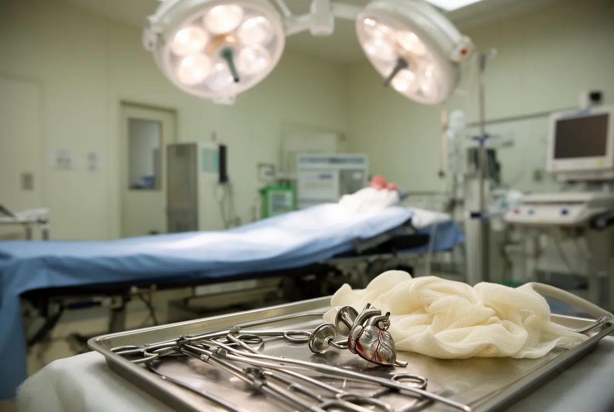
What does the Bentall procedure involve?
The Bentall procedure replaces the aortic valve, the aortic root and the first segment of the ascending aorta as a single unit, and reimplants the coronary arteries. The valved graft can be:
- Mechanical: more durable, but it requires lifelong anticoagulation.
- Biological: avoids long-term anticoagulation, but has an average lifespan of 12–18 years.
The Bentall procedure, step by step
1) Preparation before surgery
- Full assessment: consultation with the surgeon and the cardiologist, blood tests, an electrocardiogram and imaging (echo, CT or MRI) to measure the aortic root precisely.
- Medicines and fasting: you will be told which medicines to stop (anticoagulants, for example) and from when not to eat or drink.
- Admission and checks: on arrival at the hospital, the team confirms your identity, allergies and anaesthetic plan. You meet your anaesthetist and sign the informed consent.
2) Anaesthesia and preparation in the operating room
- General anaesthesia: you are deeply asleep; you will feel no pain and will not remember the operation.
- Advanced monitoring: lines and sensors are placed to monitor the heart, blood pressure, breathing and brain function.
- Sterile field: the chest is disinfected and covered with sterile drapes.
3) The incision (reaching the heart)
Sternotomy: this can be full (opening the sternum from top to bottom) or a mini-sternotomy (a partial “J” or “inverted T” incision in the upper third). A mini-sternotomy aims for less pain, less bleeding and a shorter scar, when the anatomy and the case allow it.
4) Connection to the “heart-lung machine”
The body is temporarily connected to a cardiopulmonary bypass machine, which does the work of the heart and lungs while the surgeon repairs the aorta. This allows the surgeon to work in a bloodless field with a still heart, which improves precision.
5) Protecting the heart
A special cold solution (cardioplegia) is given to “put the heart to rest” and protect it while it is stopped.
6) Exposing the diseased aortic root
The surgeon opens the first part of the aorta and assesses the aortic valve and the sinuses of Valsalva (the dilated area). The decision is made to replace the valve with a mechanical or a biological prosthesis, as agreed with you before surgery.
7) Removing the damaged tissue precisely
The dilated aortic root and the diseased valve (where applicable) are removed. Two small “coronary buttons” are created: these are the little openings where the right and left coronary arteries arise, so they can be reimplanted later without tension.
8) Preparing the “spare part”: the valved graft
A Dacron graft (a strong tube) that already contains a new valve (mechanical or biological) is used. Think of it as replacing the first stretch of a pipe and its main valve with a new, safe part.
9) Suturing (connecting) the graft to your heart
- Attachment to the aortic annulus: the lower end of the graft is sutured to the base where the original valve sat.
- Reimplanting the coronary arteries: the two coronary “buttons” are sutured to small windows made in the graft so that blood flows to the heart muscle again.
- Joining the ascending aorta: the upper end of the graft is connected to healthy aorta, above the damaged area.
In some special cases, if a coronary artery is under tension or the wall is fragile, a small extension (the Cabrol technique) is used to connect it safely.
10) Safety checks
Air is removed from the system (de-airing) to avoid bubbles, the team checks for leaks and makes sure the new valve opens and closes properly, and the body is rewarmed to its normal temperature.
11) Coming off bypass and closing
The heart starts beating again and the heart-lung machine is gradually withdrawn. Drains are placed to prevent blood or fluid collecting. The sternum is closed with special wires and the skin with sutures or staples.
12) Immediate recovery (after surgery)
You go to intensive care for 24–48 hours (depending on progress), where pain, blood pressure and heart rhythm are controlled. Then you move to the ward: early walking, deep breathing with an incentive spirometer and adjustments to your medicines. Discharge is usually at around 5–7 days if all goes well.
Surgical variants
- Cabrol technique: uses a small interposed graft to reconnect the coronary arteries when direct reimplantation is difficult.
- David or Yacoub procedure (valve reimplantation): preserves the native valve if it is structurally sound.
- Wheat procedure: a technically simpler alternative to the Bentall technique, as it avoids reimplanting the coronary ostia. It is considered an option of choice when the aortic root is of normal size and shape.
- Ross procedure: the patient's own pulmonary valve replaces the aortic valve, giving a completely biological option.
Each variant aims to tailor the operation to the patient's age, anatomy and lifestyle.

Bentall through a mini-sternotomy
In recent years, several specialist centres have adopted the Bentall procedure through a mini-sternotomy as a less invasive alternative to the traditional approach.
What does it involve?
A mini-sternotomy is a partial incision of the sternum (in a “J” or “inverted T” shape), usually limited to the upper third of the bone. It gives access to the heart and aortic root with the same safety, but with less surgical trauma.
Main advantages
- Less bleeding during the operation and less need for transfusion.
- Faster recovery, with less postoperative pain.
- A better cosmetic result, with a shorter, more discreet scar.
- A lower risk of sternal infection, especially in patients with diabetes or obesity.
- A shorter hospital stay, on average 1 to 2 days less than with a full sternotomy.
Comparative results
Recent studies show that Bentall through a mini-sternotomy has equivalent results in safety and durability, with no increase in aortic cross-clamp time or complications. In experienced centres, the technique gives excellent surgical exposure while maintaining the precision of the coronary reimplantation and the quality of the reconstruction.
Limitations
- It requires a highly specialised surgical team.
- It is not always advisable in acute dissection or very extensive aneurysms.
- It may lengthen the operation slightly during the learning curve.
In carefully selected patients, the mini-sternotomy is a modern, safe and cosmetic option, in line with the trend towards minimally invasive heart surgery.
Alternatives to Bentall
In selected cases, the following may be considered:
- Isolated valve repair, if the root is intact.
- Simple valve replacement, when there is no significant dilation.
- Transcatheter valve implantation (TAVI), indicated only in patients at very high surgical risk, although it does not correct aneurysms.
The treatment should always be decided by a Heart Team, combining the experience of surgeons, cardiologists and imaging specialists.
Surgical risks and complications
Although this is major surgery, results today are very good. The risks include:
- Bleeding or the need for reoperation.
- Neurological complications (stroke).
- Infection of the prosthesis (endocarditis).
- Temporary heart failure or prosthetic dysfunction.
In highly specialised centres, surgical mortality is around 2–5%, with success rates above 95%.
Long-term results and durability
The Bentall procedure offers excellent long-term survival, with a life expectancy close to that of the general population when it is done preventively.
- Durability: mechanical valved grafts can last a lifetime; biological ones last 12 to 20 years, depending on the patient's age and metabolism.
- Heart function: most patients recover a normal ejection fraction and return to a fully active life.
- Follow-up: yearly echocardiograms and strict blood pressure control are recommended to prevent residual dilation in other segments of the aorta.
Reoperations and future surgical options
Thanks to today's advances, reoperations after a Bentall are uncommon, but they may be needed in certain situations:
- Degeneration of a biological prosthesis: today this can be treated without reopening the chest, with a transcatheter valve-in-valve procedure (a TAVI inside the previous graft).
- Aneurysm of the remaining ascending aorta or aortic arch: may require further surgery or a hybrid repair with an endoprosthesis.
- Prosthetic infection (late endocarditis): although rare, it may require complete replacement of the graft.
Reoperations today are safer thanks to minimally invasive techniques, advanced cardiopulmonary bypass and continuous brain monitoring, with mortality below 5–8% in experienced centres.
Conclusion
The Bentall procedure is one of the great achievements of modern heart surgery: it rebuilds the root of life with millimetre precision and a view of the patient as a whole. Thanks to accumulated experience and the work of specialised teams, an aortic root aneurysm today is no longer a sentence but a treatable condition, with excellent results and the prospect of a full life.
Timely diagnosis, and treatment in highly specialised centres with experience in this type of procedure, are vitally important. If you would like more information about your own case, remember that you can request a video consultation on our Contact page.






