

Intraoperative Echocardiography
Learn how intraoperative echocardiography delivers real-time cardiac imaging during heart surgery.
What is intraoperative echocardiography?
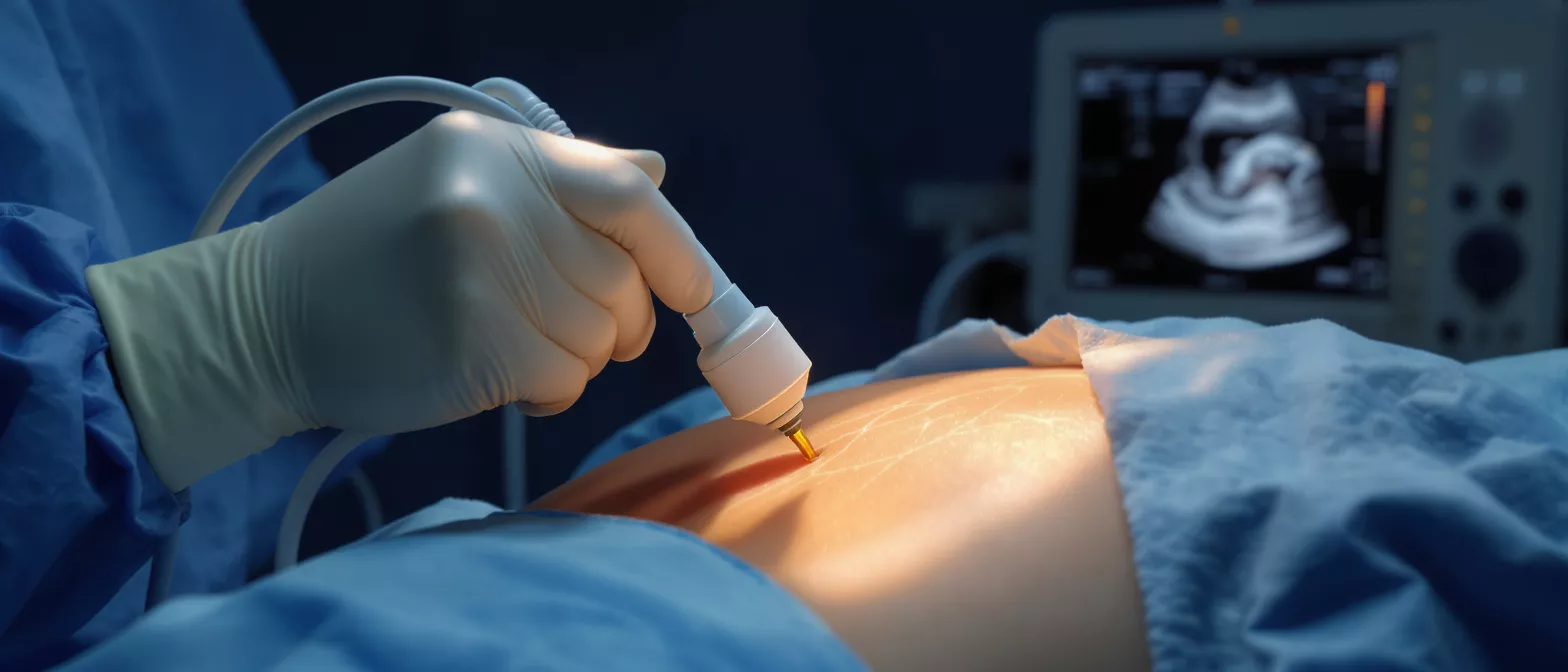
Intraoperative echocardiography is a specialized imaging test performed while a patient is undergoing heart surgery. It uses high-frequency sound waves to create real-time, moving pictures of the heart, allowing the surgical team to see its structures, valves, and chambers exactly as they appear in the operating room. Rather than relying solely on static images taken before the procedure, your surgeon gains an immediate, live view of your cardiovascular anatomy.
This technology serves as an internal navigation system during complex cardiac operations. When you or a loved member of your family are facing heart surgery, knowing that the surgical team uses advanced visual guidance can provide significant peace of mind. It effectively translates physical heart anatomy into a clear, dynamic video feed that surgeons rely on to make critical decisions in the moment.
The term itself simply breaks down into its practical components: "intraoperative" means occurring during the operation, and "echocardiography" is the use of ultrasound to examine the heart. Together, they represent a standard of modern surgical care that bridges the gap between a planned surgical repair and the actual reality inside the patient's chest.
How intraoperative echocardiography works during heart surgery
The process relies on a small device called a transducer, which sends and receives sound waves. These sound waves bounce off the different structures of the heart—such as the muscle tissue, valves, and blood—and return to the transducer. A computer then instantly translates these returning echoes into a detailed, moving image displayed on a monitor right next to the surgical field.
Doppler imaging in the operating room: advanced surgical echocardiography techniques go beyond just showing the physical shape of the heart. By using a feature called intraoperative Doppler imaging, the team can actually see and measure the speed and direction of blood flow. This allows them to detect any leaks or blockages inside the vessels and valves without having to physically touch the heart.
Because these images are generated instantly, the surgical team can continuously evaluate how the heart is behaving in real time. They do not have to pause the operation to wait for a radiologist to develop an image. Instead, the live feedback loop helps them adjust their surgical approach second by second, ensuring precision.
Types of intraoperative echocardiography
Surgeons utilize two primary approaches to capture these critical images during an operation, and the choice between them depends on the specific surgical procedure being performed. Both methods share the same goal of providing exceptional visual data, but they access the heart through entirely different pathways.
- Transesophageal echocardiography (TEE): A specialized probe is gently passed down the patient's esophagus while they are fully anesthetized. Because the esophagus sits directly behind the heart, this pathway provides remarkably clear, unobstructed images of the cardiac structures.
- Epicardial echocardiography: The surgeon places a sterile transducer directly on the surface of the beating heart during open-chest procedures. This method is highly useful for confirming physical anatomy before bypass grafts are placed or when TEE imaging is not suitable for the patient.
Both techniques are valuable diagnostic tools that keep the surgical team fully informed. Your surgeon will decide which method offers the safest and most accurate visualization for your specific heart anatomy and surgical needs.
Why is echo used in cardiac surgery?
Understanding why this technology is so crucial helps patients and their families grasp the level of precision involved in modern cardiac care. Intraoperative echocardiography acts as a constant quality control measure, ensuring that the surgical plan is executed flawlessly and that the heart is functioning properly before the operation concludes.
Pre-surgery structural assessment: Before the surgeon makes the first physical incision into the heart, the echo provides a baseline assessment. This helps confirm that the planned surgical approach still makes sense given the patient's current anatomy under anesthesia, preventing unexpected complications mid-surgery.
Post-surgery functional verification: After the primary repair—such as a valve replacement or congenital defect closure—is complete, the surgeon uses the echo to verify the success of the work. They can visually confirm that blood is flowing normally, that no new leaks have been introduced, and that the overall heart function is stable before the chest is officially closed. Finding a residual issue now, while still in the operating room, prevents the need for a secondary emergency surgery later.
Intraoperative TEE transesophageal echocardiography explained
Transesophageal echocardiography, commonly referred to as TEE, is the most widely used form of imaging during cardiac surgery. Because the esophagus passes immediately behind the heart, placing an ultrasound probe there allows for an incredibly close, high-resolution view of the cardiac chambers and valves without the interference of the ribs or lungs.
The probe used for TEE is essentially a flexible, thin tube with a miniature ultrasound camera mounted on its tip. Because the patient is deeply asleep and under general anesthesia, the anesthesiologist or a specialized cardiologist can safely guide this tube into the correct position without causing any discomfort or awareness.
Continuous TEE monitoring: advanced intraoperative cardiac monitoring equipment allows for continuous TEE monitoring throughout the entire procedure. This continuous vigilance means the surgical team can watch the heart adapt to surgical changes in real time, ensuring that heart rhythm and blood pressure remain stable even as the organ is being manipulated or temporarily stopped for a bypass.
Epicardial echocardiography and intraoperative ultrasound heart imaging
While TEE is the standard approach for many operations, epicardial echocardiography offers a direct, complementary alternative. This technique involves placing a small, sterile ultrasound probe directly on the outer surface of the heart itself, known as the epicardium, while the chest is open.
This direct approach is particularly useful for procedures involving the aorta or for evaluating specific regions of the heart that might be difficult to see clearly through the esophageal route. By scanning directly from the surface, the surgeon can gain precise anatomical information to guide the placement of surgical instruments or grafts.
Complementary imaging strategies: In complex cases, high-resolution intraoperative ultrasound heart imaging might be used in conjunction with other modalities. This multi-faceted approach aims to give the surgeon, however complex the structural heart disease, a complete, three-dimensional understanding of the tissue they are repairing.
Benefits of real-time cardiac monitoring surgery
The integration of real-time cardiac monitoring during surgery offers profound benefits for patient outcomes. By bridging the gap between preoperative imaging and physical surgical intervention, this technology eliminates guesswork and dramatically reduces the risk of incomplete repairs.
For the patient, the most significant benefit is the immediate verification of surgical success. If a valve repair has a tiny residual leak, the surgeon sees it instantly on the echo monitor and can correct it before the operation ends. This prevents the need for a second, highly risky open-heart surgery in the days or weeks following the initial procedure.
Reduced complication rates: By identifying potential issues like air bubbles in the heart chambers or sudden drops in contractility during the operation, the surgical team can address them immediately. This proactive approach greatly reduces the likelihood of postoperative complications and helps ensure a smoother, faster recovery in the intensive care unit.
Are there risks with intraoperative echocardiography?
When facing cardiac surgery, it is completely natural to worry about every additional procedure involved. Fortunately, intraoperative echocardiography is considered extremely safe. Because the patient is already under general anesthesia and fully monitored in a controlled operating room environment, the risks associated with the echo itself are remarkably low.
For TEE, the primary risks involve the insertion of the probe into the esophagus, which carries a very small chance of minor dental trauma or temporary throat soreness. However, because the patient is deeply asleep, the throat is relaxed, and the anesthesiologist carefully protects the airway, these risks are minimized and managed professionally.
Minimizing patient harm: The concept of weighing the risk of a procedure against its benefits is central to cardiac care. The incredibly detailed visual data provided by this monitoring far outweighs the minimal risks of the imaging itself, making it a universally accepted and expected standard of care in modern cardiac operating rooms.
What to expect after intraoperative echo assessment
Once the surgery is complete, the intraoperative echocardiography assessment has already played its crucial role in ensuring your heart is functioning as intended. The probe is safely removed before you wake up, and the surgical team takes over your care in the intensive care unit with full confidence in the structural repair they have achieved.
You might experience a mild sore throat upon waking, similar to the sensation after having a breathing tube placed. This is a completely normal and temporary side effect of the TEE probe, and it typically resolves within a day as you begin your healing process without any need for specific intervention.
From a medical standpoint, your postoperative care team already has a clear picture of your repaired heart based on the final echo images captured in the operating room. They can compare these images against future echocardiograms taken during your recovery to confirm that your heart continues to heal and function perfectly over time.
Key takeaways on heart surgery echocardiography
Navigating a cardiac surgery journey is an overwhelming experience, but understanding the tools your surgical team uses can bring immense reassurance. Intraoperative echocardiography is not just an extra test; it is a foundational element of modern, safe cardiac surgical care that provides a critical safety net for patients.
By allowing surgeons to see the heart's function in real time, verify repairs before closing the chest, and adapt to the immediate realities of your unique anatomy, this technology drastically improves surgical outcomes. It transforms the operating room from a space of educated estimation into a highly controlled environment of visual certainty.
Knowing that your surgical team is equipped with this level of precision means that you or your loved one are receiving the highest standard of cardiac care available today. This continuous visual guidance ensures that when you wake up, the repair to your heart is already confirmed as a success, giving you the best possible foundation for your recovery journey.






