

Janeway Lesions
Learn about Janeway lesions, their causes, symptoms, link to endocarditis, and how cardiologists diagnose and treat them.
What Are Janeway Lesions?
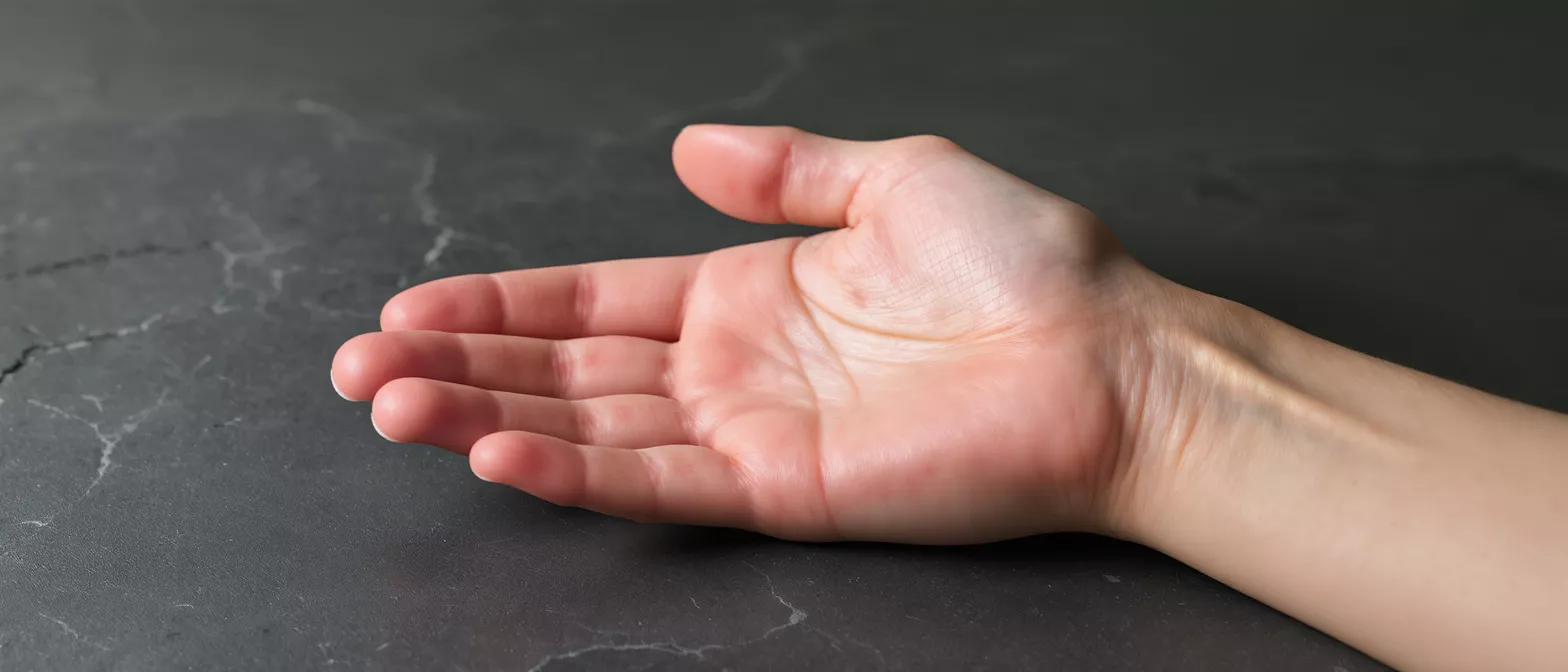
Janeway lesions are small, painless, red or purple spots that appear on the skin, most commonly on the palms of the hands or the soles of the feet. In the context of cardiac care, they serve as a crucial clinical sign that helps physicians identify a serious underlying heart infection. When a patient or their family members notice these unexpected markings, it is natural to feel anxious, but understanding what they represent is the first step toward effective treatment.
Are Janeway lesions dangerous? The spots themselves are harmless and leave no scar, but their presence signals that bacteria are circulating in the bloodstream and affecting the heart valves. They are an indicator rather than a localized skin problem. Cardiologists look for these external signs to confirm suspicions of a deeper systemic issue.
Recognizing janeway lesions early allows the medical team to expedite diagnostic testing, such as blood cultures and echocardiograms. By acting quickly, doctors can begin life-saving antibiotic therapy and protect the heart muscle from further damage.
How Do Janeway Lesions Relate to Endocarditis?
The appearance of these skin markings is heavily linked to infective endocarditis, an infection of the heart's inner lining or valves. When bacteria enter the blood, they can attach to damaged heart valves, forming clumps known as cardiac vegetations. These vegetations continuously shed small bacterial clusters into the bloodstream, which is why external signs like skin spots develop.
Why do cardiac vegetations cause skin spots? As the infected material travels through the arteries, tiny fragments can lodge in the small blood vessels of the hands and feet. This process temporarily blocks local circulation, resulting in the localized red or purple marks visible on the skin. Once the underlying heart infection is successfully treated, the fragments stop circulating, and the external spots gradually fade.
Understanding this relationship helps patients see why a heart surgeon or cardiologist asks to examine their hands and feet. The skin often provides a visible window into the body's internal cardiovascular health, guiding the medical team toward a definitive diagnosis.
What Causes Janeway Lesions on the Skin?
The root cause of these lesions is septic embolization, a process where infected material breaks off from a heart valve and travels through the bloodstream. These micro-emboli become trapped in the peripheral capillary beds of the palms or soles. The tiny blood vessels become temporarily blocked, depriving that small patch of skin of oxygen and triggering an immune response.
What bacteria most commonly cause janeway lesions? The underlying heart infection is typically caused by gram-positive bacteria, most notably Staphylococcus aureus and various species of Streptococcus. These particular strains are highly aggressive and prone to forming large, fragile vegetations on the heart valves that easily shed into circulation. The body's inflammatory reaction to these trapped bacteria results in the visible color change on the skin.
It is important to note that these spots are not caused by an allergy, a local skin infection, or poor hygiene. They are strictly a mechanical and immunological consequence of bacteria flowing from the heart to the extremities.
What Are the Symptoms and Appearance of Janeway Lesions?
Identifying these marks relies on recognizing their specific visual and physical characteristics. They present as flat or slightly raised macules or nodules, usually measuring a few millimeters to a centimeter in diameter. The color typically ranges from bright red to a deep, dark purple, resembling a small bruise or blood blister.
Do janeway lesions hurt? The defining feature of these spots is that they are entirely painless. Unlike other dermatological manifestations of endocarditis, applying gentle pressure to these spots does not cause discomfort. They also do not blanch, meaning they will not turn white or fade when you press a clear glass against them, which confirms that the small blood vessels beneath the skin have leaked.
They most frequently appear on the thenar eminence, the fleshy part of the palm at the base of the thumb, as well as the fingers and toes. Because they are painless, patients often discover them incidentally while washing their hands or showering. When recovering from a heart procedure, wearing a medical-grade compression sleeve can help manage arm swelling, though it will not interfere with identifying these marks.
Janeway Lesions vs Osler Nodes: What Is the Difference?
Patients and families often confuse janeway lesions with Osler nodes, as both are dermatological signs associated with infective endocarditis. However, they have distinct clinical features that help cardiologists tell them apart. The primary difference lies in how they feel and their underlying mechanism of formation.
- Location: Janeway lesions are found on the palms and soles, while Osler nodes typically appear on the pads of the fingers and toes.
- Sensation: Janeway lesions are painless, whereas Osler nodes are tender and often described as highly sensitive to the touch.
- Pathology: Janeway lesions are caused by micro-emboli lodging in capillaries, while Osler nodes are caused by immune complex deposition creating localized inflammation.
Which sign is more common in endocarditis? Both are relatively rare in modern medicine due to the rapid use of antibiotics, but Osler nodes are slightly more frequently reported in subacute infections. Distinguishing between the two is an essential step in the janeway lesions differential diagnosis, guiding the cardiology team's approach.
How Do Doctors Diagnose Janeway Lesions?
The diagnosis of these skin spots is primarily clinical, meaning a doctor identifies them through visual examination and patient history rather than a biopsy. A cardiologist or infectious disease specialist will inspect the patient's hands and feet, looking for the characteristic non-blanching, painless red spots. Finding them immediately prompts a clinical investigation for infective endocarditis.
What tests confirm the underlying heart infection? To confirm the diagnosis, the medical team will order blood cultures to identify the specific bacteria in the bloodstream, followed by an echocardiogram to look for cardiac vegetations. Blood tests will also show elevated inflammatory markers, indicating the body is fighting an infection. Sometimes, a small skin biopsy is performed to confirm the presence of bacteria within the capillary beds, though this is rarely necessary.
Diagnosing the underlying condition quickly is critical to preventing long-term damage to the heart valves. By piecing together the physical examination with laboratory results, the cardiology team can formulate a targeted treatment plan. Patients managing arrhythmias alongside their recovery might consider using a portable ECG monitor to share data with their specialist.
What Is the Pathophysiology of Janeway Lesions?
The pathophysiology of these spots is rooted in the mechanics of blood flow and infection. When bacteria colonize a heart valve, they multiply and form vegetative growths that disrupt normal blood flow. These fragile growths are constantly battered by the force of the heart's pumping action, causing microscopic pieces to break off and enter the systemic circulation.
How do emboli affect peripheral tissue? The detached bacterial clusters travel through the arteries until they reach vessels too narrow to allow passage, specifically the small arterioles in the palms and soles. Once lodged, they cause a tiny area of tissue ischemia, starving the skin cells of oxygen, while the bacteria directly inflame the vessel wall.
This combination of mechanical blockage and localized inflammation produces the visible red or purple hue. The body's immune system eventually breaks down the trapped material and clears the blockage, but the skin takes time to return to its normal color.
What Is the Clinical Significance of Janeway Lesions?
In clinical cardiology, recognizing these spots is highly significant because they are a hallmark of active infective endocarditis. Their presence indicates that the heart infection is severe enough to seed bacteria throughout the body. For a cardiac-surgery patient or someone with a prosthetic heart valve, finding these marks is a major red flag that requires immediate medical attention.
Do janeway lesions indicate a poor prognosis? While their presence means the infection is active, modern antibiotic therapies are highly effective, and they do not automatically mean a poor prognosis. However, it does signal to the care team that the infection is producing complications. The rapid identification of these marks correlates with faster treatment initiation, which reduces the risk of permanent valve damage or organ injury.
These spots are part of a broader set of signs that doctors look for when evaluating a heart infection. They help confirm the clinical picture alongside symptoms like a persistent fever, a new heart murmur, and unexplained weight loss. By understanding their significance, patients can seek care promptly.
How Are Janeway Lesions Managed and Treated?
The management of janeway lesions focuses entirely on treating the underlying heart infection rather than the skin spots themselves. The lesions will naturally resolve over a few weeks once the infection in the blood is cleared. The standard treatment requires a prolonged course of intravenous antibiotics to eradicate the bacteria embedded within the heart valves.
Is surgery needed for infective endocarditis? In cases where antibiotics fail to clear the infection, or if the cardiac vegetations cause severe valve leakage, heart surgery is necessary. A cardiac surgeon will operate to remove the infected tissue and repair or replace the damaged heart valve. Once the source of the bacteria is eliminated, no new embolic spots will form.
Post-operative care involves monitoring for any new signs of infection and ensuring complete healing of the surgical site. Patients are closely followed by their cardiology team to ensure the infection does not return.
What Is the Differential Diagnosis for Janeway Lesions?
When a patient presents with painless red spots on their hands or feet, doctors must consider several possible causes. The clinical process of distinguishing between these possibilities is known as the differential diagnosis. While infective endocarditis is the most serious concern, other benign conditions can mimic this appearance.
- Petechiae: Small red or purple spots caused by minor bleeding under the skin, often from physical strain or low platelets, without infection.
- Trauma or injury: Unnoticed bruises or minor physical impacts to the hands that mimic the dark coloration.
- Vasculitis: Inflammation of the blood vessels from autoimmune conditions, which can create similar localized markings.
Why is the clinical context crucial? A doctor differentiates these conditions by looking at the whole clinical picture. If a patient has a fever, an artificial heart valve, or a history of dental work, endocarditis becomes the primary suspect. The cardiology team uses laboratory tests to rule out other causes and confirm the exact nature of the skin marks.
In summary, these spots are a visible warning sign of a deeper heart infection. Recognizing their painless nature and specific location helps patients seek timely evaluation. With prompt diagnosis and modern cardiac care, the underlying infection can be treated effectively, allowing the skin to heal and the heart to recover.






