

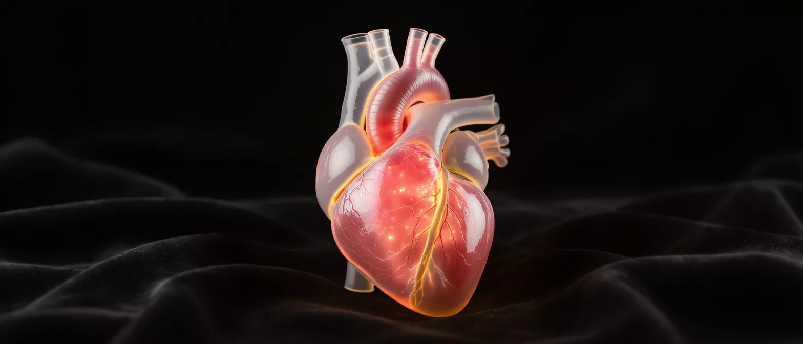
Pulmonary Valve
Discover how the pulmonary valve works, common disorders like stenosis and regurgitation, and expert surgical treatments.
What is the pulmonary valve?
The pulmonary valve is one of the four essential valves inside the human heart, acting as a critical gateway between the right lower heart chamber and the lungs. When most people think of heart valves, they immediately picture the mitral or aortic valves on the left side of the heart. However, the right side plays an equally vital role, and the pulmonary valve ensures that deoxygenated blood moves efficiently toward the lungs to pick up fresh oxygen. Understanding this structure is the first step in grasping how the heart functions as a complete circulatory pump.
Anatomically, this valve is located at the base of the pulmonary artery, the large vessel responsible for carrying blood from the heart to the lungs. It consists of three thin, flexible flaps of tissue known as leaflets or cusps. These leaflets open and close with every single heartbeat, reacting precisely to changes in blood pressure within the heart chambers. By acting as a one-way door, the valve ensures that blood flows in a single, forward direction without reversing course.
Why this valve matters to cardiac patients: For patients facing heart surgery, knowing the location and purpose of each valve helps demystify the surgical process. If a surgeon mentions a problem with the right side of your heart, they are likely referring to the pulmonary valve. Because it manages blood flow to the lungs, any dysfunction here can directly affect your breathing, energy levels, and overall cardiovascular stability, making it a primary focus during comprehensive cardiac evaluations.
How does pulmonary valve function work?
Understanding pulmonary valve function requires looking at the cardiac cycle, specifically the phase where the heart relaxes and contracts. When the right ventricle squeezes to push blood toward the lungs, the pressure forces the pulmonary valve leaflets to open fully. This opening allows a precise volume of deoxygenated blood to exit the heart and travel into the pulmonary artery. The blood then continues its journey to the lungs, where carbon dioxide is exchanged for fresh oxygen.
Once the right ventricle finishes its contraction and begins to relax, the pressure inside the chamber drops significantly. This sudden drop in pressure causes the blood in the pulmonary artery to naturally want to flow backward into the heart. To prevent this backward flow, the three leaflets of the valve snap shut tightly, creating a secure seal. This precise opening and closing mechanism ensures that blood moves in a continuous, efficient loop without wasting any energy.
What happens if the valve cannot close properly: If the leaflets fail to form a tight seal, blood can leak backward into the right ventricle, a condition known as regurgitation. This forces the heart to work harder with every beat to pump the same blood forward twice. Over time, this extra workload can cause the heart muscle to thicken or enlarge, which is why evaluating pulmonary valve function is a standard part of diagnosing right-sided heart issues.
What is pulmonary valve stenosis?
Pulmonary valve stenosis is a specific condition where the valve leaflets become stiff, thickened, or fused together, restricting the normal flow of blood. Because the opening is narrowed, the right ventricle must pump with much greater force to push blood through the tight space and into the lungs. This condition is most often congenital, meaning it develops before birth, but it can also occur as a result of other medical conditions or rare infections later in life.
The severity of the narrowing determines how the condition affects the body. In mild cases, the restriction is so slight that the heart compensates easily, and patients may go years without noticing any symptoms. However, in severe stenosis, the right ventricle faces extreme resistance. The heart muscle must strain against this obstruction continuously, which can eventually lead to noticeable fatigue, chest discomfort, and fluid retention if the heart begins to tire.
How surgeons address severe narrowing: When the narrowing becomes dangerous, interventions are required to widen the valve opening and restore normal blood flow. Cardiologists often use a minimally invasive balloon procedure to stretch the valve open, but in complex cases, surgical repair or replacement is necessary. Surgeons may choose tissue heart valve replacements if the native valve cannot be salvaged, ensuring blood flows freely to the lungs once again.
What is pulmonary valve regurgitation?
Pulmonary valve regurgitation occurs when the valve leaflets do not close completely, allowing blood to leak backward into the right ventricle after it has been pumped out. This backflow means that with every heartbeat, a portion of the blood simply washes back into the chamber instead of moving forward toward the lungs. While a tiny amount of backflow is common and harmless, significant regurgitation places a continuous strain on the heart.
This dysfunction is often a secondary issue, meaning it happens as a result of another problem, such as pulmonary hypertension or a previous heart procedure. When the pressure in the pulmonary artery is unusually high, it pushes blood backward through a valve that otherwise might close normally. Over time, the right ventricle expands to accommodate the extra volume of returning blood, which can weaken the heart muscle and disrupt the carefully timed rhythm of the cardiac cycle.
Signs that regurgitation is worsening: Patients with progressive regurgitation often experience shortness of breath, especially during physical exertion. The heart struggles to keep up with the body's demand for oxygen because a significant fraction of its pumping effort is wasted on moving blood backward. Monitoring the size of the right ventricle through imaging is crucial, as an expanding chamber is a clear warning sign that surgical intervention may soon be required.
What are symptoms of pulmonary valve disease?
Recognizing the symptoms of pulmonary valve disease can be challenging because they often develop gradually and mimic other respiratory conditions. In the early stages, many patients experience no symptoms at all, discovering the issue only during a routine examination when a doctor hears a heart murmur. As the disease progresses and the right ventricle struggles to function, the body begins to send clearer signals that oxygen delivery is compromised.
Because this valve directly impacts blood flow to the lungs, the most prominent symptoms are respiratory and circulatory. Patients frequently report unexplained fatigue, a heavy feeling in the chest, and a decreased ability to tolerate exercise. You might find yourself gasping for air after climbing a single flight of stairs, or noticing swelling in your lower legs and ankles as the heart fails to circulate blood efficiently.
When to seek immediate medical evaluation: If symptoms like fainting, severe dizziness, or sudden chest pain occur, immediate medical attention is necessary. These signs indicate that the heart is failing to pump adequate blood to meet the body's baseline needs. If you are awaiting surgery or monitoring a known valve issue, reporting these changes to your cardiac care specialists promptly ensures your surgical plan is adjusted before an emergency arises.
How is pulmonary valve disease diagnosed?
Diagnosing pulmonary valve dysfunction begins with a thorough physical examination and a careful review of the patient's medical history. Using a stethoscope, a cardiologist listens for a heart murmur, which is a specific sound caused by turbulent blood rushing through a narrowed or leaking valve. While a murmur strongly suggests a valve problem, advanced imaging and diagnostic testing are required to confirm the exact cause and measure the severity of the dysfunction.
Echocardiography is the primary tool used to visualize the valve in action. This painless ultrasound test uses sound waves to create detailed moving images of the heart, allowing doctors to see the leaflets opening and closing in real time. It also measures the speed of blood flow across the valve, helping cardiologists calculate the pressure gradient and determine exactly how much blood is leaking backward or facing resistance.
Additional tests for comprehensive evaluation: To build a complete picture of the heart's health, doctors may order a cardiac MRI or an exercise stress test. These tools show how the heart performs under physical exertion and provide highly detailed images of the right ventricle's size and shape. This diagnostic data is vital for surgeons to plan precise pulmonary valve repair or replacement procedures tailored to the patient's unique anatomy.
When is pulmonary valve repair needed?
Surgery is considered when the valve's dysfunction causes severe symptoms or begins to weaken the heart muscle. If diagnostic tests show that the right ventricle is enlarging or losing its ability to pump efficiently, intervening before permanent damage occurs is the primary goal. Pulmonary valve repair is often preferred over replacement because it preserves the patient's natural tissue, reducing the need for long-term blood thinners and maintaining the structural integrity of the heart.
The decision to repair depends heavily on the specific anatomy of the valve and the underlying cause of the dysfunction. In cases of stenosis, a surgeon might perform a procedure to separate fused leaflets or widen the valve opening. In situations involving regurgitation, the surgeon may rebuild the supporting structures of the valve to ensure the leaflets meet tightly. Repair aims to restore normal function while keeping the natural valve intact whenever possible.
Balancing repair versus replacement: Surgeons evaluate multiple factors, including the patient's age, the severity of the leakage, and the condition of the surrounding tissue. Sometimes, a planned repair must be converted to a replacement in the operating room if the tissue is too damaged to support a lasting fix. Having an open discussion with your surgical team about the advanced heart valve surgery options available helps set realistic expectations for the procedure.
What happens during pulmonary valve replacement?
Pulmonary valve replacement is a surgical procedure performed to remove a diseased or dysfunctional valve and insert a new, functional one in its place. This operation is typically recommended when the native valve is too severely damaged to be repaired. The surgery is conducted under general anesthesia, and in many cases, the surgical team utilizes a heart-lung bypass machine to temporarily take over the pumping and oxygenation of the blood, allowing them to work on a still heart.
Surgeons choose between two main types of replacement valves: biological tissue valves and mechanical valves. Biological valves are made from animal tissue and behave very similarly to natural human valves. Mechanical valves are constructed from durable synthetic materials. The choice depends on the patient's age, medical history, and ability to manage blood-thinning medications, which are often required with mechanical valves to prevent clots from forming.
The surgical approach and recovery timeline: The surgeon makes an incision to access the heart, carefully removes the faulty valve, and securely sews the new prosthetic valve into position. Once the heart-lung bypass machine is disconnected, the heart resumes its normal rhythm with the new valve in place. Patients are then closely monitored in the intensive care unit for a day or two before beginning their recovery journey on a standard hospital ward.
What is recovery like after pulmonary valve surgery?
Recovery from pulmonary valve surgery is a gradual process that requires patience, careful monitoring, and a commitment to cardiac rehabilitation. Immediately after the procedure, patients spend time in the intensive care unit where medical staff manage pain, monitor heart rhythms, and ensure the new valve is functioning perfectly. As strength returns, patients transition to a regular hospital room and begin light, supervised physical activity.
Once discharged, the recovery continues at home, typically lasting several weeks to a few months. It is entirely normal to feel tired and sore during the first few weeks, but energy levels steadily improve as the heart heals. Cardiac rehabilitation plays a central role during this phase, offering supervised exercise routines and education on heart-healthy living to help patients safely return to their daily routines.
Key milestones in post-operative healing: Patients are usually encouraged to start walking short distances within days of the surgery to prevent complications and promote circulation. Over the following weeks, lifting restrictions are enforced to protect the chest bone as it heals, and driving is restricted until the surgeon gives clearance. Regular follow-up appointments are essential to ensure the repaired or replaced valve continues to perform optimally.
- Week 1-2: Focus on gentle walking at home and managing post-operative pain.
- Week 3-6: Gradual increase in stamina, often beginning outpatient cardiac rehabilitation.
- Week 8+: Resumption of most normal activities, pending final surgical clearance.
Long-term outlook for pulmonary heart valve conditions
The long-term outlook for patients with pulmonary heart valve conditions has improved dramatically over the past few decades. With advances in surgical techniques and post-operative care, most patients who undergo valve repair or replacement experience a significant restoration of their quality of life. The heart's workload is immediately decreased, allowing the right ventricle to heal and return to a normal size, which dramatically improves overall cardiovascular function.
Living with a repaired or replaced valve does require a commitment to ongoing medical care, but it does not mean the end of an active lifestyle. Many patients return to the sports, hobbies, and daily activities they enjoyed before their symptoms began. The key to long-term success is consistent monitoring, maintaining heart-healthy habits, and strictly adhering to any prescribed medication regimens to protect the longevity of the heart and the new valve.
Maintaining heart health after surgery: Regular echocardiograms become a routine part of life to monitor the function of the repaired or replaced valve. Patients are encouraged to adopt comprehensive lifestyle changes, including a balanced diet and regular exercise, to support their overall cardiovascular health. By partnering closely with their cardiology team, patients can look forward to a vibrant, active future free from the shadow of valve dysfunction.
- Routine Check-ups: Annual imaging ensures the valve remains structurally sound and functional.
- Heart-Healthy Diet: Focus on low-sodium, nutrient-rich foods to reduce strain on the heart muscle.
- Consistent Exercise: Maintaining an active lifestyle keeps the cardiovascular system strong and resilient.






