

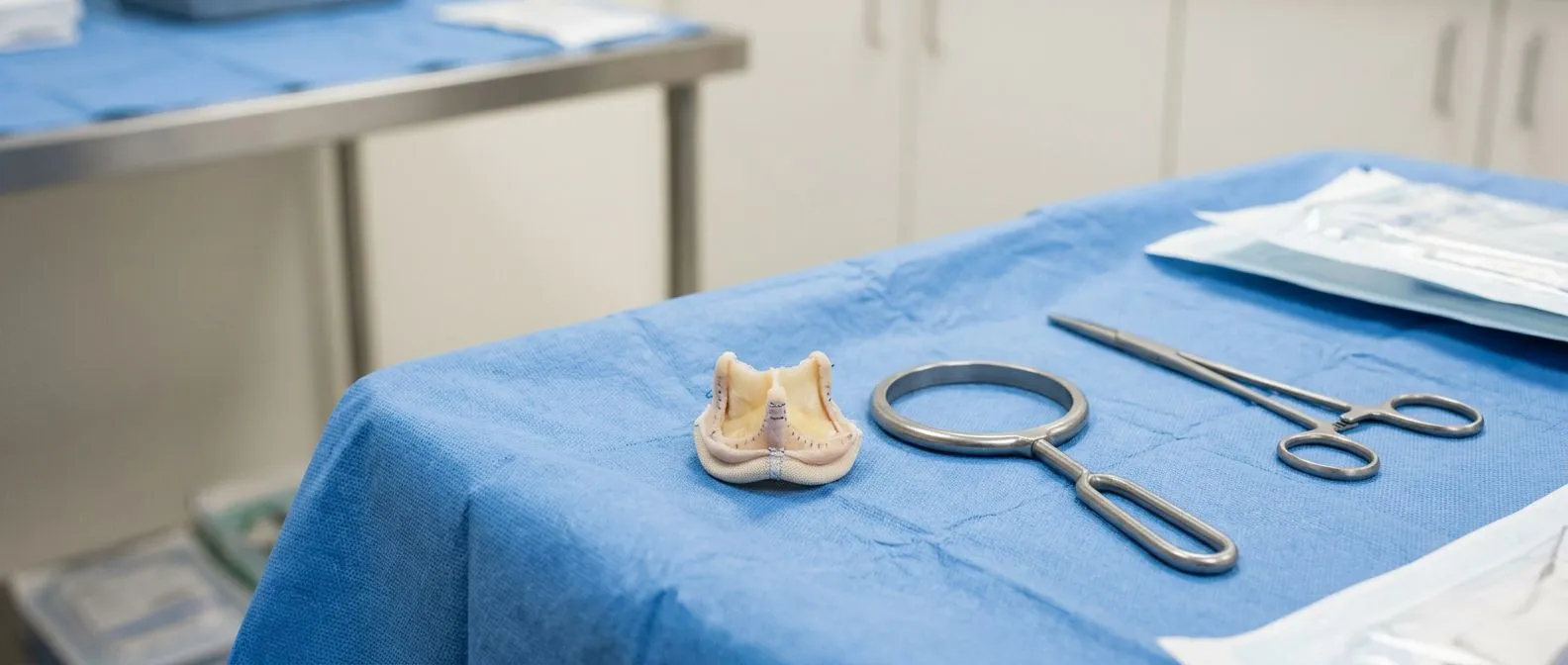
Pulmonary Valve Replacement: Surgery and Catheter Options
Learn about pulmonary valve replacement options in Guadalajara.
If you or someone in your family has been told the pulmonary valve needs attention, the first thing to know is this: you are dealing with one of the most treatable problems in heart care. The right path may be a pulmonary valve replacement through open surgery, or it may be a catheter-based procedure that avoids opening the chest at all. This guide walks you through what the valve does, what goes wrong with it, the surgical and catheter options, what recovery actually looks like, and how to decide with confidence. It is written for patients and families, in plain language, with the clinical terms explained as they come up.
What Is the Pulmonary Valve and What Does It Do?
The pulmonary valve sits between the right ventricle, the pumping chamber that sends blood to the lungs, and the pulmonary artery, the large vessel that carries that blood. It has three thin, flexible leaflets, like a three-flap doorway. Every time the right ventricle squeezes, the valve opens to let blood through. When the chamber relaxes, the leaflets snap shut so blood does not leak backward. That simple one-way action is what keeps blood moving in a single direction, from the heart to the lungs, where it picks up oxygen.
Because the pulmonary valve works at much lower pressures than the valves on the left side of the heart, problems there often develop slowly. The heart can compensate for years before symptoms appear. That is a blessing and a trap: a leaky or narrowed valve can quietly change the shape and function of the right ventricle while you feel mostly fine. Regular echocardiogram checks, which use ultrasound to image the heart, are how cardiologists track whether the valve is still doing its job.
People are often surprised to learn that the pulmonary valve is one of the four heart valves, alongside the aortic, mitral, and tricuspid valves. It gets less attention than its left-sided neighbours, yet when it fails, the whole chain of oxygen delivery is affected. Understanding its role is the first step to understanding why replacing it, at the right time, protects the heart muscle itself.
What Are the Symptoms of a Bad Pulmonary Valve?
Two main problems affect the pulmonary valve. Stenosis means the valve is narrowed and does not open fully, so the right ventricle has to push harder to force blood through. Regurgitation, sometimes called leaks or insufficiency, means the valve does not close properly, so blood flows backward into the right ventricle with every beat. Both problems make the right ventricle work overtime, and over the years that extra workload can stretch and weaken it.
Early symptoms are easy to miss. Many people feel only vague fatigue, or they notice they cannot keep up with friends during exercise. Others feel nothing at all, and the problem is picked up on a routine echocardiogram or a heart murmur heard through a stethoscope. Because the right side of the heart feeds the lungs, some patients notice breathlessness on exertion before anything else.
Advanced symptoms demand attention. As the right ventricle enlarges, you may feel fullness or palpitations in the chest, swelling in the ankles or abdomen, and breathlessness with increasingly modest activity. In severe cases, the rhythm of the heart can be disturbed. If you experience fainting, marked swelling, or a racing heartbeat, those are reasons to be evaluated promptly, not reasons to wait. The honest picture, confirmed by cardiology guidelines, is that symptoms tend to arrive late in pulmonary valve disease, which is why imaging and follow-up matter so much.
What Is Another Name for the Pulmonic Valve?
If you read medical reports, you will see the valve called by several names, and it helps to know they all mean the same thing. Pulmonic valve is the most common alternative; it is simply the adjective form used in North American medical writing. Your echocardiogram may say "pulmonic regurgitation" while your surgeon says "pulmonary leak." Neither is more correct, and neither signals a different condition.
Less often, you may see it described as the "right ventricular outflow tract valve," because it guards the exit channel of the right ventricle, a region doctors abbreviate as the RVOT. In congenital heart disease clinics, you will also encounter terms like "RVOT conduit" or "homograft," which refer to replacements implanted in that same position. When a report uses any of these words, it is describing the same doorway between the right ventricle and the pulmonary artery.
This naming matters in a practical way: when you seek a second opinion or compare reports from different hospitals, do not assume different names mean different diagnoses. Bring every report to your consultation and ask the team to explain the findings in plain words. We do exactly that in our own consultations; we take the time to explain what the studies actually show, in everyday language, and to answer every question before any decision is made. In our consultation we take the time to explain what the studies actually show, in plain words, and to answer every question before any decision.
When Is a Pulmonary Valve Replacement Needed?
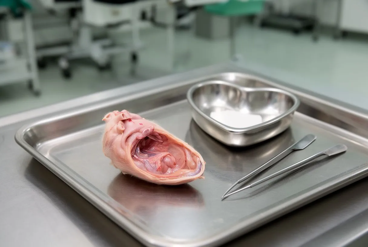
Not every pulmonary valve problem leads to surgery. Some patients with mild narrowing or a small leak are followed with imaging for years, or even indefinitely. A pulmonary valve replacement enters the conversation when the valve damage is significant enough to affect the right ventricle, or when symptoms are clearly caused by the valve. The decision rests on measurements from your echocardiogram and, often, cardiac MRI, which is the most accurate test for judging right ventricular size and function.
Several situations commonly lead to valve replacement. They are worth listing, because they show how varied the underlying stories are:
- After tetralogy of Fallot repair: adults born with this congenital heart condition often had their pulmonary valve patched or removed in childhood, and decades later the leak catches up with the right ventricle, making a pulmonary valve replacement after tetralogy of Fallot one of the most common reasons for this operation today.
- Severe stenosis: a valve narrowed by birth defect, rheumatic disease, or calcification that raises the pressure the right ventricle must generate.
- Severe regurgitation: a leak causing the right ventricle to enlarge progressively, even if you feel few symptoms.
- Worn-out replacement: a biological conduit or tissue valve implanted years earlier that has degenerated, requiring an RVOT conduit replacement.
Treatment of pulmonary valve regurgitation, in particular, follows a careful balance. Operating too early exposes you to surgical risk you may not have needed to accept; operating too late can mean the right ventricle has already lost function it will not fully recover. Cardiology guidelines therefore lean on objective markers, especially right ventricular volumes on MRI, rather than symptoms alone. A pulmonary valve replacement vs repair discussion is also part of this conversation, because in some valves, especially those affected by mitral valve prolapse on other valves, repair may preserve your own tissue, while in the pulmonary position, replacement is usually the definitive answer once the valve is beyond repair.
How Does Pulmonary Valve Replacement Surgery Work?
Conventional pulmonary valve replacement surgery is performed through an opening in the chest, usually a median sternotomy, which is the vertical incision down the breastbone. In many pulmonary cases, a smaller incision between the ribs is possible, which is the minimally invasive approach. The surgeon opens the pulmonary artery, removes the diseased or absent valve, and sews in a substitute. The heart-lung machine may or may not be needed depending on the approach; in many pulmonary operations it is used briefly.
What replaces the valve? The two families of substitutes are biological valves and mechanical valves. A biological valve replacement in the pulmonary position is by far the most common choice: it may be a homograft, which is human donor tissue, or a porcine valve, made from pig tissue, or a bovine one. These integrate well, work naturally, and do not require lifelong blood thinners. A mechanical pulmonary valve replacement is rarer in this position; mechanical valves are extremely durable but require permanent anticoagulation medication, and most teams reserve them for specific circumstances.
The published clinical literature on tissue valves in this position is substantial, including a decade-long clinical experience with decellularized fresh homografts for pulmonary valve replacement, which reported favorable outcomes for donor tissue prepared in ways that reduce immune reaction (Decellularized fresh homografts for pulmonary valve replacement: a decade of clinical experience). The practical takeaway for patients is that surgeons have more than one good tissue option, and the choice depends on your anatomy, your age, and the plan for future procedures, because biological valves eventually need replacing again.
Steps of the procedure, in plain terms, look like this: anesthesia and preparation, the chest incision, connection to support if needed, removal of the old valve or conduit, implantation and sewing of the new one, closure, and transfer to recovery. Our team performs conventional, minimally invasive, robotic, and transcatheter procedures, and we recommend the less invasive route only where the evidence supports it for that particular patient. Our team performs conventional, minimally invasive, robotic, and transcatheter procedures, and we recommend the less invasive route only where the evidence supports it for that patient.
How Does Transcatheter Pulmonary Valve Replacement Work?
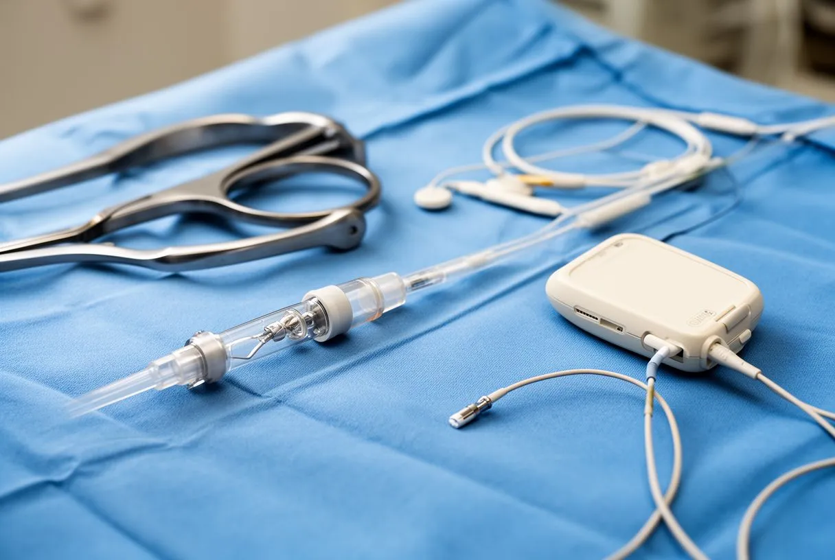
Transcatheter pulmonary valve replacement, often called TPVR, replaces the valve without opening the chest. Instead, a folded replacement valve is mounted on a balloon catheter, a thin flexible tube, and delivered through a vein, usually in the groin. The catheter is guided to the pulmonary position, the balloon inflates to expand the new valve into place inside the old conduit or ring, and the catheter is withdrawn. The whole procedure is done through a small puncture rather than an incision.
Two devices dominate this field. The Melody valve is a bovine jugular vein valve mounted in a stent, and it was the first transcatheter pulmonary valve approved for patients with failing RVOT conduits. The balloon-expandable pulmonary valve adapts the balloon-expandable valve widely used in TAVR, transcatheter aortic valve replacement, to the pulmonary position. Both are suited to patients who already have a conduit or surgical ring to anchor the new valve; a native pulmonary artery without an existing structure is generally not a suitable landing zone, which is why TPVR is most often offered to adults with repaired congenital heart disease.
The access-site technique has matured considerably. Achieving reliable closure of the puncture after transcatheter valve procedures is a well-documented challenge, and dedicated closure techniques have been described even in complex anatomies (Achieving Hemostasis Posttranscatheter Aortic Valve Replacement Using Perclose Device). In everyday terms, this means the entry site is managed with the same care as the valve itself, and most patients go home the next day or the day after.
For patients, the trade-offs are straightforward. TPVR avoids sternotomy, usually means one or two nights in hospital, and has a much faster recovery. Its limits are anatomy and durability: it needs a suitable landing zone, and the delivered valves are smaller in diameter than surgical ones. Valve-in-valve procedures, where a transcatheter valve is placed inside a degenerated surgical valve, are a natural fit for this approach and are used both in the pulmonary position and in Transcatheter Mitral Valve Replacement programs elsewhere in the heart.
How Long Does a Pulmonary Valve Last?
This is one of the most frequent questions families ask, and the honest answer is: it depends on the type of valve and your age when it is implanted. Biological valves, including homografts and porcine or bovine tissue, gradually degenerate. Tissue valves in the pulmonary position commonly serve well for many years, but younger patients wear them out faster, and a teenager or young adult may need a second replacement sooner than someone implanted in their sixties. Mechanical valves last far longer but carry the trade-off of lifelong blood thinner medication, which is why they are uncommon in the pulmonary position.
Because no valve lasts forever, the modern strategy is often staged: implant a good biological valve when it is needed, monitor it with yearly echocardiograms, and when it degenerates, consider a transcatheter valve-in-valve procedure rather than another open operation. This is a major reason TPVR has changed the outlook for adults with congenital heart disease. A pulmonary valve replacement placed surgically in your twenties may well be refreshed by a catheter procedure decades later, without ever opening the chest again.
What you can do to protect your valve is mostly about follow-up. Attend every scheduled echocardiogram. Report new breathlessness or palpitations rather than assuming they are just tiredness. Avoid infections and treat dental infections promptly, since bacteria can settle on prosthetic valves; most patients with replacement valves are advised on antibiotic prophylaxis before certain dental procedures. And keep the conversation open with your cardiology team, because the timing of the next intervention is a shared decision, not a calendar date.
What Is the Pulmonary Valve Replacement Recovery Time?
Recovery differs sharply between the two approaches, and knowing what to expect helps families plan realistically. The figures below describe typical, uncomplicated courses; your own team will adjust them to your situation.
- Open surgical replacement: one to three days in intensive care or a monitored unit, roughly five to seven days in hospital, then four to eight weeks of gradual recovery at home, with sternal precautions, meaning no lifting over a few kilograms and no driving, for about six weeks.
- Transcatheter replacement: usually one night of observation after the procedure, most people back to desk work and light activity within a week, and full activity within two to four weeks, with the main restriction being no strenuous exercise for a short period while the access site heals.
Pulmonary valve replacement recovery is generally easier than recovery from left-sided valve surgery, partly because the pulmonary position is more accessible and often allows a smaller incision. Still, recovery is not just physical. Many patients describe an emotional arc, a few low days in the second or third week, and worry about every twinge. That is normal. A structured cardiac rehabilitation program, where one is available, speeds the return to confidence as much as the return to fitness.
Follow-up matters as much as the operation itself. Expect an early postoperative check, an echocardiogram at around one to three months, and then yearly monitoring for the life of the valve. Our multidisciplinary team reviews every case against international practice guidelines before a recommendation is given, and patients receive a clear proposal with the alternatives, with the final decision always theirs. Every case is reviewed by our multidisciplinary team against international practice guidelines before a recommendation is given; the patient receives a clear proposal with the alternatives, and the final decision is always theirs.
Is Pulmonary Valve Replacement Serious?
Yes, it is heart surgery, and it deserves respect. But context matters. Pulmonary valve operations are performed on the lower-pressure, right side of the heart, which generally makes them less demanding than aortic or mitral procedures, and outcomes for appropriately selected patients are consistently good in the published experience. Serious complications are uncommon in centers that do this work regularly, but they are not zero: bleeding, infection, arrhythmias, and problems with the new valve itself are the risks your surgeon will discuss with you specifically.
For the transcatheter route, the risk profile shifts rather than disappears. Because there is no sternotomy, wound complications and prolonged recovery are far less likely. The trade-offs include the possibility that the valve migrates or needs adjustment, rhythm disturbances requiring a pacemaker in a small proportion of patients, and access-site complications at the groin. Candidly, the question is not "which option is riskless," because none is, but "which option carries the lowest risk for your situation," and that depends on your anatomy, your age, and your prior surgeries.
One more honesty note for international patients: pulmonary valve replacement cost in Mexico varies widely between hospitals, implant types, and insurance arrangements, and we do not quote figures generically because a number without your specific clinical plan would be misleading. What we can say is that a transparent itemized proposal, covering the hospital, the device, the surgeon's fee, and expected hospital stay, should be part of any consultation, and you should never accept an estimate that cannot show its components.
How Do You Choose the Right Team for Pulmonary Valve Replacement?
Choosing where and with whom to have this procedure may matter as much as choosing the procedure itself. Experience with both surgical and transcatheter techniques is valuable, because the right answer for you may be either one, and a team that only offers one of them has an obvious bias. Ask directly: how many pulmonary valve procedures does this center perform per year, and what proportion are transcatheter? A confident team welcomes these questions.
Our practice is built around that multidisciplinary model. We are a team of cardiac surgeons led by Dr Alejandro Molina, with consulting rooms in Guadalajara, Querétaro and Hermosillo, and our work covers valve disease, coronary disease, the aorta, and structural heart conditions. Dr Molina trained in cardiothoracic surgery at the Universidad de Guadalajara, in cardiac, minimally invasive, robotic, and structural surgery at the University of Montreal, and in structural cardiology and TAVR at CHU Bordeaux; the team has also trained in Jena, Bad Berka, and Buffalo. Dr Molina trained in cardiothoracic surgery at the Universidad de Guadalajara, in cardiac, minimally invasive, robotic and structural surgery at the University of Montreal, and in structural cardiology and TAVR at CHU Bordeaux; the team also trained at Jena, Bad Berka and Buffalo.
The first step for most patients is simple and free of pressure. We offer a first contact as a free video consultation: we get to know you, talk through your condition, and identify exactly which studies are still needed so no unnecessary tests are ordered; a face-to-face visit at the consulting rooms follows only when you decide to go ahead. The first contact is a free video consultation: we get to know the patient, talk through the condition, and say which studies are still needed so no unnecessary ones are ordered; a face-to-face visit at the consulting rooms follows when the patient decides to go ahead. We also encourage second opinions and treat seeking one as a normal part of deciding about heart surgery, not as a betrayal of your doctor.
To close: a failing pulmonary valve is a serious problem, but it is also one of the problems modern heart care handles best. Whether your path is a conventional pulmonary valve replacement or a transcatheter valve, the goal is the same, to protect your right ventricle and restore your energy for the life you actually live. Do not self-diagnose from articles, including this one. Bring your reports, your questions, and your doubts to a qualified cardiac team, ask about both surgical and catheter options, and make the decision with people who will explain every step. If you are considering care in Guadalajara, Querétaro, or Hermosillo, booking a consultation with a team that offers both approaches is the sensible next move.






