

Surgical Exposure
Learn how surgical exposure affects heart surgery outcomes, key techniques, approaches, and recovery details.
What is surgical exposure in heart surgery?
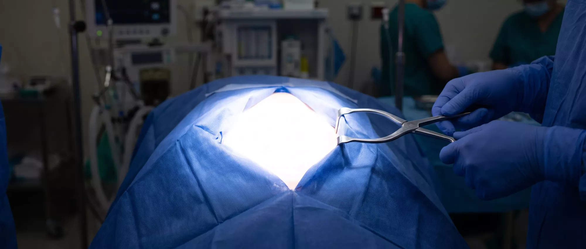
Surgical exposure refers to the physical access and clear visualization a surgeon requires to safely reach, inspect, and repair the heart. In cardiac procedures, the chest wall naturally protects the heart, meaning the surgical team must carefully navigate through layers of skin, muscle, and bone to create a working window. This access is the critical first step of any operation.
Defining the operative field: The operative field is the three-dimensional space created by the surgeon. By gently holding surrounding tissues aside with specialized instruments, the surgeon establishes a stable, unobstructed view of the target blood vessels or heart chambers. This careful preparation dictates how effectively the rest of the procedure can proceed.
For patients and families, understanding this step demystifies the beginning of the operation. The approach chosen by your surgeon depends entirely on the specific condition being treated, ensuring the medical team has the safest and most direct route to the heart.
Why surgical exposure matters for cardiac patients
The quality of surgical exposure directly influences the safety, precision, and overall success of the operation. When a surgeon has a clear, stable view of the heart, they can perform delicate repairs with absolute confidence, minimizing trauma to surrounding healthy tissues.
Impact on surgical precision: The difference between a good outcome and a great one often comes down to millimeters. Excellent cardiac surgical exposure allows the surgeon to navigate complex anatomy, place grafts accurately, or repair valves seamlessly. Poor visualization can lead to longer operative times or incomplete repairs, which is why the initial access strategy is planned so meticulously.
Patients often worry about the size of an incision, but the primary goal is always balance. The surgical team aims to provide the necessary access while respecting the body's structural integrity, ensuring you receive the most effective and durable repair possible.
How surgical exposure techniques work
Creating the surgical window involves a highly coordinated sequence of steps. After administering anesthesia, the surgeon makes precise incisions to separate superficial tissues. To access the heart, they often need to temporarily divide the sternum (breastbone) or spread the ribs, depending on the chosen approach.
Retraction and stabilization: Once the pathway is clear, mechanical devices called retractors are positioned to hold the chest walls open. These tools apply gentle, even pressure to keep the operative field visible without causing unintended fractures or tissue damage. The surgical team then secures the surrounding structures to prevent them from shifting during the procedure.
Throughout this process, maintaining a bloodless and still environment is crucial. The heart-lung machine may be introduced to take over circulation, allowing the heart to rest. This combination of physical retraction and physiological support creates the ideal conditions for complex repairs.
Types of surgical exposure approaches
Cardiac surgeons utilize several distinct methods to access the heart, each tailored to the specific disease and the patient's anatomy. The choice of approach is a major determinant of the recovery timeline and the extent of postoperative discomfort.
Comparing standard and alternative routes: The traditional method involves a full vertical incision down the chest, while alternative approaches use smaller incisions between the ribs or on the side of the chest. Each route offers different advantages regarding visibility and postoperative pain management.
- Median Sternotomy: The most common approach, providing comprehensive access to the entire heart for complex, multi-vessel, or emergency procedures.
- Mini-Thoracotomy: A smaller incision between the ribs, often used for mitral valve repairs or solitary bypasses, offering a faster recovery for suitable candidates.
- Sub-mammary Incision: An approach that curves beneath the breast, used for specific congenital repairs to optimize cosmetic outcomes while maintaining necessary access.
Deciding which method to use requires a detailed evaluation of your medical history and the technical requirements of the planned operation. Your surgeon will explain why a particular route is recommended for your specific case.
Median sternotomy exposure for open heart surgery
The median sternotomy remains the gold standard for open heart exposure. By dividing the breastbone vertically down the middle, the surgeon gains an unobstructed, direct view of the entire heart and major vessels. This approach is highly reliable and allows for rapid access in urgent situations.
Healing the sternum: After the heart repair is complete, the breastbone is brought back together and secured using stainless steel wires. These wires remain in the body permanently, acting like an internal cast. The bone heals over several weeks, much like any other fracture, requiring patients to follow specific precautions to protect the chest.
While the recovery from a sternotomy requires patience, the unparalleled visualization it provides makes it the safest option for extensive operations like multiple bypasses or complex aortic work. Patients can take comfort in knowing this method has been refined over decades.
Thoracic surgical exposure through thoracotomy
A thoracotomy creates access by making an incision between the ribs on the side of the chest, avoiding the breastbone entirely. This thoracic surgical exposure is frequently utilized for less invasive valve surgeries or to access specific arteries on the back of the heart.
Benefits of avoiding the sternum: Because the breastbone remains intact, patients generally experience less overall trauma to the chest wall. This can translate to a quicker return to normal activities and a reduced need for pain medication during the early recovery phase. It is important to use a postoperative chest support cushion to stabilize the ribcage during movement and coughing, ensuring the intercostal spaces heal securely.
However, this method is not suitable for every patient. The surgeon must carefully plan the angle of entry to ensure the target area is reachable through a smaller, lateral window without compromising the quality of the repair.
Surgical visualization and operative field management
Once physical access is established, the surgical team focuses on optimizing the operative field. Visualization is not just about seeing the heart; it is about creating a magnified, well-lit, and dry environment where tiny structures can be manipulated with absolute accuracy.
Modern visual aids: Surgeons often wear surgical loupes, which are specialized magnifying glasses, to enhance their view of delicate coronary arteries and valve leaflets. In some minimally invasive cases, a tiny camera is inserted into the chest to project a high-definition image onto a monitor. This technology allows the surgeon to navigate tight spaces with exceptional clarity.
Managing blood flow is equally critical. By using specialized suction devices and stabilizing platforms, the team keeps the immediate area clear.
Surgical site exposure risks and considerations
Every surgical exposure carries inherent considerations. Creating an opening into the chest cavity naturally introduces a risk of infection or bleeding, which is why the surgical environment is strictly controlled and sterilized. The team takes extensive precautions to protect the exposed tissues throughout the operation.
Protecting surrounding structures: When spreading the ribs or dividing the sternum, great care is taken to avoid damaging nearby nerves or the pleural lining around the lungs. Minor irritation to these structures can cause temporary discomfort during deep breathing after surgery, which is managed with tailored pain relief strategies.
Patients should be aware that the size of the incision directly relates to the extent of the operation, not necessarily the severity of the disease. Your surgeon will discuss the specific risks associated with your chosen surgical site exposure, ensuring you understand both the benefits and the potential complications before giving your consent.
Are minimally invasive exposure techniques safe?
Many patients ask if smaller incisions are as safe as traditional open approaches. Minimally invasive surgical access to the heart has been extensively studied and is considered highly safe for specific, carefully selected procedures. The safety relies entirely on the surgeon's ability to achieve adequate visualization through the restricted window.
Evaluating candidacy for smaller incisions: Not every heart condition can be treated through a minimal approach. Complex blockages, extensive valve damage, or emergency situations often require the full visibility provided by a sternotomy. The surgical team evaluates each patient individually, weighing the benefits of a smaller scar against the need for uncompromised surgical precision.
When a minimally invasive route is deemed appropriate, patients often enjoy shorter hospital stays and less noticeable scarring.
Recovery and healing after surgical exposure
The way the chest was opened dictates the recovery process. Healing from surgical exposure is a gradual journey that requires patience and adherence to medical guidance. The superficial skin layers generally heal within a few weeks, but the deeper structural tissues take longer to regain full strength.
Protecting the access site: During the first few weeks, patients must avoid heavy lifting and sudden pulling motions to allow the bone and muscle to knit together securely. Your medical team will provide clear guidelines on how to safely perform daily tasks, such as getting out of bed or supporting your chest when coughing, without stressing the healing incision.
Ultimately, the temporary discomfort of the healing process is a necessary step toward long-term cardiovascular health. By following your surgeon's instructions and attending all follow-up appointments, you allow your body the time it needs to fully recover from the procedure and restore your heart's function.






