

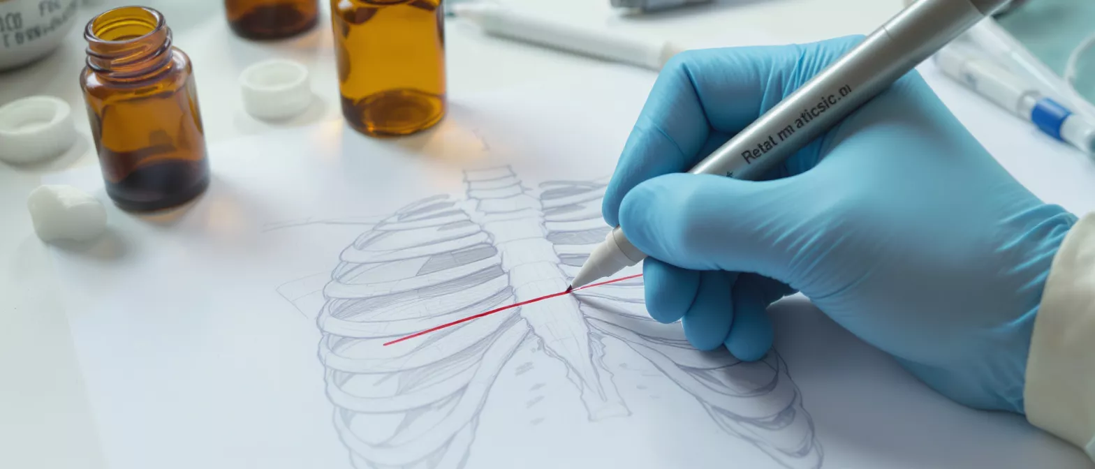
Thoracotomy
Learn what a thoracotomy is, how it works in heart surgery, recovery, risks, and post-op care.
What is a thoracotomy?
A thoracotomy is a major surgical procedure that involves making a large incision into the chest wall to access the organs inside the thoracic cavity, primarily the heart and lungs. For a patient facing heart surgery, hearing this term can naturally feel overwhelming. However, this approach is a fundamental and well-established technique that allows surgeons to clearly see and repair vital structures that are otherwise impossible to reach. It is a deliberate, controlled entry into the chest to fix critical problems.
Why the chest must be opened: The ribcage is a rigid, protective cage of bone designed to shield the heart and lungs from trauma. While this protection is essential for daily life, it creates a physical barrier during a medical emergency or planned operation. A thoracotomy provides the necessary physical space and direct visibility required to perform intricate repairs on the heart, major blood vessels, or lung tissue.
The difference between an incision and the procedure: The thoracotomy incision itself is simply the pathway created by the surgeon. Depending on the specific cardiac condition being treated, the cut may be made between the ribs or through the breastbone. Understanding this distinction helps patients realize that the incision is just the first step in a highly choreographed operation aimed at restoring cardiovascular health.
Why thoracotomy is performed in heart surgery
When it comes to cardiac care, a thoracotomy procedure is performed to give surgeons direct access to the heart and the great vessels surrounding it. The heart is located in the mediastinum, the central compartment of the chest. Accessing it requires careful navigation through the chest wall. This surgical route is often necessary for repairing congenital heart defects, replacing damaged heart valves, or performing coronary artery bypass surgery.
Emergency versus planned operations: In trauma situations, a thoracotomy might be performed as an emergency life-saving measure to relieve pressure around the heart or repair a ruptured blood vessel. In the context of planned heart surgery, it is a calculated decision made by your surgical team to provide the safest and most effective environment for complex repairs. Both scenarios rely on the same fundamental principle of gaining clear access to the chest cavity.
The specific approach chosen depends heavily on the exact nature of the heart condition. A surgeon will evaluate diagnostic imaging to determine whether a traditional opening through the sternum or an incision between the ribs offers the best chance for a successful outcome. Every decision is tailored to the individual patient's anatomy and the surgical goals.
How thoracotomy surgery works
Understanding the thoracotomy surgery process can demystify the experience for patients and their families. Once you are under general anesthesia, the surgical team prepares the chest area and makes the planned incision. To reach the heart, the surgeon must carefully move through layers of skin, muscle, and sometimes bone. Specialized retractors are used to gently spread the ribs or separate the sternum, creating a wide enough window to operate.
Once the chest is open, the surgeon gains a direct view of the heart and surrounding structures. Depending on the operation, you may be placed on a heart-lung bypass machine. This machine temporarily takes over the job of pumping blood and oxygenating it, allowing the surgeon to stop the heart safely and perform delicate repairs on a still, bloodless surface.
- Incision and access: Careful entry through tissue and bone to expose the heart.
- Bypass initiation: Diverting blood circulation to keep the heart still during repair.
- Surgical repair: Fixing valves, clearing blockages, or correcting structural defects.
After the cardiac repair is complete, the heart is allowed to resume its normal beating. The surgeon then meticulously closes the incision, wiring the sternum back together if it was divided, and suturing the muscle and skin layers. The entire process is a highly coordinated effort between the surgeon, anesthesiologist, and perfusionist.
Thoracotomy vs thoracostomy key differences
Patients often confuse similar medical terms, and the comparison of thoracotomy vs thoracostomy is a common source of confusion. While they sound almost identical, they represent very different medical procedures. A thoracotomy is a major open surgery that involves cutting into the chest wall to access the organs inside, typically requiring an operating room and general anesthesia.
Understanding a thoracostomy: A thoracostomy, commonly known as a chest tube insertion, is a much smaller procedure. It involves placing a thin, flexible tube through the chest wall into the pleural space to drain fluid, blood, or air. It does not involve opening the chest to expose the organs. It is frequently used in emergency departments to treat a collapsed lung or pleural effusion.
The primary difference lies in the scale and purpose of the intervention. A thoracotomy is designed for direct surgical intervention on internal organs, while a thoracostomy is designed for drainage and decompression. Knowing the difference can help patients better understand their medical records and the specific procedure their doctor is recommending.
Types of thoracotomy incisions
The thoracotomy incision is not a one-size-fits-all approach. Surgeons use several different types of cuts depending on the specific heart or lung condition being treated. The location and size of the incision are carefully chosen to provide the best access while minimizing trauma to the surrounding healthy tissue. Your surgeon will select the most appropriate method based on your anatomy and surgical needs.
Median sternotomy: This is the most common incision for heart surgery. The surgeon makes a vertical cut down the middle of the chest and divides the sternum, or breastbone. This approach provides excellent exposure to the heart and is routinely used for valve replacements and bypass surgeries.
Posterolateral and anterolateral incisions: These approaches involve making an incision along the side of the chest and entering between the ribs. They are often used for lung surgeries or specific heart procedures like repairing certain congenital defects. Because the sternum remains intact, recovery from the bone itself is different than a median sternotomy.
Minimally invasive thoracotomy approaches
Advancements in surgical technology have led to minimally invasive thoracotomy options for certain heart patients. Instead of a large, open incision, surgeons can sometimes perform procedures through much smaller cuts. These techniques utilize specialized instruments and small cameras to navigate the chest cavity, reducing the overall trauma to the body.
Video-assisted thoracoscopic surgery (VATS): VATS involves making several small incisions between the ribs. A tiny camera is inserted to project images onto a monitor, guiding the surgeon as they use specialized tools. This is more commonly used for lung biopsies or specific lung resections, but minimally invasive cardiac techniques are expanding rapidly.
Not every patient is a candidate for a minimally invasive approach. The surgeon must evaluate the complexity of the required repair, the patient's overall health, and their specific anatomy. For many, minimally invasive heart surgery tools offer a quicker return to normal activities, but traditional open methods remain the gold standard for complex reconstructions.
Thoracotomy recovery and hospital stay
The thoracotomy recovery process is a critical phase that requires patience and careful monitoring. Immediately after surgery, patients are transferred to the intensive care unit (ICU). Here, the medical team closely watches vital signs, manages pain, and ensures the heart and lungs are functioning properly as the anesthesia wears off. The initial hospital stay typically lasts several days to a week, depending on the complexity of the operation.
Initial healing and monitoring: During the first few days, patients will have several tubes and monitors attached to them. These include chest tubes to drain excess fluid, intravenous lines for medication, and monitors to track heart rhythm. The nursing staff will encourage gentle movement, such as sitting up in bed or taking short walks, to promote circulation and prevent complications.
Before being discharged from the hospital, the care team ensures the patient is stable, pain is manageable, and basic mobility is restored. It is completely normal to feel fatigued and sore during this time. The chest wall has undergone significant trauma, and the body needs energy to heal the deep tissues and bone.
Post thoracotomy care and pain management
Effective post thoracotomy care is essential for a smooth recovery at home. Pain management is a top priority, because deep breathing and movement are necessary for healing but can be uncomfortable after a chest incision. Your medical team will prescribe a regimen of pain relievers to keep discomfort at a tolerable level. It is crucial to take these medications as directed rather than waiting for the pain to become severe.
Breathing exercises and incision care: You will be given a spirometer, a small device that helps you practice taking deep breaths. Using this regularly prevents pneumonia by keeping the lungs fully inflated. You must also keep the incision site clean and dry, watching for any signs of infection such as redness, swelling, or unexpected discharge. Utilizing proper post-operative recovery supports can also ease the strain on the chest during movement.
- Deep breathing: Use the spirometer hourly while awake to keep lungs clear.
- Gradual walking: Increase walking distance daily to rebuild stamina safely.
- Incision protection: Avoid heavy lifting and sudden twisting to protect the healing bone.
Recovery is a gradual journey. Patients should expect to feel easily tired for several weeks. Following the surgeon's specific guidelines regarding activity restrictions, wound care, and medication schedules is the best way to ensure the chest heals completely.
Thoracotomy risks and potential complications
As with any major operation, there are inherent thoracotomy risks and potential complications. Because this procedure involves opening the chest and stopping the heart in some cases, it is a serious medical event. However, it is important to remember that these operations are performed by highly trained specialists who take extensive precautions to minimize danger and ensure patient safety.
Common surgical risks: General risks include bleeding, infection at the incision site, and adverse reactions to anesthesia. Because the chest wall is opened, there is a specific risk of air leaking from the lung into the chest cavity, which may require a chest tube to resolve. Irregular heart rhythms, known as arrhythmias, are also relatively common after heart surgery and are typically managed with medication.
Patients can actively reduce certain risks by quitting smoking before surgery, maintaining good nutrition, and closely following pre-operative instructions. The surgical team will conduct a thorough evaluation beforehand to weigh the risks of the procedure against the risks of leaving the heart condition untreated. In almost all cases, the danger of the underlying disease far exceeds the risks of the surgery itself.
Life after thoracotomy and long-term outlook
Life after a thoracotomy generally involves a period of adjustment, but the long-term outlook is highly positive for most patients. The primary goal of the surgery is to correct a life-threatening or debilitating heart condition. Once the initial recovery phase is complete, many patients experience a significant improvement in their symptoms, such as reduced chest pain and increased exercise tolerance.
Resuming normal activities: Full recovery of the chest tissues and bone can take several months. Patients are usually advised to avoid strenuous activities, such as heavy lifting or driving, for the first few weeks. Gradually, as the body heals, you will be able to return to your normal routine. Cardiac rehabilitation is often recommended to safely rebuild cardiovascular strength under supervised conditions.
It is natural to have concerns about the physical changes after surgery. The thoracotomy scar will fade over time, serving as a permanent reminder of a successful medical intervention.






