

Haemodynamic Monitoring in Cardiac Intensive Care
Haemodynamic monitoring cardiac ICU: understand how continuous blood flow tracking guides safe recovery after heart surgery.
Haemodynamic monitoring in the cardiac ICU is the continuous measurement of how blood is flowing through your body after heart surgery. If your family member has just come out of the operating room, you will see screens beside the bed filled with numbers, waveforms and colours. Those numbers are not decoration. They are the team's way of watching the heart work minute by minute, so that problems are corrected before they become complications. This guide explains what is being measured, what each number means, and how it shapes decisions about recovery.
What Is Haemodynamic Monitoring in Medical Terms?
In medical terms, "haemodynamic" refers to the forces that move blood through the circulation: the pressure the heart generates, the resistance it meets in the vessels, and the volume of blood it pumps with each beat. Haemodynamic monitoring is the practice of measuring these forces continuously so that treatment can be adjusted in real time. In the Intensive Care Unit, it is the difference between reacting to a crisis and preventing one.
Why the heart needs watching after surgery. A heart that has just been operated on is temporarily tired. The muscle has been handled, cooled, and given medications to slow it down during the operation. The first hours after surgery are exactly when the heart may not pump as well as it did before. Continuous monitoring lets the team see this early, while the response to treatment is still simple.
What the screens actually show. The monitors display blood pressure waveforms, heart rate, oxygen saturation, and the derived values calculated from them. Each has a role. The team does not treat numbers in isolation; they read patterns over time. We tell families the same thing we tell patients: a single number rarely tells the story, but the trend does.
What Are the Basic Principles of Haemodynamics?
Four basic questions guide every haemodynamic assessment in the cardiac ICU. Is there enough blood volume? Is the pump strong enough? Are the vessels open and responsive? Is the blood actually delivering oxygen where it is needed? Every monitor, catheter and measurement in the unit answers one of these questions.
Preload, contractility, and afterload. These are the three words you will hear most often. Preload is how much blood fills the heart before it beats. Contractility is the strength of the squeeze. Afterload is the resistance the heart has to pump against. When a patient's blood pressure is low, the team works out which of these three is the problem, because the treatment differs completely for each.
From measurement to action. The point of monitoring is not the data itself but the decisions it enables. A consensus published by the European Society of Intensive Care Medicine Consensus on circulatory shock and hemodynamic monitoring emphasises that monitoring is only valuable when the results lead to a defined, prompt treatment plan. That principle shapes cardiac ICU protocols worldwide, including the ones we follow: every case is reviewed by the multidisciplinary team against international practice guidelines before any decision is made.
What Are the Main Methods for Measuring Cardiac Output?
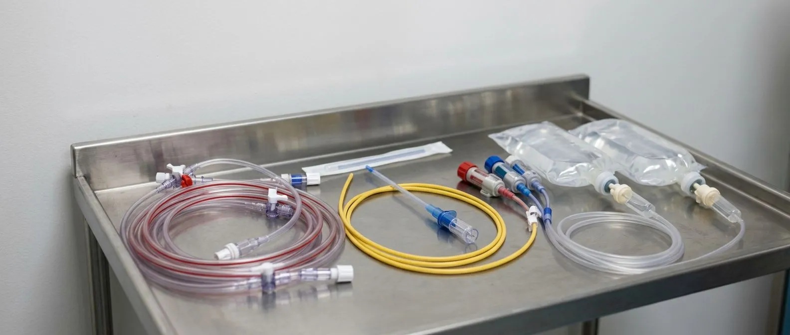
Cardiac output is the volume of blood the heart pumps each minute, and knowing it is central to haemodynamic monitoring. Several cardiac output monitoring methods exist, and cardiac ICUs commonly use more than one at the same time. The main techniques include:
- Pulmonary artery catheter (thermodilution): a thin catheter measures blood temperature changes to calculate flow directly. The classic reference method.
- Pulse contour analysis: an arterial line's waveform is analysed to estimate each heartbeat's output continuously, without extra catheters.
- Transoesophageal echocardiography: ultrasound via a probe in the oesophagus shows the heart's chambers and valves directly, common during and immediately after cardiac surgery.
- Mixed venous oxygen saturation: indirect flow measurement, since blood returning to the heart reveals how much oxygen the body has used.
- Oesophageal Doppler: sound waves track blood velocity in the aorta to estimate flow.
Which method is chosen and why. The choice depends on the type of surgery, the patient's condition, and how invasive the team judges it is reasonable to be. Simpler patients are managed with non-invasive devices; complex cases get direct measurement. The aim is always the least invasive method that still answers the clinical question.
What Is an Arterial Line in the Cardiac ICU?
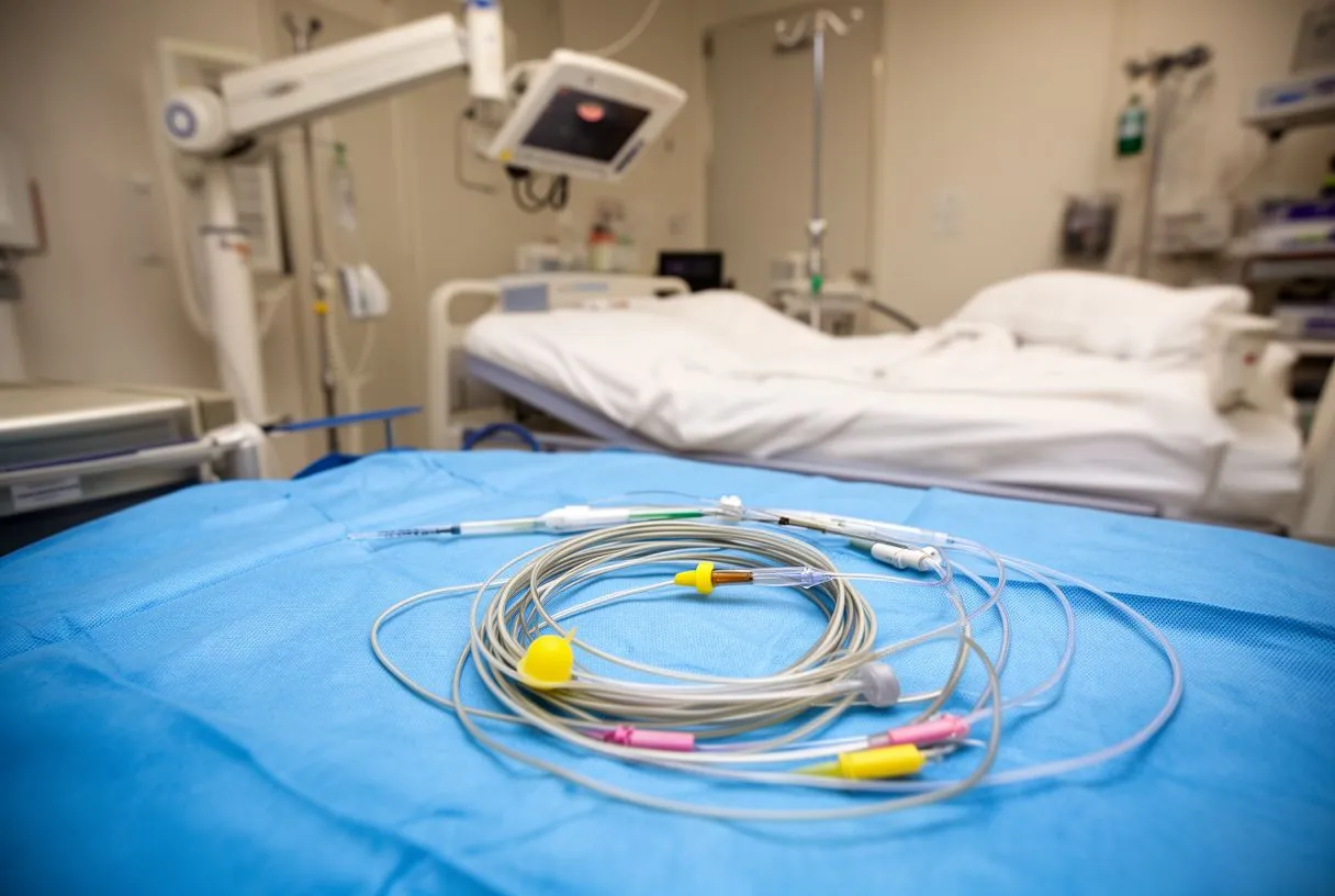
An arterial line is a very thin catheter placed into an artery, usually at the wrist, connected to a pressure sensor. It gives a beat-to-beat blood pressure reading on the monitor, far more responsive than an arm cuff, which inflates every few minutes. In an arterial line cardiac ICU setup, this waveform is also the raw data used to calculate stroke volume and fluid responsiveness.
What it feels like and how long it stays. For the patient, an arterial line is usually painless once placed; the wrist may feel slightly bruised afterwards. It typically stays in for one to three days after surgery, removed once medications are stopped and blood pressure is stable on intermittent cuff readings.
Why invasive haemodynamic monitoring matters early on. After open-heart surgery, blood pressure can swing quickly, especially during the first night when the effects of anaesthesia wear off. A cuff-based reading that updates every five minutes is simply too slow. In our practice, we explain to patients before surgery exactly which lines will be placed and why, so the ICU looks familiar rather than frightening when they wake up.
When Is Pulmonary Artery Catheter Monitoring Used?
Pulmonary artery catheter monitoring involves a longer catheter, guided through a neck vein into the pulmonary artery, the vessel running from the right side of the heart to the lungs. It measures pressures inside the heart and lungs and allows direct cardiac output measurement by thermodilution. It is the most complete form of invasive haemodynamic monitoring available at the bedside.
When the team reaches for it. It is reserved for complex situations: patients with severe pulmonary hypertension, significant valve disease, weak heart muscle before surgery, or unexpected instability after surgery. For routine cases, less invasive tools are sufficient. The decision is a judgement about how much information the situation truly requires.
Accuracy versus risk. The catheter carries small risks of arrhythmia during placement and of infection with prolonged use, which is why modern practice uses it selectively rather than routinely. A large review presented at the 36th International Symposium on Intensive Care and Emergency Medicine International Symposium on Intensive Care and Emergency Medicine reflects this shift: cardiac output is now often measured with less invasive devices, reserving the pulmonary artery catheter for cases where its detail genuinely changes management.
What Do CVP and Mixed Venous Oxygen Saturation Tell Us?
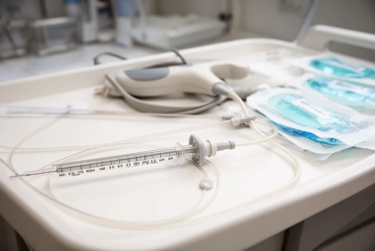
CVP monitoring is standard in every cardiac ICU. CVP stands for central venous pressure, and it estimates the filling pressure of the right side of the heart. It is measured through the central line already placed before surgery, so it adds no new procedure. A low CVP often points to low blood volume; a high one may suggest the heart is struggling to handle the blood it receives, or that fluid is backing up.
Mixed venous oxygen saturation explained. Mixed venous oxygen saturation, written SvO2, measures how much oxygen remains in the blood returning to the heart after the body has used it. If the body is taking too much oxygen out of the blood, the returning saturation drops. This can happen when cardiac output is low, when the patient is feverish, or when pain and shivering increase the body's demand. It is an early warning signal, often changing hours before blood pressure does.
Interpreting the two together. Neither number is conclusive alone. A high CVP with a low SvO2 tells a different story from a low CVP with a normal SvO2. Reading them as a pair, alongside the arterial line and cardiac output, is how the team distinguishes between "give fluid," "support the heart," or "reduce the workload."
What Is a Bad Cardiac Output Number?
A normal cardiac output in an adult at rest is roughly 4 to 8 litres per minute. But raw output depends on body size, so clinicians use cardiac index, which is cardiac output divided by body surface area. A normal cardiac index is about 2.5 to 4.0 litres per minute per square metre. A bad number is not a single value but a pattern: a cardiac index falling below about 2.2, combined with signs that the organs are not getting enough blood, defines a genuinely concerning situation.
Context changes the threshold. A patient who is warm, awake, urinating and breathing comfortably can tolerate a lower index for a while than one who is cold, confused and producing no urine. This is why the team always reads the number against the patient in front of them rather than against the screen alone. When we review a postoperative course, our team looks first at the trend and the clinical picture together, then at the isolated figure.
What happens when the number is bad. The response is stepwise: check volume status, correct anaemia or rhythm problems, then support the heart with medication. Persistent low output despite these steps may require returning to the operating room, which is why early detection matters so much.
How Is Fluid Responsiveness Checked After Heart Surgery?
Fluid responsiveness answers a deceptively simple question: if we give this patient extra fluid, will the heart actually pump more, or will the fluid simply pool in the lungs? Giving fluid to a patient whose heart cannot use it causes congestion; withholding it from a patient who needs it causes poor perfusion. The measurement helps the team get this right.
Stroke volume variation. The arterial line allows continuous calculation of stroke volume variation, the change in each beat's output caused by the natural rise and fall of pressure during breathing. When the variation is large, the heart is likely to respond to extra fluid. When it is small, the heart is already filling well and more fluid will not help. Patients on a ventilator with a regular heart rhythm give the most reliable values.
Other bedside tests. Where variation cannot be used, the team may perform a small, controlled fluid challenge, watching how the pressures and output respond over ten to fifteen minutes. It is a careful, measured trial rather than a guess, and it is repeated only when needed.
How Are Vasopressors Titrated in the Cardiac ICU?
Vasopressors are medications that tighten blood vessels to raise blood pressure. Vasopressor titration in the cardiac ICU means adjusting their dose continuously to hit a defined target pressure while using the smallest possible amount. Too little leaves the organs under-perfused; too much squeezes the small vessels, including those feeding the kidneys and gut. The goal is the balance, not the maximum.
What titration looks like in practice. The team sets a target range, often based on the patient's own baseline blood pressure rather than a fixed number for everyone. They adjust the infusion every few minutes against the arterial line, checking urine output, lactate levels and, where available, cardiac output. As the heart recovers its strength over the first day or two, the medication is weaned step by step.
Lactate as a guide. Lactate is a substance released when tissues are short of oxygen. A falling lactate on repeat blood tests is one of the most reassuring signs that perfusion is adequate, even if the blood pressure itself is not yet where the team wants it. Rising lactate despite treatment signals that something deeper needs investigating.
What Is Low Cardiac Output Syndrome and How Is It Caught Early?
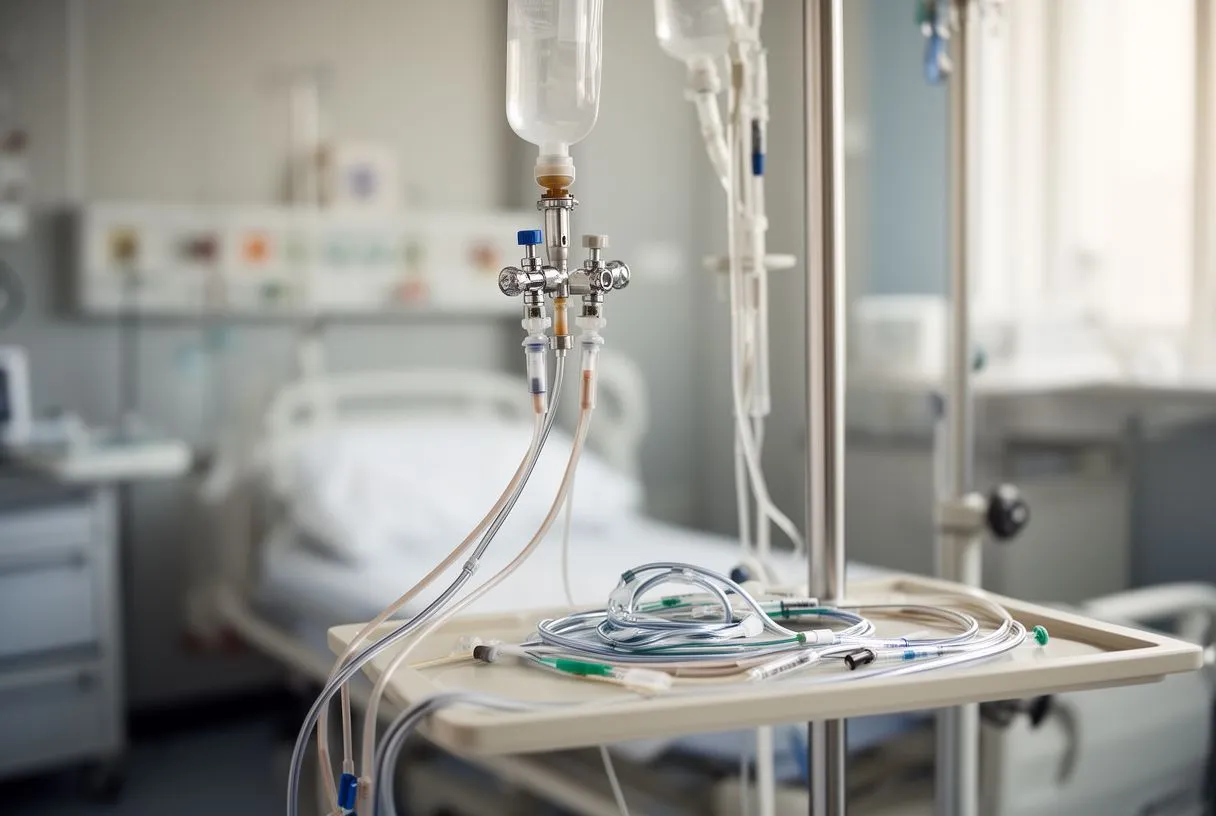
Low cardiac output syndrome is the most significant haemodynamic complication after cardiac surgery: the heart simply does not pump enough to meet the body's needs. It typically appears in the first hours to days after surgery, while the heart is still recovering from the operation itself. Continuous haemodynamic monitoring exists largely to catch this syndrome early, because the treatments work best when applied promptly.
The warning signs. The syndrome rarely announces itself with one number. Instead, several findings drift in the same direction over a few hours. The signs the team watches for are:
- A cardiac index steadily falling toward or below 2.2 litres per minute per square metre.
- Cool hands and feet with prolonged capillary refill, showing the body prioritising vital organs.
- Urine output dropping below what the kidneys should be producing.
- Lactate rising on repeat blood tests despite adequate oxygen levels.
- New rhythm disturbances such as atrial or ventricular arrhythmias.
How it is treated. Treatment follows the same ladder: optimise volume, correct the rhythm, support the heart with inotropes (medications that strengthen the squeeze), and escalate if the numbers and the patient do not improve. The overwhelming majority of episodes resolve with medication as the heart recovers. In our experience, when patients and families understand what the monitors are watching for, the first night in the ICU feels far less frightening — and we make a point of explaining it before the operation, during the consultation, in plain words, answering every question before any decision.
Choosing care that monitors well. If you are weighing where to have surgery, the quality of postoperative monitoring deserves as much attention as the operation itself. Ask which monitoring devices the ICU uses routinely, whether a cardiac output measurement will be available for your case, and how the team defines its targets for the first night. Before surgery with us, the first contact is a free video consultation, where we review the studies already done and say which ones are still needed, so nothing unnecessary is ordered. We also encourage second opinions and treat them as a normal part of deciding about heart surgery.
Haemodynamic monitoring in the cardiac ICU is best understood as a set of questions asked continuously: is there enough blood, is the pump strong, are the vessels responsive, and is oxygen reaching the tissues? Each device on the bedside screen answers one of them. If you or a family member is preparing for heart surgery, bring your questions about the ICU to the team before the operation. A qualified cardiac team — ideally one that reviews every case against international guidelines, as ours does — can explain exactly which monitoring will be used for your situation and why. Book a consultation to discuss your own circumstances rather than relying on general information; the details of your heart, your operation and your recovery are the ones that matter.






