

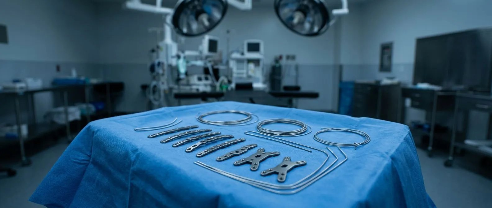
Inverted T-Incision in Upper Sternotomy Valve Surgery
Inverted t-incision sternotomy offers faster recovery with less chest trauma for heart valve surgery.
An inverted t-incision sternotomy is a way of opening the chest for heart valve surgery that spares most of the breastbone. Instead of a full cut down the entire sternum, the surgeon makes a shorter upper incision and divides the bone in a T shape — across the top and down only partway. For patients facing aortic or mitral valve surgery, this can mean less chest trauma, a faster return to daily life, and a smaller scar. This guide walks through what the technique involves, how it compares with a full sternotomy, who qualifies, what recovery really looks like, and the honest risks — so you can discuss your own situation with a cardiac team from a position of understanding.
What Is an Inverted T-Incision Sternotomy?
The sternum is the flat bone at the front of your chest. In traditional open heart surgery, it is divided completely from top to bottom so the surgeon can reach the heart. In an inverted T approach, the incision starts at the top of the sternum and the bone is cut across, with a short vertical extension — like a letter T turned upside down.
Why the shape matters: the aortic valve and the upper part of the heart sit directly behind the top of the breastbone. A partial upper cut gives the surgeon a direct window to exactly that area, without disturbing the lower half of the bone or the rib attachments below it.
Sternal sparing cardiac surgery: keeping most of the sternum intact preserves the chest wall's strength. The bone heals faster because less of it was divided, and the lower chest — where the diaphragm and abdominal muscles attach — keeps its normal mechanics. This is the core idea behind all sternal-sparing techniques, of which the inverted T is one of the most established.
Inverted T Sternotomy vs Full Sternotomy: What Changes?
Understanding the comparison helps you weigh the trade-off. A full sternotomy gives the surgeon the widest possible view and access to every part of the heart. It has been the standard for decades, and it remains the right choice for many operations — especially multi-valve or combined procedures.
Access and visibility: with a partial upper sternotomy, exposure is more limited. The surgeon works through a smaller window, which demands more experience and better instruments. Some patients are not ideal candidates because of their anatomy, body habitus, or the specific problem being treated.
Recovery differences: because less bone is divided, patients generally mobilize earlier, breathe more comfortably, and avoid the full six to eight week sternal precautions that a completely divided sternum requires. Wound care is simpler and the visible scar is shorter.
- Full sternotomy: maximum access, standard for complex or emergency cases, longer bone healing.
- Inverted T (upper sternotomy): smaller window, suited to isolated valve work, faster chest wall recovery.
Which Heart Valves Can Be Treated Through an Inverted T-Incision?
The upper sternum sits directly over the aortic valve, so this approach was developed first for aortic valve replacement. It is now used more broadly as surgical experience has grown.
Aortic valve surgery: the aortic valve — the valve between the left pumping chamber and the main artery to the body — is the natural fit. Aortic stenosis (narrowing) and aortic regurgitation (leaking) are both treated through this window, and the approach pairs well with a less invasive aortic valve replacement using any standard prosthetic valve.
Mitral valve access: the mitral valve — the valve between the left collecting chamber and the left pumping chamber — sits slightly deeper and to the left. Some surgeons repair or replace it through an upper sternotomy, though many prefer a right minithoracotomy (a small cut between the ribs on the right side) for that valve. Conditions like Mitral Valve Prolapse, where the valve leaflets billow backward, can be repaired through small incisions when the team has the right experience.
How the Inverted T-Incision Procedure Works, Step by Step
Knowing what actually happens in the operating room removes much of the fear. The heart-lung machine is still used, and the operation is still genuine open heart surgery — only the access is different.
The incision and exposure: you are under general anesthesia. The surgeon makes a skin incision roughly 8 to 10 centimeters at the top of the chest, then divides the upper sternum transversely and partway down — the T shape. A small retractor gently opens this window.
The valve operation: the heart is connected to the cardiopulmonary bypass machine, which takes over circulation so the heart can be temporarily stopped and still. The diseased valve is removed or repaired, and the new valve — whether a tissue valve such as a Bovine Pericardial Valve or Porcine Valve, or a mechanical one — is sewn into place. A Stented Valve includes a cloth-covered frame that helps it seat securely; a Stentless Valve, used mainly in the aortic position, has no frame and may fit more naturally.
Closure: the heart is restarted, the small window is closed with wires or plates for the divided portion of bone, and the skin is closed in layers. Many patients are surprised that the visible incision is shorter than a standard collarbone-to-abdomen scar.
Who Is a Candidate for Upper Sternotomy Minimally Invasive Surgery?
Candidacy depends on the valve involved, your anatomy, and your overall health — not on age alone.
Good candidates usually: need isolated aortic valve surgery, have no severe calcification extending beyond the upper sternum, have reasonable lung function, and have not had previous chest surgery through the same area. Body build matters; very broad or very small chests can change the geometry of access.
Patients who may need a different approach: those needing multiple valves repaired at once, those with an enlarged aorta requiring full replacement, or patients whose calcified aorta makes handling risky. In those situations, a full Median Sternotomy, a Mini-sternotomy, or a transcatheter route may be safer. Knowing your heart: the aortic valve and its geometry is precisely why the team studies your CT scan before recommending any incision.
- Reviewed individually: no one should be told "you can have minimally invasive surgery" based on a phone call alone.
- Imaging first: a CT scan of the chest and aorta drives the incision choice more than any preference.
At our practice, every case is reviewed by the multidisciplinary team against international practice guidelines before a recommendation is given, and the patient receives a clear proposal with the alternatives — the final decision is always theirs.
Inverted T-Incision Sternotomy Recovery: What to Expect Week by Week
The hospital stay: most patients stay three to five days. Because the lower sternum is intact, you can typically sit up, cough, and walk with less discomfort than after a full sternotomy. Chest drainage tubes come out within a day or two.
The first weeks: you will still follow sternal precautions — no lifting, pushing, or pulling with your arms — but usually for a shorter period, since only the upper portion of bone was divided. The inverted T incision healing time varies by person, bone quality, and factors like diabetes or smoking, but the divided segment is small and generally knits faster than a fully split sternum.
Back to life: light activity resumes within weeks under your cardiac rehabilitation team's guidance, with driving and work returning once your surgeon confirms adequate bone healing on follow-up imaging. Recovery is a partnership: the operation is the start, and rehab is where the outcome is secured.
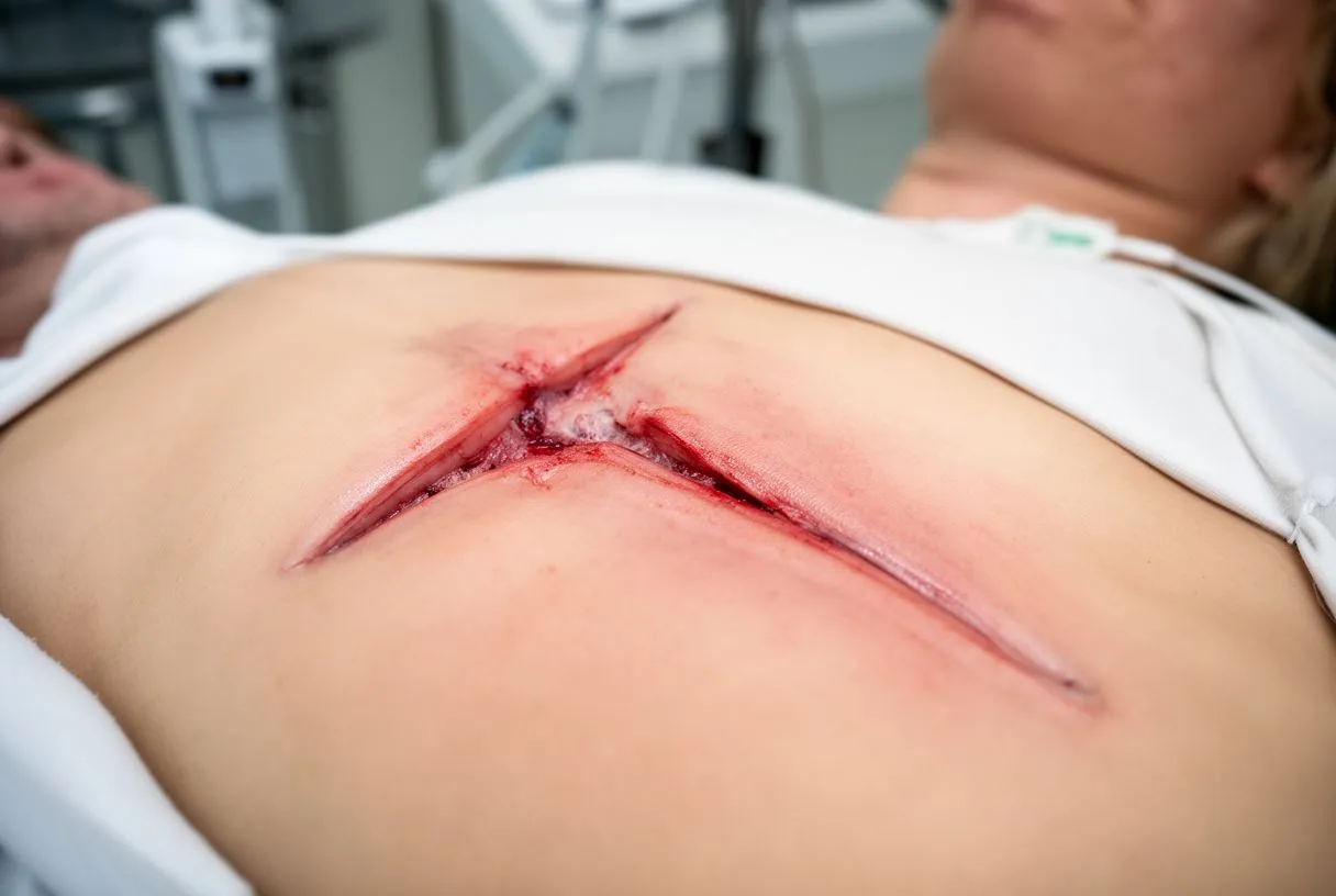
In our practice, patients recovering from these procedures tell us the smaller incision made coughing, sleeping, and early walking noticeably easier — and that shorter precautions made the day-to-day recovery feel less restrictive. We still insist on the same discipline with rehab and follow-up.
Inverted T Sternototomy Complications and Risks
Honesty about risk is part of good medical advice. The overall complication profile of a partial upper sternotomy is comparable to — and in selected patients favorable against — a full sternotomy, but the risks are not zero.
Surgical risks to discuss: bleeding, infection of the wound or bone, stroke, heart rhythm disturbances, and the need to convert to a full sternotomy if exposure proves inadequate. Conversion is not a failure; it is a planned fallback every experienced team keeps ready.
How outcomes are measured: modern valve surgery research uses standardized endpoint definitions, such as the Valve Academic Research Consortium 3: Updated Endpoint Definitions for Aortic Valve Clinical Research (https://pure.eur.nl/en/publications/c616e4fd-9912-4322-8400-da97c8169414), which define complications like bleeding, stroke, and Structural Valve Deterioration in consistent ways so results from different centers can be compared honestly. When a team quotes you complication rates, ask which definitions they used.
Access-site care: precise management of vessels and closure devices matters in minimally invasive work; techniques for achieving reliable closure are an active area of surgical refinement, as described in published case reports such as Achieving Hemostasis Posttranscatheter Aortic Valve Replacement in a Patient with Aortobifemoral Bypass Graft Using Perclose Device: A Novel Technique (http://dx.doi.org/10.1155/2021/4253570). Small details like this are where surgical experience shows.
Because we perform conventional, minimally invasive, robotic and transcatheter procedures, we are honest with patients about when the inverted T route is genuinely the lowest-risk option for their situation — and when it is not.
Less Invasive Aortic Valve Replacement: TAVR and When It Fits
What TAVR is: Transcatheter Aortic Valve Implantation — called TAVR in the United States and here — replaces the aortic valve through a catheter, usually inserted through an artery in the groin, with no chest incision at all. A Sutureless Valve design makes rapid deployment possible inside the catheter system. For many older or higher-risk patients, TAVR has become the preferred route.
How TAVR compares with an inverted T approach: TAVR avoids the sternum entirely, but it is currently best established for older patients and those at higher surgical risk, and long-term durability data continues to mature through ongoing studies — you can search the current landscape of such trials at clinicaltrials.gov (https://clinicaltrials.gov/). An inverted T sternotomy offers direct surgical repair or replacement with decades of prosthetic valve track record, which may matter more for a younger, active patient.
Neither is "better" in the abstract: the right choice depends on your age, valve anatomy, calcification pattern, life expectancy, and goals. A Sutureless Valve deployed surgically through an upper sternotomy is one middle path some teams offer; a Valve-in-valve procedure can extend the life of a previous surgical valve without reoperation.
In our consultations we take the time to explain what the studies actually show, in plain words, and to answer every question before any decision — including what is not yet known about newer options.
Choosing a Heart Surgeon for Small Incision Cardiac Valve Surgery
The surgeon's experience with the specific approach matters as much as the technique itself. A small incision operation performed rarely is riskier than a full sternotomy performed routinely.
Questions that reveal experience: ask how many upper sternotomy or minimally invasive valve operations the surgeon performs per year, what their conversion rate is, and what their published or tracked outcomes are for your specific valve problem. A confident team will answer without hesitation.
Training and team depth: look for formal training in minimally invasive and structural heart techniques, and a team that can offer the full spectrum — so the recommendation is not biased toward the one approach they happen to do.
Dr Molina trained in cardiothoracic surgery at the Universidad de Guadalajara, in cardiac, minimally invasive, robotic and structural surgery at the University of Montreal, and in structural cardiology and TAVR at CHU Bordeaux — and our team performs the full range of approaches precisely so the recommendation fits the patient, not the surgeon's comfort zone.
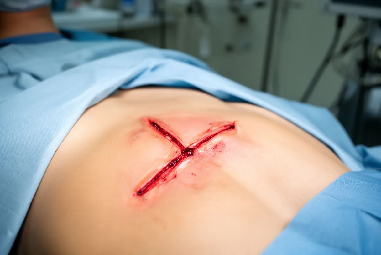
Questions to Ask Before Your Valve Surgery
Coming to a consultation with the right questions changes the conversation. Write your list down; good surgeons welcome it.
Start with these: What exactly is wrong with my valve, and how severe is it? What are all my options — surgical, minimally invasive, and catheter-based — and what are the trade-offs of each? What incision do you recommend for me, and why that one over the alternatives?
Then dig into practicalities: How long will I be in hospital? What restrictions will I have, and for how long? What follow-up imaging and appointments are planned? Who do I call if something worries me at home?
And about the team: Who else reviews my case? We encourage second opinions and treat seeking one as a normal part of deciding about heart surgery — a good team will never resist it. If the answers feel rushed or incomplete, that itself is information.
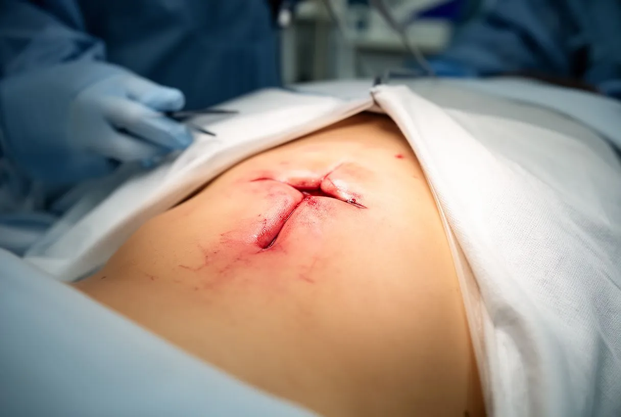
Conclusion: Making Your Decision with Confidence
An inverted T-incision sternotomy is a genuine, well-established option for many patients facing aortic — and in some cases mitral — valve surgery. It trades a smaller operative window for less chest trauma and a faster return to normal life, and in the right patient, selected by a team that reviews every case carefully, it can be the lowest-risk route available. It is not the only route, and it is not automatically the right one: TAVR, a full sternotomy, a Mini-sternotomy, or robotic repair may each be the better answer depending on your anatomy and goals.
The single most useful step you can take is not to self-diagnose from articles like this one, but to sit down with a qualified cardiac team, bring your questions, and understand all your options in plain language before deciding. An inverted t-incision sternotomy is one powerful tool among several — and the right decision is the one made with full information, with your family present, and with the final word always yours. If you are in Guadalajara, Querétaro, Hermosillo or anywhere in Mexico, a free video consultation with a heart surgeon is a sensible, no-pressure first step: we get to know your situation, explain which studies are genuinely needed, and make sure no unnecessary ones are ordered.






