

Left Atrial Appendage and Stroke Risk in AFib
Left atrial appendage stroke risk rises with AFib when blood clots form.
Experiencing an irregular heartbeat can feel unsettling, especially when you learn it carries a serious risk of blood clots. Understanding the link between the left atrial appendage and stroke is the first step toward protecting your health. For many patients navigating AFib stroke risks, the sheer volume of medical terminology can feel overwhelming, but breaking down the anatomy and treatment options brings clarity and calm. By focusing on what happens inside the heart and why clots form, you can approach your care with confidence rather than fear.
What Is the Left Atrial Appendage Stroke Connection?
Understanding the anatomy: The left atrial appendage is a small, ear-like pouch located on the upper left chamber of the heart, known as the atrium. In a normally beating heart, this pouch contracts along with the rest of the heart muscle, squeezing blood out and emptying efficiently. However, when an irregular rhythm develops, the mechanical squeezing action weakens, and blood can pool inside this small, dead-end space. This stagnation is the root cause of the left atrial appendage stroke connection.
The danger of stagnant blood: When blood sits still instead of flowing briskly, the body's natural clotting proteins activate. This is the same process that causes a scab to form on a cut, but inside the heart, it is incredibly dangerous. If a clot forms in the appendage and then escapes into the circulating blood, it can travel directly to the brain. Once it lodges in a brain artery, it blocks oxygen delivery, causing an ischemic stroke.
Targeting the source: Recognizing that the appendage is the origin of most clots in atrial fibrillation is a major medical breakthrough. Because the majority of these dangerous emboli originate in this one tiny pouch, cardiac specialists can focus stroke prevention strategies directly at the source. By sealing or managing this specific area, doctors can drastically reduce the risk of stroke without needing to treat the entire heart with high doses of medications.
How Does Atrial Fibrillation Cause Blood Clots?
The electrical disruption: Atrial fibrillation is an electrical disorder that causes the upper chambers of the heart to quiver rapidly instead of beating forcefully. A related rhythm issue, Atrial Flutter, can also produce similar risks, though the quivering is often more organized. When the atrium fibrillates, the lack of a strong, coordinated mechanical contraction means blood moves sluggishly through the heart, especially in the appendage.
Clot formation mechanics: This sluggish movement creates the perfect environment for clot formation. As blood pools, platelets stick together and clotting factors weave a mesh of protein fibers. According to clinical guidance from heart.org, effectively managing this erratic rhythm or preventing clots with medication is essential to minimizing brain injury. The process is gradual but dangerous, as a newly formed clot may not cause symptoms until it dislodges.
Structural changes over time: As fibrillation persists over months or years, the walls of the atrium may stretch and remodel, creating an even larger space for blood to stagnate. This structural change explains why long-term, consistent management of atrial fibrillation blood clots is vital for patient safety. Restoring a normal rhythm, or effectively preventing the stagnant blood from causing harm, is the primary goal of cardiology interventions.
What Are the Main AFib Stroke Risk Factors?
Calculating personal risk: Not everyone with an irregular heartbeat faces the same likelihood of a stroke. Medical professionals use scoring systems, like the CHA2DS2-VASc score, to quantify AFib stroke risk factors. This clinical tool considers your age, sex, and the presence of other medical conditions such as high blood pressure, diabetes, or previous heart failure. A higher score indicates a greater need for intervention.
Compounding vascular conditions: Other anatomical and structural issues also contribute to your overall risk profile. The presence of an Atheromatous Plaque in the arteries, which is a buildup of cholesterol and fats, can independently increase stroke risks. Similarly, disease or enlargement in the Ascending Aorta can compound the danger, making a thorough cardiovascular evaluation essential. Your cardiology team will look at all these elements together.
The cumulative nature of risk: It is important to remember that stroke risk is cumulative. A younger patient with isolated, infrequent irregular rhythms has a significantly lower baseline risk compared to an older patient with multiple health conditions. By accurately calculating these factors, doctors can decide whether simple monitoring, rhythm control, or aggressive stroke prevention with blood thinners is necessary for your specific situation.

How Is Left Atrial Appendage Thrombus Diagnosed?
Advanced imaging techniques: Finding a clot inside the heart requires specialized imaging because the appendage is small and deeply situated. The most common diagnostic tool is a transesophageal echocardiogram (TEE). This test uses a specialized ultrasound probe placed gently down the esophagus to get a clear, close-up view of the heart's back structures, allowing the cardiologist to see a left atrial appendage thrombus directly.
Other diagnostic methods: While Auscultation with a stethoscope can easily reveal an irregular heartbeat, it cannot detect a physical clot inside the heart. Other imaging methods might include a standard transthoracic echocardiogram or a cardiac CT scan. These tools help rule out the presence of stagnant blood or formed clots, though a TEE remains the gold standard for direct visualization before any intervention.
Why diagnosis precedes treatment: Diagnosing a clot is crucial before starting certain medications or procedures. If a clot is already present, starting blood thinners or attempting to mechanically close the appendage could accidentally dislodge the clot and send it to the brain. Identifying a left atrial appendage thrombus ensures that your care team chooses the safest path forward, which might mean a period of blood thinners to dissolve the clot first.
What Are the Options for AFib Stroke Prevention?
Traditional blood thinners: Preventing a stroke from atrial fibrillation generally falls into two main categories: anticoagulant medications and mechanical closure. Blood thinners, such as Aspirin (used only for very low risk cases) or more potent prescription anticoagulants, are the traditional standard of care. These medications alter the blood's chemistry so that clots cannot form easily, though they require strict adherence and carry a lifelong risk of bleeding.
Mechanical closure alternatives: For patients who cannot tolerate long-term blood thinners, mechanical closure is a highly effective alternative. The 2020 ESC Guidelines for the diagnosis and management of atrial fibrillation developed in collaboration with the European Association for Cardio-Thoracic Surgery (EACTS) strongly support the use of occlusion devices for patients with bleeding contraindications. This procedure physically seals off the appendage, permanently removing the source of the clots.
- Anticoagulants: Reduce clotting chemically but require lifelong adherence and carry bleeding risks.
- Device Closure: Physically seals the appendage, offering an option for those with bleeding risks.
- Rhythm Control: Medications or ablation therapies to restore a normal heartbeat and reduce blood stagnation.
Weighing the trade-offs: Every approach has distinct trade-offs. Blood thinners are non-invasive but require constant management. Mechanical closure involves a one-time procedure but requires careful follow-up imaging to ensure the device heals properly. Weighing these options is a critical conversation you must have with your cardiologist to find the lowest risk for your situation.
How Does the LAA Occlusion Procedure Work?
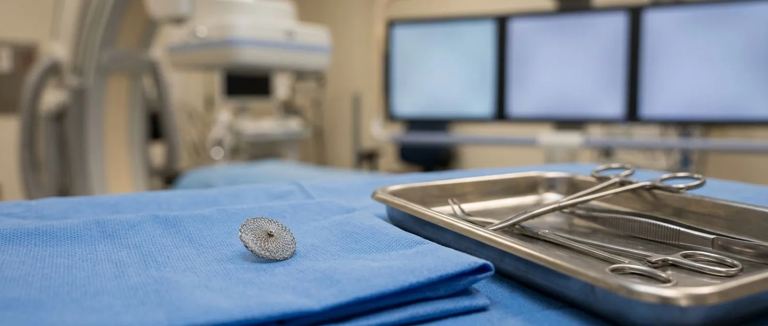
Minimally invasive approach: The LAA occlusion procedure is a targeted, minimally invasive technique designed to seal the appendage without open chest surgery. A specialist inserts a long, thin tube called a catheter through a vein in the leg and carefully guides it up through the blood vessels directly to the heart. Once positioned at the opening of the appendage, a small, umbrella-like mesh device is deployed to close it off.
The occlusion device: The most common form of this technology is a left atrial appendage occlusion device. Once the device is released, it expands to fit the shape of the appendage opening. Over the following weeks, the body's own heart tissue grows over the mesh, permanently walling off the pouch from the rest of the heart. This prevents any future clots that form inside from escaping into the bloodstream.
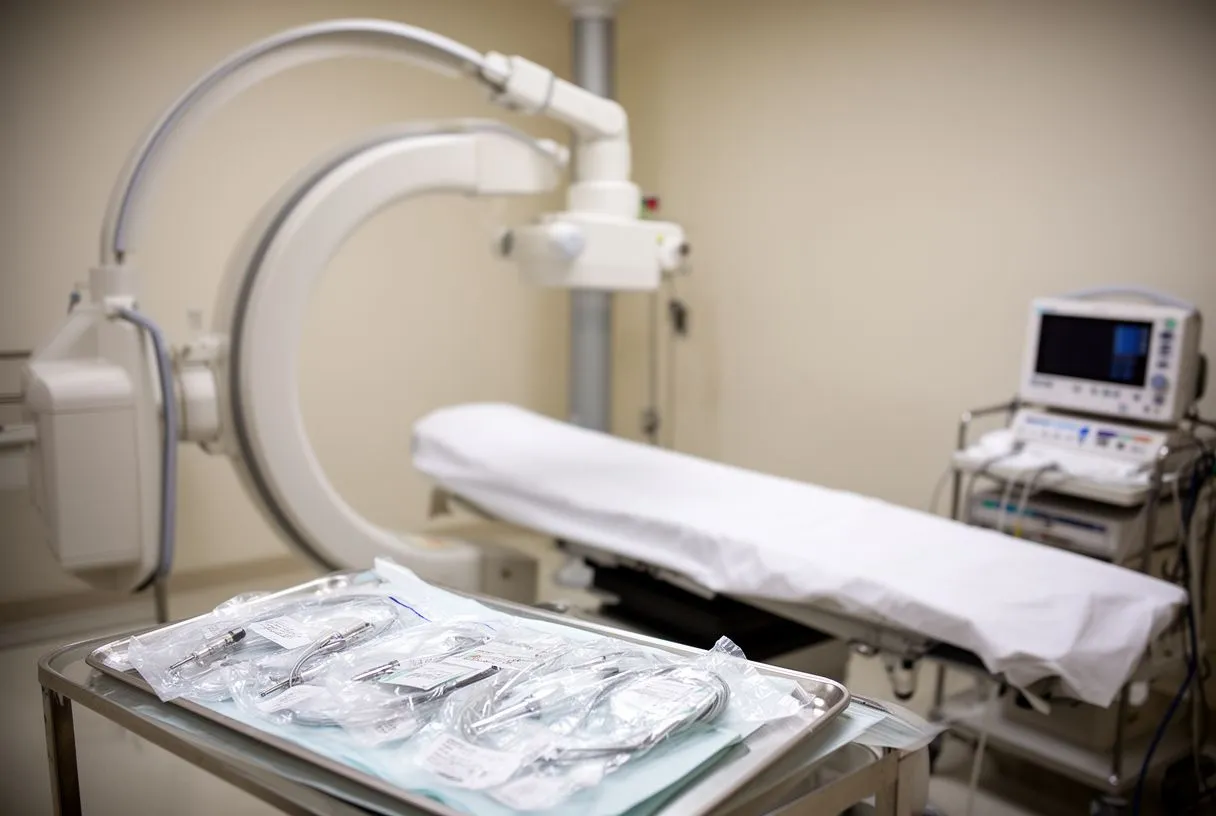
Ensuring a sterile environment: The procedure usually takes about an hour, and patients are typically under general anesthesia. During this process, maintaining a completely sterile field is vital to prevent severe systemic infections. Preventing these profound systemic responses, which are categorized by clinical standards such as The Third International Consensus Definitions for Sepsis and Septic Shock (Sepsis-3), is a top priority in the catheterization lab. This targeted approach addresses the root cause of left atrial appendage embolization safely.
What Is Recovery Like After Appendage Occlusion?
Immediate post-procedure care: Recovery from an appendage occlusion is generally straightforward and brief. Patients usually spend one night in the hospital for observation to ensure the insertion site in the leg heals properly and the heart rhythm remains stable. You may experience some mild discomfort or bruising at the insertion site, but significant Arm Pain or severe chest pain is not typical and should be reported to your medical team immediately.
The healing timeline: For the first 45 days after the procedure, you will likely need to take a blood thinner and a mild antiplatelet medication. This ensures a smooth, thin layer of tissue grows over the mesh device. After about 45 days, your cardiologist will perform a follow-up scan, usually a TEE, to confirm the seal is complete. If it is fully closed, you may be able to stop taking prescription blood thinners.
Resuming daily activities: It is important to maintain gentle activity during recovery to promote healthy circulation. You should avoid heavy lifting or strenuous exercise for a week or two, but daily walking is highly encouraged. Following the specific timeline and activity restrictions provided by your care team ensures the safest and most complete healing process, allowing you to return to a normal routine quickly.
Are Left Atrial Appendage Sealing Devices Safe?
Procedural versus long-term safety: Safety is the primary concern for any patient considering a cardiac implant. While no medical procedure is entirely without risk, left atrial appendage sealing devices have been studied extensively and are generally considered safe for appropriate candidates. The most common risks involve the procedure itself, such as bleeding at the access site or rare instances of fluid accumulating around the heart.
Structural integrity: Once the device is implanted, its structural integrity is highly robust. The mesh is designed to conform to the unique shape of your appendage, and once endothelialized, it becomes a permanent part of your heart. Device migration is extremely rare after the initial healing period, making it a durable solution for long-term stroke prevention. The materials are biocompatible, meaning they do not degrade or cause harmful reactions inside the body.
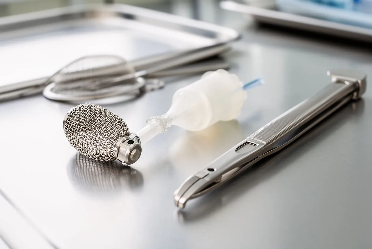
Comparing risks and benefits: Long-term clinical data shows that the risk of stroke is significantly reduced compared to patients who stop taking blood thinners without any alternative therapy. For patients who have experienced bleeding complications from medications, the safety profile of a mechanical device often outweighs the ongoing danger of unmanaged atrial fibrillation blood clots. It provides a permanent shield against embolization.
When Should You Consider AFib Stroke Anticoagulation Alternatives?
High bleeding risk scenarios: Deciding to pursue a mechanical closure over medication depends heavily on your personal bleeding history. If you have experienced gastrointestinal bleeding, frequent unexplained bruising, or have a high risk of falls, continuing blood thinners may be more dangerous than the AFib stroke itself. In these specific cases, finding an alternative to daily anticoagulation becomes essential for your overall safety.
Complex medication regimens: Other cardiac conditions can complicate medication choices. If you have undergone a Balloon Angioplasty to place stents in your coronary arteries, your doctor must carefully balance the medications needed to keep the stents open against the medications used to prevent atrial clots. Managing multiple blood thinners simultaneously increases bleeding risks exponentially, making a mechanical closure an attractive option to simplify your daily regimen.
- High Bleeding Risk: History of internal bleeding, ulcers, or severe anemia.
- High Fall Risk: Elderly patients or those with ongoing balance and mobility issues.
- Medication Fatigue: Difficulty maintaining strict daily anticoagulation routines or affording prescriptions.
Finding the lowest risk for your situation: Ultimately, the decision to transition to an occlusion device is about minimizing harm. You should discuss this option with your doctor if your daily life is disrupted by the side effects of blood thinners, or if routine lab monitoring proves too difficult to maintain safely. A tailored approach ensures you get the protection you need without undue suffering.
How to Talk to Your Doctor About AFib Stroke Risk?
Preparing for your consultation: Your consultation is the time to bring all your questions to the table. Be prepared to discuss your symptoms, your daily routine, and any family history of cardiac events or strokes. If you are experiencing palpitations, shortness of breath, or dizziness, describe exactly when they happen, what you are doing at the time, and how long they last. This information is crucial for an accurate diagnosis.
Asking the right questions: Ask your cardiologist to explain your personal CHA2DS2-VASc score and what it means for your specific risk profile. Discuss whether a simple course of medication is sufficient, or if the left atrial appendage stroke connection in your case warrants a structural intervention. Bring a complete list of all medications, over-the-counter drugs, and supplements you currently take to avoid any dangerous interactions.

Partnering with your cardiac team: Remember that modern cardiac care is highly individualized. What works for one patient may not be appropriate for another. Whether you are visiting our practice in Guadalajara, Querétaro, or Hermosillo, Dr Alejandro Molina and our team are here to help. By engaging in an open, honest dialogue with a qualified cardiac team, you can build a plan that protects your brain health while fitting your lifestyle. We encourage you to book a consultation to discuss your specific situation rather than self-diagnosing.






