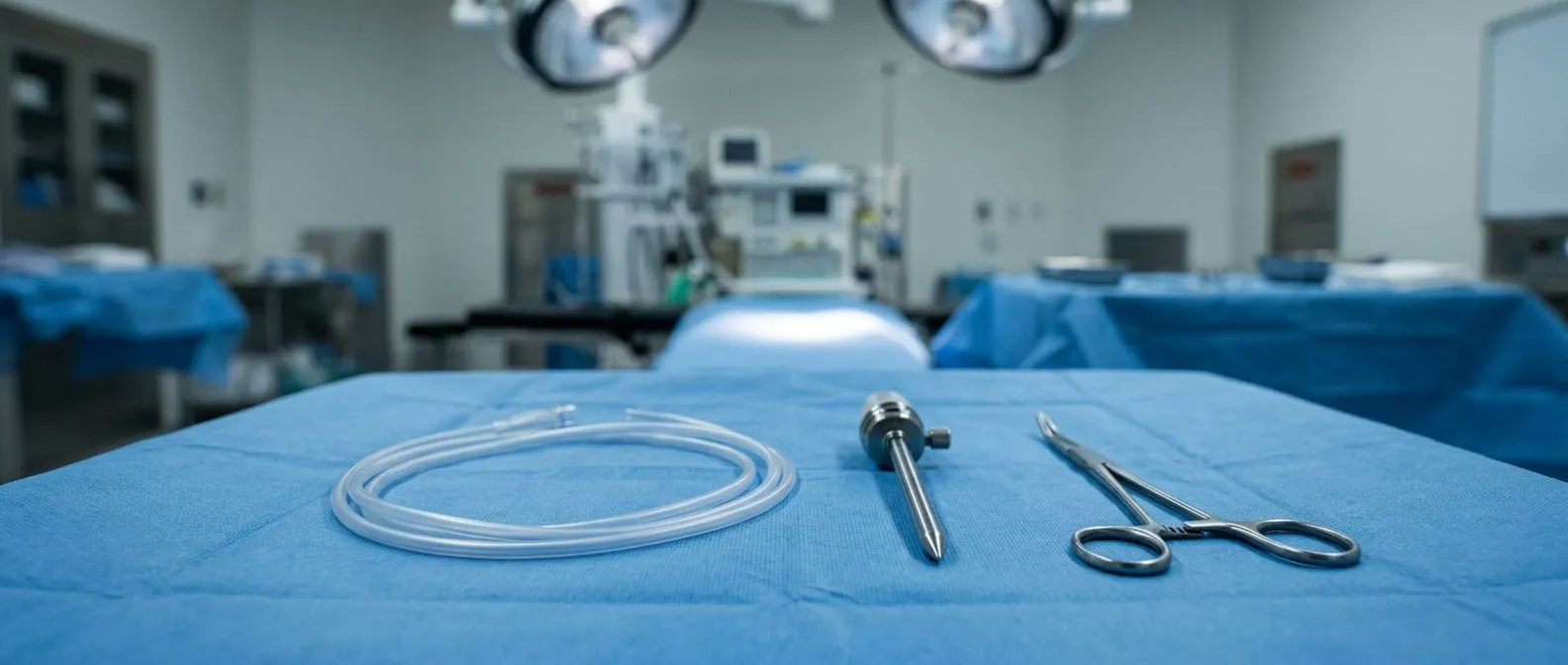
Pericardial Sac Surgery: Pericardiectomy and Drainage
Pericardial sac surgery relieves pressure on your heart through pericardiectomy or drainage.
Pericardial sac surgery encompasses vital procedures designed to relieve pressure around the heart and restore normal cardiac function. When the protective lining around the heart becomes diseased, patients often face shortness of breath, fatigue, and chest discomfort. Our team at Cirugía de Corazón, led by Dr Alejandro Molina in Guadalajara, Querétaro, and Hermosillo, provides calm, precise, and evidence-based surgical solutions for these exact challenges. Understanding the anatomy, the symptoms, and the surgical options empowers you to make informed decisions about your care.
What is Pericardial Sac Surgery?
Understanding the pericardium's role: The pericardium is a double-layered, fluid-filled sac that surrounds the heart, anchoring it in the chest and protecting it from infection. When this sac becomes inflamed, thickened, or filled with excess fluid, it restricts the heart's ability to pump efficiently. Pericardial sac surgery refers to a group of operations—ranging from simple fluid drainage to complete sac removal—aimed at restoring normal heart mechanics.
Identifying the clinical signs: Patients often arrive at the clinic reporting a distinct chest pressure that worsens when lying flat. During a physical exam, auscultation—listening to the chest with a stethoscope—might reveal a high-pitched, scratching sound known as a pericardial friction rub. This clinical clue, combined with imaging, helps our cardiac team determine if the pericardium is the source of the problem.
Navigating the surgical options: The specific approach depends entirely on the underlying pathology. A patient with acute fluid buildup might only need a needle drainage or a small pericardial window, whereas someone with chronic scarring might require a complete pericardiectomy. We evaluate your unique situation to recommend the lowest risk option for your specific anatomy and medical history.
How Does Pericardial Effusion Affect the Heart?
The mechanics of fluid accumulation: A pericardial effusion occurs when excess fluid collects between the two layers of the pericardium. Because the sac is fibrous and cannot stretch easily, even a moderate amount of fluid can rapidly increase pressure. This pressure squeezes the heart chambers, preventing them from filling properly between beats. The pericardium encases the heart and the roots of the great vessels, including the ascending aorta, meaning pressure here can have systemic effects.
Recognizing tamponade physiology: When the pressure inside the sac exceeds the pressure inside the heart, a dangerous condition called cardiac tamponade occurs. The heart's output drops drastically, leading to a drop in blood pressure, dizziness, and extreme fatigue. This is a medical emergency that requires immediate intervention to drain the fluid and relieve the compression on the heart muscle.

Differentiating from other chest pains: The discomfort from a pericardial effusion is often sharp and positional, differing from the crushing pain of a heart attack or the chronic tightness associated with an atheromatous plaque in the coronary arteries. Echocardiography is used to distinguish between fluid outside the heart and issues within the heart muscle itself, so the correct diagnosis is made before any surgical plan is finalized.
What is a Pericardial Window Procedure?
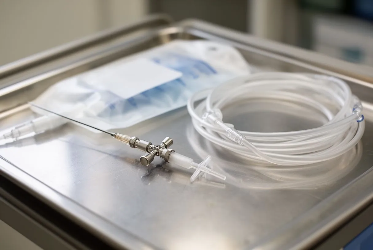
Creating a drainage pathway: A pericardial window is a surgical procedure designed to create a permanent opening in the pericardium, allowing fluid to drain continuously into the pleural space (the chest cavity) where the body can absorb it safely. This is often recommended for patients with recurring effusions caused by inflammation, autoimmune conditions, or certain infections. The fluid that is drained is then sent for laboratory analysis to identify the underlying cause.
Surgical approaches available: Surgeons can create a pericardial window using a subxiphoid approach (just below the breastbone) or via video-assisted thoracoscopic surgery (VATS). VATS uses small camera ports, offering a minimally invasive route. According to comprehensive surgical texts published by Springer, thoracoscopic approaches often yield faster recovery times and less postoperative pain compared to open surgical techniques.
When a window is insufficient: While a pericardial window is highly effective for managing fluid, it does not remove the thickened, scarred pericardium itself. If the sac has become calcified and is physically squeezing the heart—a condition known as constrictive pericarditis—a simple window will not relieve the symptoms. In those cases, a more extensive removal of the sac is required.
How is Constrictive Pericarditis Diagnosed?
Tracking the progression of scarring: Constrictive pericarditis occurs when the pericardium loses its elasticity and becomes a rigid, scarred shell around the heart. This is often the result of chronic or recurrent pericarditis. The rigid sac prevents the heart from expanding normally, meaning less blood enters the heart with each beat, and the heart cannot pump enough blood to meet the body's needs.
Diagnostic imaging and catheterization: Diagnosis requires a combination of imaging and pressure measurements. CT scans and MRIs are excellent for showing the thickness of the pericardium and any calcification. A right heart catheterization is often necessary to measure the specific pressure waveforms inside the heart chambers, which shows the classic "square root sign" indicating constriction.
- Echocardiography: Visualizes heart movement and how the ventricles interact during breathing.
- CT or MRI Imaging: Identifies thickening or calcium deposits in the pericardium.
- Hemodynamic Cath: Measures intracardiac pressures to confirm restrictive filling.
When is a Pericardiectomy Recommended?
Indications for surgical removal: A pericardiectomy, or the surgical removal of the pericardium, is the definitive treatment for severe constrictive pericarditis. It is recommended when medical management with anti-inflammatory drugs fails to relieve symptoms, or when imaging shows the heart's filling is mechanically restricted by a thickened, calcified sac. The goal is to free the heart chambers, allowing them to expand and fill with blood normally.
Managing comorbidities before surgery: Patients facing a pericardiectomy often have other underlying conditions that must be managed. For instance, if a patient has a history of arrhythmias like atrial flutter, or if they are on daily aspirin for secondary cardiovascular prevention, these medications and conditions must be carefully coordinated. We also evaluate for valve issues; if a patient needs a transcatheter aortic valve replacement (TAVR) alongside pericardial disease, the sequence of interventions is meticulously planned.

The goal of complete resection: During a pericardiectomy, the surgeon aims to remove as much of the diseased pericardium as possible, focusing on the areas over the left and right ventricles. Removing this restrictive barrier immediately changes the heart's workload. In some cases, the right atrium may also be heavily restricted, requiring careful dissection to ensure the upper chambers can fill adequately without tearing delicate tissue.
Minimally Invasive Pericardiectomy vs Traditional Surgery
Traditional open approaches: A traditional pericardiectomy is typically performed through a median sternotomy, an incision down the center of the chest that splits the breastbone. This provides the surgeon with excellent, direct visualization of the entire heart and pericardium, which is crucial when dealing with dense, calcified adhesions that can easily stick to the underlying heart muscle or coronary arteries.
Evolving minimally invasive techniques: For select patients with less severe adhesions, a minimally invasive pericardiectomy using smaller incisions or robotic assistance may be an option. While endovascular techniques have advanced significantly—as seen in complex aortic repairs like the use of a modified proximal scalloped aortic arch endograft—pericardial removal still heavily relies on direct surgical access to safely peel the thickened tissue away from the beating heart without damaging it.
Choosing the right surgical route: The decision between a minimally invasive approach and a traditional open surgery depends on the extent of the calcification, the patient's anatomy, and previous surgical history. At our practice, we prioritize safety and complete removal over a smaller incision, ensuring the constriction is fully relieved rather than partially addressed.
What to Expect During Pericardial Sac Removal
Anesthesia and monitoring preparation: Pericardial sac removal is performed under general anesthesia. Because the heart is being physically manipulated and the restrictive layer is being peeled away, blood pressure can fluctuate significantly. To monitor this precisely, an arterial line is placed in a wrist artery, providing real-time, beat-to-beart blood pressure readings throughout the operation.
The process of decortication: Once the chest is open, the surgeon begins the meticulous process of dissecting the thickened pericardium away from the epicardium (the outer layer of the heart muscle). This is known as decortication. It requires extreme precision, as the pericardium may be fused tightly to the heart. To minimize the need for donor blood, autologous blood salvage techniques are often used, collecting and recycling the patient's own blood during the procedure.
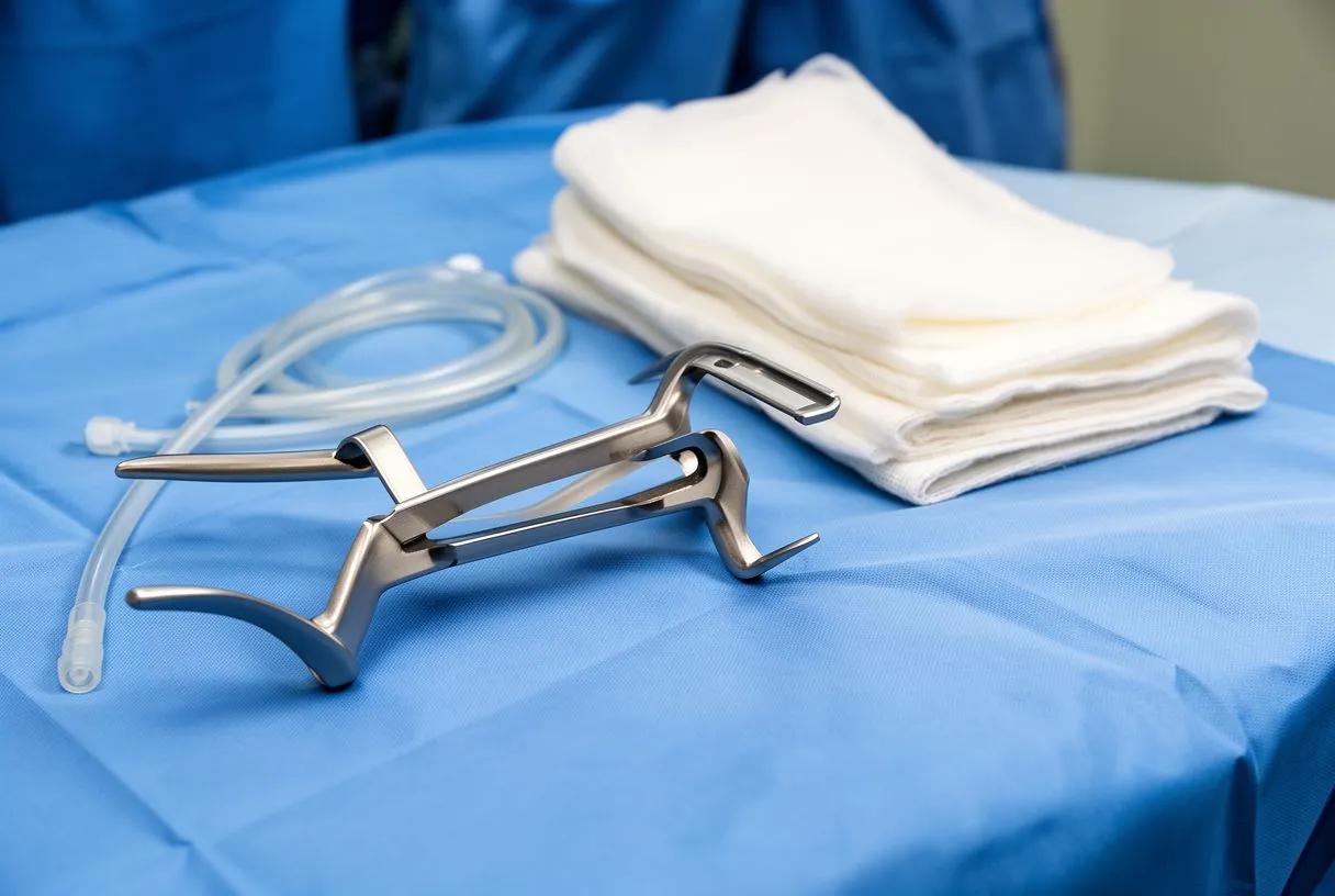
Immediate hemodynamic changes: As the restrictive sac is lifted away, you can often see the heart chambers expand visibly, filling with blood that was previously blocked. This immediate release of pressure is the surgical endpoint. Once the surgeon is satisfied that the heart is free, the chest is closed with wires, and the patient is transferred to the intensive care unit for initial recovery.
Recovery Timeline After Pericardial Sac Surgery
Initial intensive care stay: After pericardial sac surgery, the first 24 to 48 hours are spent in the intensive care unit. Here, the medical team closely monitors heart rhythms, fluid balance, and chest tube output. Because the heart is suddenly able to fill and pump larger volumes of blood, the kidneys and cardiovascular system need time to adapt to this new hemodynamic reality, which can temporarily cause fluid shifts.
Transitioning to the cardiac ward: Once stable, patients are moved to a standard cardiac ward. Early mobilization is encouraged; sitting up in a chair and walking short distances with assistance helps prevent complications like blood clots or pneumonia. Pain management is a priority, and patients are taught how to splint their chest when coughing to protect the healing sternum.
Returning home and long-term healing: Most patients spend a week in the hospital before being discharged. Full recovery from a sternotomy takes about 6 to 8 weeks. During this time, heavy lifting and driving are restricted. A structured rehabilitation plan and regular check-ups help monitor your heart function and ensure the constriction does not return.
Are There Risks with Pericardial Drainage Surgery?
General surgical risks: Like any cardiac procedure, pericardial drainage and pericardiectomy carry risks. Bleeding and infection are standard surgical risks, but because the heart is involved, there is also a risk of arrhythmias. The physical manipulation of the heart muscle can irritate the electrical pathways, leading to temporary rhythm disturbances that may require medication or a brief period of monitoring.
Risks specific to decortication: The most significant risk during a pericardiectomy is inadvertently tearing the heart muscle or a coronary artery while peeling away the densely calcified sac. Extreme physiological states—such as the massive hemodynamic shifts seen in conditions like aortic dissection in pregnancy—show how fragile the cardiovascular system can be under stress. Our surgical team uses meticulous techniques to avoid these structures, but sometimes small bleeds occur and are immediately controlled in the operating room.
- Bleeding: From the surface of the heart or the incision site.
- Arrhythmias: Irregular heartbeats due to surgical irritation.
- Pneumonia: A risk associated with general anesthesia and chest surgery.
Long-Term Outlook After Heart Lining Surgery
Improving quality of life: For the vast majority of patients with constrictive pericarditis, a successful pericardiectomy offers a dramatic improvement in symptoms. The shortness of breath, swelling in the legs, and profound fatigue gradually resolve as the heart regains its ability to pump effectively. Most patients can return to their normal daily activities and enjoy a significantly better quality of life without the constant weight of a compressed heart.
Monitoring for recurrence: While the pericardium is removed, a small amount of tissue remains. In rare cases, scar tissue can reform on the surface of the heart, causing recurrent constriction. Regular echocardiograms are used to confirm the heart remains free and functioning well. Addressing underlying inflammatory conditions is key to preventing this recurrence.
If you are experiencing symptoms of pericardial disease, do not wait for the discomfort to worsen. Early diagnosis and intervention are crucial to protecting your heart function. We encourage you to discuss your specific symptoms, concerns, and diagnostic history with a qualified cardiac team. Contact our practice to book a consultation with Dr Alejandro Molina and take the first step toward a comprehensive, patient-centered treatment plan.