Breathing Tube After Heart Surgery: Endotracheal Tube Care
Endotracheal tube after heart surgery: learn what to expect with breathing tube care, ventilator weaning, extubation timeline, and recovery.
Waking up in the intensive care unit after a major cardiac procedure can be a disorienting experience, especially when you realize you cannot speak or breathe on your own. The endotracheal tube is a critical bridge between the operating room and your independent recovery. Understanding its purpose, how long it stays in place, and what to expect during its removal can ease anxiety for both patients and their families. This guide explains the entire process, from initial intubation to ventilator weaning and respiratory recovery.
What Is an Endotracheal Tube After Heart Surgery?
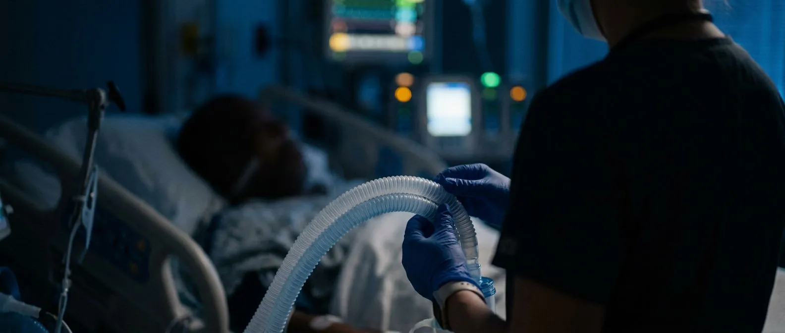
Understanding the breathing circuit: An endotracheal tube is a flexible plastic conduit inserted through the mouth or nose, passing through the vocal cords, and resting securely in the windpipe or trachea. After complex operations like a coronary artery bypass or a valve replacement, your chest muscles and diaphragm are temporarily weak. The tube connects directly to a mechanical ventilator, a machine that pushes oxygen-rich air into your lungs and removes carbon dioxide. This setup ensures your body receives adequate oxygen while your heart muscle heals from the surgical trauma.
Distinguishing from other ICU lines: Families often see multiple tubes and wires attached to their loved one and feel overwhelmed. It is important to distinguish the breathing apparatus from other monitoring tools. For instance, while an arterial line measures your blood pressure continuously, and an arterial graft might have been used to bypass a blocked vessel, the endotracheal tube serves solely to manage your airway. It is not a feeding tube or a drainage device; its exclusive job is respiratory support, keeping your airway open and protected while you are deeply sedated.
The psychological impact of intubation: Patients frequently have no memory of the tube being placed in the operating room, but waking up with it can cause panic. Because the tube passes through the vocal cords, speaking is impossible. Patients rely on nodding, hand squeezes, or writing on a clipboard to communicate. The nursing staff is highly trained to anticipate needs and reassure patients during this vulnerable phase. Knowing that this device is temporary and designed to protect your lungs helps reduce the distress associated with waking up intubated.
Why Is a Breathing Tube Needed During Cardiac Surgery Recovery?
Anesthesia and muscle relaxation: During open-heart surgery, powerful anesthetic medications paralyze the muscles, including the diaphragm. Because you cannot breathe independently under these conditions, the surgical team relies on the ventilator to take over the work of breathing. Even after the operation concludes, it takes time for the anesthesia to clear from your system. Keeping the endotracheal tube in place ensures your airway remains secure until your muscle function returns to a safe baseline.
Protecting the heart and lungs: The heart and lungs work closely together, and sudden drops in oxygen can stress a freshly repaired heart. If you were to breathe on your own immediately after surgery, the effort required might strain the myocardium. By controlling your respiration, the medical team minimizes the workload on your heart, allowing it to recover from procedures such as balloon angioplasty or complex repairs of the ascending aorta. The controlled environment prevents dangerous fluctuations in your oxygen saturation.
Managing fluid and secretions: Cardiac surgery often involves the use of a heart-lung bypass machine, which can cause temporary inflammation and fluid accumulation in the lungs. This fluid can make breathing difficult and lead to a buildup of secretions. While intubated, the respiratory therapist can easily suction out these secretions directly through the endotracheal tube, keeping your airways clear. This proactive clearance prevents pneumonia and ensures that your lung function remains optimal as your body rebalances its fluids.
How Long Will the Endotracheal Tube Stay In After Bypass?
Standard extubation timelines: For most uncomplicated cardiac surgeries, the goal is to remove the breathing tube within four to six hours after you arrive in the intensive care unit. This fast-track extubation approach helps reduce the risks associated with prolonged mechanical ventilation, such as infections or lung weakness. Your surgical team in Guadalajara, Querétaro, or Hermosillo will continuously assess your readiness. Once you are awake, following commands, and demonstrating stable blood gases, the tube can be safely removed.
Factors that delay removal: Several variables can extend the time you spend on the ventilator. If you undergo a highly complex procedure, have pre-existing lung disease like chronic obstructive pulmonary disease (COPD), or experience complications like atrial flutter, your medical team will keep the tube in longer to ensure your stability. Older age or a high EuroSCORE II—a tool used to predict surgical risk—may also justify a more cautious, prolonged approach to ventilator weaning to ensure your heart and lungs are fully ready.
The concept of awake ECMO: In rare, highly complex scenarios where the heart or lungs need extended rest, advanced support might be used instead of a standard ventilator. For example, in awake ECMO (Extracorporeal Membrane Oxygenation), blood is oxygenated outside the body, sometimes allowing the patient to be awake and even breathing without a standard endotracheal tube. However, this is a specialized therapy. For the vast majority of routine recoveries, the standard endotracheal tube is the primary tool for short-term airway management.
- Awake and responsive: You must be able to open your eyes and follow simple commands, indicating the anesthesia has worn off sufficiently.
- Stable blood work: Arterial blood gas tests must show that your oxygen and carbon dioxide levels are within a safe range without ventilator assistance.
- Adequate muscle strength: You need to demonstrate a strong hand grip and the ability to lift your head off the bed, proving the paralytic medications have worn off.
What to Expect When Waking Up With a Breathing Tube
The sensation of the tube: As you wake from anesthesia, you will likely feel pressure in your throat and a strong urge to cough. The tube rests against your vocal cords, which can feel uncomfortable but is not typically painful, as pain is managed with targeted medications. You may feel the machine pushing air into your lungs at a rhythm that does not match your natural breathing pattern. It is crucial to relax and let the ventilator do the work, fighting the machine makes breathing more difficult and delays removal.
Communication strategies: Because you cannot speak with the endotracheal tube in place, frustration is a common emotion. The ICU staff will provide alternative ways to communicate. You might use a communication board, write on a notepad, or use simple hand gestures. If your hands are lightly restrained to prevent accidental pulling of the tube, a nurse will unbind one hand to assist you. Establishing a reliable method for communication early on greatly reduces anxiety for both you and your family.
Sedation and pain management: While the tube itself is not severely painful, the surgical incision and chest drainage tubes are. Your team will continuously administer sedatives and pain relievers through your intravenous lines. The goal is to keep you calm and comfortable enough to tolerate the breathing tube but awake enough to breathe effectively. According to Verywell Health, finding this delicate balance is a priority for ICU teams, as excessive sedation can prolong recovery and weaning times.
Endotracheal Tube Care and Airway Management in the ICU
Securing the airway: The endotracheal tube must remain perfectly positioned in your trachea. If it shifts, it could enter a lung bronchus or slip out entirely. To prevent this, nurses secure the tube to your face with specialized adhesive tape or a commercial tube holder. They regularly check the depth marking on the tube at your lip line to ensure it has not moved. This careful securing prevents accidental extubation, which can be a dangerous emergency requiring rapid reinsertion.
Humidification and suctioning: The ventilator includes a humidification system to keep the air entering your lungs warm and moist, preventing the drying of mucous membranes. Despite this, secretions can still accumulate. Nurses perform suctioning by passing a thin catheter through the endotracheal tube to clear out mucus. This process may cause a brief coughing spell and a transient drop in oxygen, but the ventilator quickly replenishes your levels. Regular suctioning is a vital component of endotracheal tube care, preventing blockages and infections.
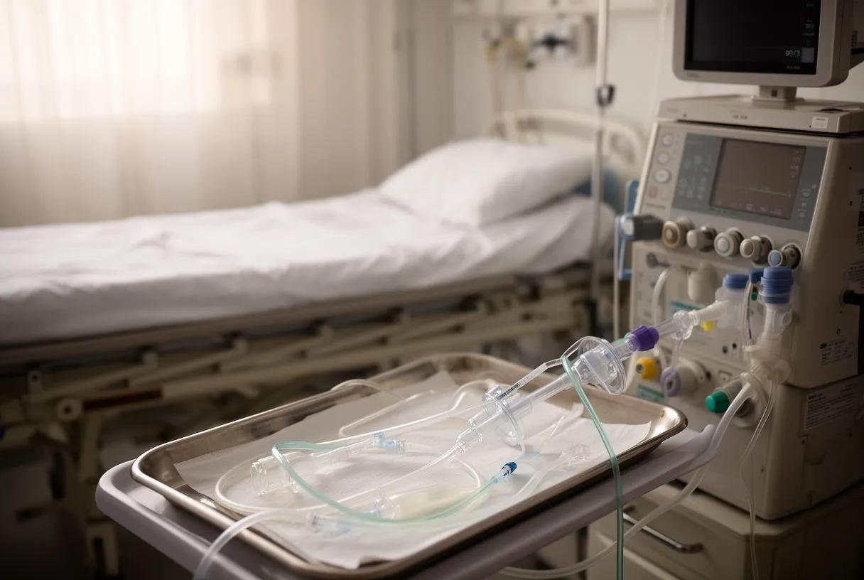
Preventing ventilator-associated pneumonia: A significant focus of airway management is preventing ventilator-associated pneumonia (VAP). The ICU team follows strict protocols, including elevating the head of your bed to at least 30 degrees, maintaining meticulous oral hygiene every few hours, and assessing your readiness for extubation daily. These evidence-based practices minimize the risk of bacteria traveling from the mouth into the lungs, ensuring your respiratory system remains healthy enough for a successful recovery.
Ventilator Weaning for Heart Patients
Reducing ventilator support: Before the endotracheal tube is removed, you must undergo a weaning process. The respiratory therapist gradually reduces the amount of work the ventilator does, lowering the pressure and oxygen concentration. This process, often done in a spontaneous breathing trial, tests whether your lungs and diaphragm can sustain the effort of breathing independently. Your heart rate, blood pressure, and oxygen saturation are closely monitored for any signs of distress during this transition.
Monitoring cardiac stability: Weaning is not just a respiratory test; it is a cardiac one. The effort of breathing increases the workload on the heart, which must pump blood to the respiratory muscles. If your heart is not yet strong enough, you might develop abnormal rhythms or a drop in cardiac output. In complex scenarios, such as those involving an atheromatous plaque in the aorta, the team must be exceptionally cautious. A study on complex aortic interventions, titled Intramural haematoma of the thoracic aorta: who's to be alerted the cardiologist or the cardiac surgeon?, highlights how critical hemodynamic stability is during delicate postoperative phases.
The spontaneous breathing trial: The final test before endotracheal tube removal is the spontaneous breathing trial. The ventilator is set to a minimal support mode, essentially allowing you to breathe through the tube with little machine assistance. If you can maintain good oxygen levels, stable carbon dioxide levels, and a calm respiratory rate for 30 to 120 minutes, you have passed the trial. This success indicates that your respiratory muscles are ready, and the medical team will proceed with the actual removal.
Endotracheal Tube Removal and Extubation Timeline
The extubation procedure: Once you pass the spontaneous breathing trial, the team prepares for extubation. A nurse or respiratory therapist will suction your airway one final time to ensure it is clear of secretions. They will then ask you to take a deep breath. As you exhale, they rapidly deflate the small balloon (cuff) that seals your trachea and gently slide the endotracheal tube out. The entire process takes only a few seconds and is often accompanied by a strong urge to cough.
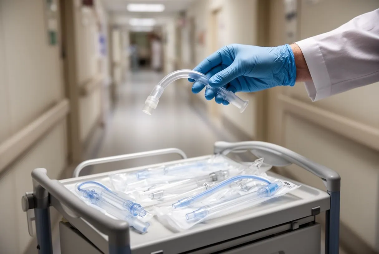
Immediate post-extubation care: Right after the tube is removed, you will receive supplemental oxygen through a mask or nasal cannula. You may feel a scratchy throat and experience hoarseness, which is entirely normal. The team will monitor your breathing pattern and perform auscultation, listening to your lungs with a stethoscope to ensure air is moving well in all lung fields. Your voice will likely be raspy for a short period as your vocal cords recover from the pressure of the tube.
Resuming normal breathing: The transition from mechanical ventilation to independent breathing requires conscious effort at first. You might feel tired, but it is essential to take deep breaths to re-expand your lungs fully. The nursing staff will encourage you to use an incentive spirometer, a simple device that measures how deeply you can inhale. Using this device frequently helps prevent the lower parts of your lungs from collapsing, a common issue after cardiac surgery.
Managing Tracheal Tube Complications and Sore Throat Relief
Common throat discomfort: A sore throat and a raspy voice are the most frequent complaints after endotracheal tube removal. The vocal cords are delicate structures, and the tube resting against them causes temporary irritation and swelling. This discomfort usually peaks on the first day after removal and gradually subsides over the next 48 to 72 hours. Swallowing may feel slightly painful, but it is important to stay hydrated to help soothe the throat tissues.
Vocal cord dysfunction: In rare cases, the vocal cords might become temporarily weakened or paralyzed, leading to a weak voice or difficulty coughing forcefully. This condition usually resolves on its own as the swelling decreases. However, if hoarseness persists for more than a few weeks, an ear, nose, and throat specialist may examine the vocal cords. While intubation is necessary for surgery, advanced techniques in airway management aim to minimize this trauma, an approach supported by research on complex vascular procedures like the Physician-modified endograft for symptomatic zone 2 penetrating ulcer of the aortic arch without bridging stenting of the left subclavian artery for vertebral preservation.
Relief strategies for patients: Simple interventions can provide significant sore throat relief. Drinking cool water or warm herbal tea helps soothe the irritated tissues. Throat lozenges or numbing sprays may be offered once you are cleared to swallow safely. Avoiding clearing your throat aggressively is also recommended, as this can further irritate the vocal cords. Instead, take a sip of water to wash down the urge to cough. These strategies help manage the discomfort while your throat heals naturally.
- Hydration is key: Sipping cool or warm fluids frequently helps lubricate the throat and reduces the friction caused by the endotracheal tube.
- Rest your voice: Avoid whispering or shouting, as both actions strain the vocal cords more than normal speaking. Let your throat rest.
- Use a humidifier: Adding moisture to the air you breathe prevents your throat from drying out, especially important in the dry climate of hospital rooms.
Breathing Exercises After Heart Surgery Recovery
The importance of incentive spirometry: After the endotracheal tube is removed, your lungs are vulnerable to collapsing, a condition known as atelectasis. An incentive spirometer is a crucial tool to prevent this. You inhale deeply through the device, holding a small ball or piston in an elevated position for a few seconds. This deep inflation forces the tiny air sacs in your lungs to remain open, improving oxygen exchange and clearing out residual anesthesia gases from your body.
Deep breathing and coughing: Along with using the spirometer, you must practice deliberate deep breathing exercises. You will be taught to hold a pillow firmly against your chest incision to provide support and reduce pain when you cough. Coughing is necessary to clear secretions that have pooled in your lungs during surgery. Failing to clear these secretions can lead to postoperative pneumonia, a complication that can significantly delay your discharge and overall recovery.
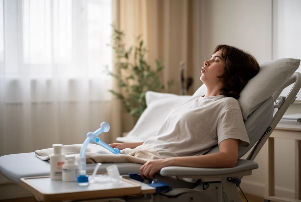
Early mobilization and lung expansion: Getting out of bed and walking is one of the most effective breathing exercises. Sitting upright and walking allows your diaphragm to move fully, which does not happen when you are lying flat in bed. The combination of early ambulation, incentive spirometry, and structured deep breathing helps restore your lung capacity to its pre-surgery baseline. Your cardiac team will guide you through a progressive mobility plan tailored to your specific heart condition and surgical approach.
When to Seek Help After Endotracheal Tube Removal
Recognizing respiratory distress: While most patients recover smoothly, it is vital to know the warning signs of respiratory complications. If you experience sudden shortness of breath that does not improve with rest, a rapid heart rate, or a persistent drop in your oxygen saturation levels, you must alert your medical team immediately. These could be signs of fluid building up around your lungs, a collapsed lung, or a pulmonary embolism. Timely intervention is crucial to prevent a minor issue from becoming a major setback.
Monitoring for aspiration and infection: After intubation, your swallowing reflex may be temporarily impaired, increasing the risk of aspirating food or liquid into your lungs. If you notice coughing or choking when drinking, inform your nurse. A speech therapist might assess your swallowing before you eat solid foods. Additionally, watch for signs of infection, such as a fever, increased yellow or green sputum, or worsening chest pain. These symptoms require prompt evaluation to rule out ventilator-associated pneumonia.
Conclusion and next steps: The presence of an endotracheal tube after heart surgery is a temporary but essential phase of your healing journey. It protects your airway, rests your heart, and ensures your body receives the oxygen it needs during the critical hours following your operation. By understanding the care process, the weaning timeline, and the exercises required for recovery, you can approach this phase with confidence. We encourage you to discuss your specific cardiac history and surgical risks with a qualified cardiac team. Book a consultation with us to receive a personalized recovery plan and ensure the lowest risk for your situation.