Cardioplegia: How the Heart Is Stopped and Restarted
Cardioplegia in heart surgery explained: how surgeons stop and restart the heart.
What Is Cardioplegia in Heart Surgery?
Cardioplegia heart surgery depends on one elegant principle: to repair a heart precisely, the surgeon must first bring it to a controlled, temporary standstill. Cardioplegia is the deliberate, reversible arrest of the heart using a specialised solution that stops electrical activity in the cardiac muscle. This is not the same as a clinical cardiac arrest — the heart is stopped intentionally and protectively, not because of disease or injury. The word itself comes from Greek roots meaning "heart striking" or "heart paralysis," and it describes a technique that has been refined over decades of cardiac surgical practice.
Why the heart must be stopped: A beating heart moves constantly, filling and emptying roughly sixty to a hundred times per minute. Operating on a moving target — suturing a valve, bypassing a blocked artery, or repairing a congenital defect — would be nearly impossible with the precision open-heart surgery demands. By arresting the heart in a controlled fashion, the surgeon gains a still, bloodless field. The challenge is that the heart, like any living muscle, needs a constant supply of oxygen and nutrients. Stopping it without protecting it would cause irreversible damage within minutes. Cardioplegia solves this by both stopping the heart and preserving it during the period of arrest.
For patients and families preparing for cardiac surgery, understanding cardioplegia helps demystify what happens behind the operating room doors. When your surgeon or perfusionist — the specialist who operates the heart-lung machine — mentions "arresting the heart," they are describing a carefully orchestrated process. The ascending aorta, the large vessel carrying blood from the heart to the body, is clamped, and the cardioplegia solution is infused directly into the coronary circulation. The heart relaxes, stops, and remains protected while the surgical repair is completed. Once the repair is done, the clamp is removed, the heart is reperfused with oxygenated blood, and it typically resumes beating on its own or with gentle electrical stimulation.
How Cardioplegia Heart Surgery Works
The mechanism behind cardioplegia heart surgery rests on manipulating the electrical and chemical environment inside cardiac muscle cells. The heart beats because of a wave of electrical depolarisation that sweeps across the atrium and ventricles in a tightly coordinated rhythm. Cardioplegia solutions achieve arrest primarily by using a high concentration of potassium, which raises the resting membrane potential of cardiac cells above the threshold needed to fire an action potential. When the cells cannot depolarise, they cannot contract, and the heart stops in diastole — its relaxed phase. This is the foundation of every cardioplegia technique in use today.
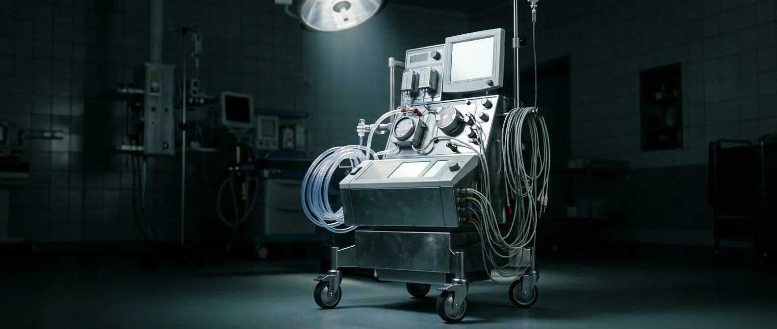
The role of the heart-lung machine: While the heart is arrested, the body still needs circulation. An arterial cannula is placed in a major blood vessel, and the heart-lung machine takes over, oxygenating blood outside the body and returning it through an arterial line to maintain perfusion of the brain, kidneys, and other organs. The perfusionist manages this circuit throughout the operation, continuously monitoring pressure, flow, and temperature. This parallel system is what makes prolonged cardiac arrest safe — the heart is protected by cardioplegia, and the rest of the body is supported by cardiopulmonary bypass. Research registered on clinicaltrials.gov continues to evaluate refinements in how these systems are managed to improve outcomes.
Temperature also plays a central role. Most cardioplegia protocols lower the heart's temperature, which reduces its metabolic demand dramatically. A cold heart at ten to fifteen degrees Celsius consumes only a fraction of the oxygen a warm, beating heart requires. This combination — chemical arrest plus hypothermia — extends the safe operating window from mere minutes to potentially hours, depending on the protocol and the specific solution used. The surgeon can then work methodically on a still, protected heart, whether performing a coronary artery bypass graft using an arterial graft, replacing a damaged valve, or repairing complex congenital defects.
Types of Cardioplegia Solutions
Cardioplegia solutions are not all identical. Over the years, cardiac surgeons and researchers have developed several formulations, each with distinct advantages depending on the type of surgery, the patient's condition, and the anticipated duration of arrest. The two broad families are crystalloid solutions and blood-based solutions, and within each there are further variations in composition, temperature, and delivery strategy. Understanding these differences helps explain why your surgical team may choose one approach over another for your specific case.
Crystalloid cardioplegia: These solutions are composed of a balanced electrolyte mixture — typically including potassium, magnesium, and sometimes procaine or lidocaine — dissolved in a crystalloid carrier. The best-known examples are the St. Thomas and Bretschneider solutions. Crystalloid solutions are simple to prepare, easy to store, and deliver predictable potassium concentrations. They were the standard for many years and remain in use in many centres worldwide. Their main limitation is that they carry no oxygen, so they provide chemical arrest and some metabolic protection but lack the oxygen-carrying capacity of blood-based alternatives.
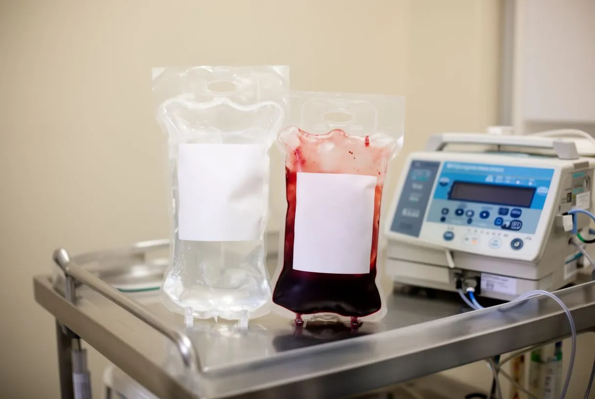
Blood cardioplegia: This approach mixes the patient's own blood with a concentrated electrolyte solution, typically in a ratio of four parts blood to one part crystalloid. Blood cardioplegia delivers oxygen directly to the myocardium, buffers acid buildup, and provides natural oncotic pressure that helps reduce tissue oedema. Many cardiac centres now prefer blood cardioplegia, especially for longer or more complex operations, because it more closely mimics the heart's natural physiological environment. Studies such as one examining a Lebanese family with congenital cardiac defects — A Novel Role for CSRP1 in a Lebanese Family with Congenital Cardiac Defects — illustrate how genetic factors can influence cardiac structures and why tailored surgical protection matters in complex cases.
Cold vs Blood Cardioplegia
The choice between cold cardioplegia and blood cardioplegia is one of the most discussed topics among cardiac surgeons and perfusionists. Both approaches achieve the same primary goal — safe, reversible arrest of the heart — but they differ in how they protect the myocardium during the arrest period. Cold cardioplegia relies heavily on hypothermia, infusing a chilled solution at four to ten degrees Celsius to rapidly lower the heart's temperature. The metabolic rate drops steeply, and the heart remains protected for longer intervals between doses. Blood cardioplegia, by contrast, may be delivered cold, tepid, or warm, and it provides ongoing oxygen delivery that cold crystalloid solutions cannot.
When cold cardioplegia is preferred: Cold crystalloid cardioplegia is often chosen for shorter procedures where the arrest time is expected to be relatively brief. It is straightforward, cost-effective, and provides excellent visualisation because the solution is clear rather than blood-tinged. Some surgeons also favour it in specific patient populations where avoiding the complexity of blood mixing circuits simplifies the operative setup. However, repeated doses are typically needed every twenty to thirty minutes, and the very low temperatures can occasionally contribute to reperfusion-related issues when the heart is rewarmed.
When blood cardioplegia is preferred: Blood-based cardioplegia tends to be favoured for longer, more complex operations — valve replacements, combined procedures, or surgery on patients with compromised heart function. The oxygen-carrying capacity of autologous blood means the myocardium receives some oxidative support even during arrest, which may reduce the risk of subtle injury. Many centres also use warm blood cardioplegia for the initial dose, followed by cold maintenance doses, a strategy known as "warm induction, cold maintenance." The specific protocol chosen depends on institutional experience, the surgeon's preference, and the patient's individual cardiac and metabolic status. Ongoing assessment of how different catheterisation and surgical strategies affect outcomes — as explored in research such as A multi-region assessment of population rates of cardiac catheterization and yield of high-risk coronary artery disease — helps teams refine these decisions.
Cardioplegia Delivery Systems Explained
Delivering cardioplegia solution to the heart requires specialised equipment designed to ensure precise flow, temperature, and pressure. The delivery system is more than a tube and a pump — it is a carefully engineered circuit that the perfusionist operates in coordination with the surgeon. The solution must reach the coronary arteries, which supply the heart muscle itself, in a controlled and uniform manner. Too much pressure can damage the coronary endothelium; too little means uneven distribution and inadequate protection of the myocardium.
Antegrade and retrograde delivery: There are two primary routes for administering cardioplegia. Antegrade delivery infuses the solution into the aortic root or directly into the coronary ostia, allowing it to flow forward through the coronary circulation just as blood normally does. Retrograde delivery, by contrast, introduces the solution through a catheter placed in the coronary sinus — the vein that collects deoxygenated blood from the heart muscle — so it flows backward through the capillary bed. Many surgeons use both methods in combination, especially when there is significant coronary artery disease or atheromatous plaque that might prevent even antegrade distribution. This dual approach ensures that all regions of the heart receive protection, even when some coronary vessels are blocked or narrowed.
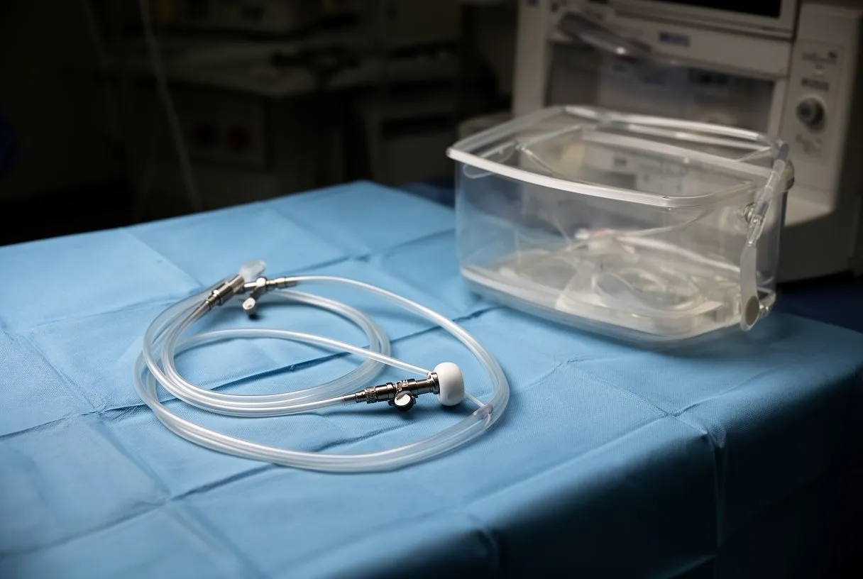
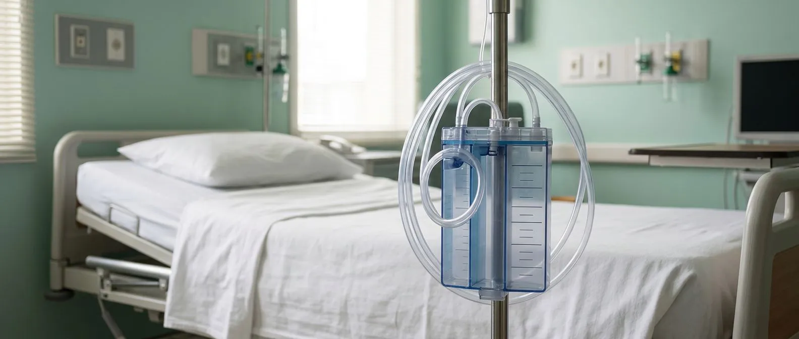
The delivery system also includes temperature control modules, which can cool or warm the solution as needed, and pressure-monitoring lines that feed real-time data back to the perfusionist. Modern systems can switch between blood and crystalloid, adjust ratios automatically, and provide alarms if pressure thresholds are exceeded. For the patient, this all happens invisibly — but understanding that a dedicated specialist is managing the protection of your heart throughout the operation can provide reassurance during an anxious time. The delivery system is, in many ways, the lifeline that makes the entire concept of cardioplegia heart surgery possible.
How Surgeons Stop the Heart
The actual process of arresting the heart is one of the most carefully choreographed moments in any cardiac operation. After the patient is anaesthetised and the chest is opened, the surgeon places the patient on cardiopulmonary bypass — the heart-lung machine takes over circulation. Once bypass is established and stable, the surgeon clamps the ascending aorta to isolate the heart from the systemic circulation. This is the critical step that allows cardioplegia solution to be infused into the coronary circulation without it being washed away into the rest of the body.
The infusion itself: The cardioplegia solution is delivered under controlled pressure, typically through a catheter placed in the aortic root. The high potassium concentration immediately begins to depolarise the cardiac muscle cells, and within seconds the electrical activity visible on the ECG monitor flattens. The heart relaxes in diastole and becomes completely still. The surgeon can visually confirm arrest — the motionless, soft, bloodless heart is the sign that the field is ready for the planned repair. For patients with significant aortic valve disease, such as those who might be candidates for a balloon-expandable valve or a surgical valve replacement, retrograde delivery through the coronary sinus may be used to ensure full protection.
During this process, the perfusionist monitors the pressure in the delivery line and the temperature of the heart. Cooling may begin immediately, and the solution may be re-dosed at intervals depending on the protocol. The entire induction — from clamp to confirmed arrest — usually takes only a minute or two, but every second is managed with precision. If the heart shows any residual electrical activity, additional cardioplegia is given until complete silence is achieved. This is why having an experienced, well-coordinated surgical and perfusion team matters so much: the safety of the arrest depends on timing, pressure, and dose all being correct.
Heart Restart After Surgery
Once the surgical repair is complete, the heart must be carefully brought back from its arrested state. This is the phase surgeons call reperfusion — the restoration of blood flow to the heart muscle after a period of ischaemia. The aortic cross-clamp is removed, and oxygenated blood from the heart-lung machine begins to flow back through the coronary arteries. The heart gradually rewarms, and the metabolic environment inside the cells normalises. In many cases, the heart begins to beat spontaneously as potassium is washed out and normal electrical conduction resumes. In other cases, a brief, low-energy electrical shock from internal paddles — much smaller than the external shock delivered by an automated external defibrillator — is used to initiate a coordinated rhythm.
What can affect the restart: The quality of the restart depends on several factors: how long the heart was arrested, how well it was protected, the underlying condition of the myocardium, and whether any reperfusion injury occurs. Most hearts resume a normal sinus rhythm within minutes of reperfusion, but some may develop atrial flutter or other arrhythmias that require temporary pacing or medical management. The surgical team watches the ECG closely during this phase, and the perfusionist continues to support circulation until the heart is beating strongly enough to take over from the bypass machine. This transition is gradual and carefully managed — it is not a sudden handoff but a staged weaning process.
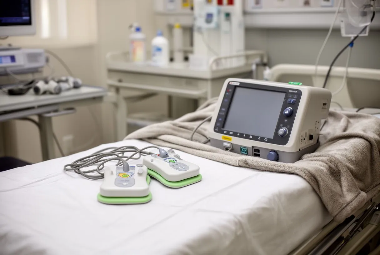
Patients sometimes worry that restarting the heart is the most dangerous part of the operation. In reality, it is a well-practised phase of cardiac surgery, and teams are prepared for a range of scenarios. Pacing wires are often placed on the surface of the heart before closing the chest, providing a backup electrical system if needed in the early postoperative period. The heart-lung machine remains on standby until the team is fully satisfied that the heart is functioning independently. For the patient's family waiting outside, this is typically the point at which the surgeon will come to the waiting area to report that the repair is complete and the heart is beating again.
Reperfusion Injury Prevention
Reperfusion injury is a paradoxical but well-recognised phenomenon: when blood flow is restored to a tissue that has been ischaemic, the very act of reoxygenation can cause additional damage. This is not unique to cardioplegia heart surgery — it occurs in any setting where blood supply is interrupted and then restored — but it is a particular concern in cardiac surgery because the heart has been deliberately arrested and must be reperfused. The injury is driven by a cascade of events including calcium overload, generation of reactive oxygen species, and inflammation. The goal of reperfusion injury prevention is to blunt this cascade and protect the myocardium during the vulnerable transition period.
How cardioplegia protocols address this: Modern cardioplegia strategies incorporate several protective mechanisms. Blood cardioplegia provides a more physiological reoxygenation than crystalloid solutions because the blood contains natural antioxidants and buffers. Controlled reperfusion — gradually restoring flow rather than suddenly flooding the coronary bed — helps prevent the abrupt calcium influx that triggers much of the damage. Some protocols add specific additives such as lidocaine, magnesium, or buffer agents to the cardioplegia solution to stabilise cell membranes and reduce the metabolic stress of reperfusion. Temperature management also matters: slowly rewarming the heart, rather than rapid rewarming, gives the cells time to recover their metabolic balance.
For patients, this means that the period of arrest and restart is not simply a matter of turning the heart off and on. It is a carefully managed physiological transition, and the quality of that transition has real implications for how well the heart recovers. A heart that has been well protected during arrest and gently reperfused is more likely to resume strong, coordinated contractions and less likely to suffer subtle injury that could affect long-term function. This is one reason why experienced centres with dedicated perfusion teams tend to report smoother recoveries — the details of cardioplegia management, though invisible to the patient, directly influence the outcome.
Cardioplegia Risks and Safety
No surgical technique is without risk, and cardioplegia is no exception. The risks associated with cardioplegia heart surgery fall into several categories: risks related to the arrest itself, risks related to the delivery system, and risks related to the reperfusion phase. Understanding these risks — and how experienced teams mitigate them — is important for patients making informed decisions about their care. The overall safety of cardioplegia has improved enormously over decades of refinement, but it remains a technique that demands precision and vigilance.
Specific risks and mitigations:
- Inadequate protection: If cardioplegia distribution is uneven — due to coronary disease or atheromatous plaque — some regions of the heart may not be fully protected. Surgeons mitigate this with combined antegrade and retrograde delivery.
- Temperature-related issues: Excessive hypothermia can impair enzyme function; insufficient cooling can leave the heart metabolically active. Precise temperature monitoring throughout the procedure keeps the heart in the optimal range.
- Arrhythmia on restart: The heart may develop atrial flutter or other rhythm disturbances during reperfusion. Pacing wires, antiarrhythmic medications, and temporary pacing are standard backup measures.
- Reperfusion injury: As discussed above, controlled reperfusion protocols and protective additives in the cardioplegia solution help minimise this risk.
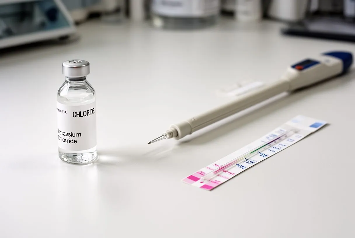
It is worth noting that these risks are managed continuously by the surgical team. The perfusionist monitors every parameter in real time, and the surgeon adjusts the approach based on direct visual assessment of the heart. If a problem is detected — for example, persistent electrical activity despite cardioplegia delivery — the team acts immediately. This is why the duration of cardioplegia arrest, while important, is only one factor among many. The quality of protection and the skill of the team matter at least as much as the raw time on bypass. Patients should feel empowered to ask their surgical team about their experience with cardioplegia protocols and their approach to myocardial protection.
Recovery After Cardioplegia Heart Surgery
Recovery after cardioplegia heart surgery is a process that begins in the operating room and continues for weeks to months afterward. In the immediate postoperative period, the patient is typically cared for in an intensive care unit where heart rhythm, blood pressure, and cardiac enzyme levels are closely monitored. The heart that has been arrested and restarted needs time to fully recover its normal metabolic and electrical function, even if it is beating strongly. Most patients are extubated within hours, and early mobilisation — sitting up, standing, and walking — begins within the first day or two in uncomplicated cases.
What to expect in the weeks ahead: The first few weeks at home involve gradual increases in activity, wound care, and medication management. Patients are often prescribed aspirin or other medications to support healing and prevent complications. Cardiac rehabilitation — a structured programme of supervised exercise and education — typically begins four to six weeks after surgery and continues for several months. During this time, the heart continues to heal, and most patients report a steady improvement in energy and exercise tolerance. Persistent symptoms such as arm pain, chest discomfort, or unusual shortness of breath should be reported to the cardiac team promptly, as they may indicate a problem that needs evaluation.
The long-term outlook after cardioplegia heart surgery is generally excellent when the underlying condition has been properly addressed and the heart has been well protected during the operation. The cardioplegia itself leaves no lasting effect — once the potassium is washed out and normal blood flow is restored, the heart resumes its natural rhythm and function. What shapes the long-term outcome is the quality of the surgical repair, the patient's overall cardiovascular health, and their commitment to follow-up care and lifestyle modifications. For patients in Mexico and those travelling to centres like those in Guadalajara, Querétaro, and Hermosillo, having a clear understanding of the recovery timeline and knowing when to seek help is essential. We encourage every patient to discuss their specific situation with a qualified cardiac team and to book a consultation rather than relying on general information alone — your individual anatomy, condition, and goals should guide every decision.