


12-Lead ECG: What the Full Heart Tracing Reveals
12-lead ECG results explained: how to read your heart tracing, what normal and abnormal findings mean, and when to seek cardiac care in Guadalajara.
When you or a loved one faces a potential heart problem, the physician often orders a 12-lead ECG. This simple, non-invasive test is the frontline tool for evaluating heart electrical activity. A 12-lead ECG provides a comprehensive view of the heart's electrical system from twelve different angles, allowing cardiac specialists to detect irregularities quickly. We know that seeing medical jargon on a chart can feel overwhelming, but understanding your 12-lead ECG results empowers you to make informed decisions alongside your cardiac team.
What is a 12-Lead ECG and Why Do Doctors Use It?
Understanding the resting ECG test meaning. A 12-lead ECG is a non-invasive recording of the electrical signals that travel through the heart muscle with each beat. Small sensors placed on the skin pick up these electrical impulses and translate them into a wavy line on a printout. Doctors use this test to evaluate the resting state of the heart, checking the rate, rhythm, and structural orientation before any intervention. It acts as an electrical map, showing the precise pathway of the heartbeat.
Why twelve leads are necessary. The heart is a three-dimensional organ, and a single wire cannot capture the full picture. Each of the twelve leads acts like a different camera angle, looking at the electrical pathway from a unique vantage point. According to Johns Hopkins Medicine, this multi-angle view is critical for pinpointing the exact location of damage or rhythm disturbances. Weaving in clinical understanding helps patients feel more grounded in their care.
The role of the test in initial screening. If you experience vague symptoms like dizziness or palpitations, this tracing acts as a baseline. It helps differentiate between benign causes and serious structural issues. By mapping the electrical sequence, your cardiologist can see if the upper chambers, known as the atrium, are conducting impulses correctly to the lower pumping chambers. This initial snapshot guides all subsequent diagnostic decisions.
How Does ECG Tracing Analysis Work on the Skin?
How the heart generates electricity. The heart has its own natural pacemaker called the sinoatrial node, which fires an electrical impulse that spreads across the heart muscle. This electrical wave causes the muscle cells to contract and pump blood. ECG tracing analysis captures this microscopic electrical leak through the skin, amplifying it so we can see the P waves, QRS complexes, and T waves on paper. The skin acts as a conductor, allowing the external machine to visualize the internal rhythm.
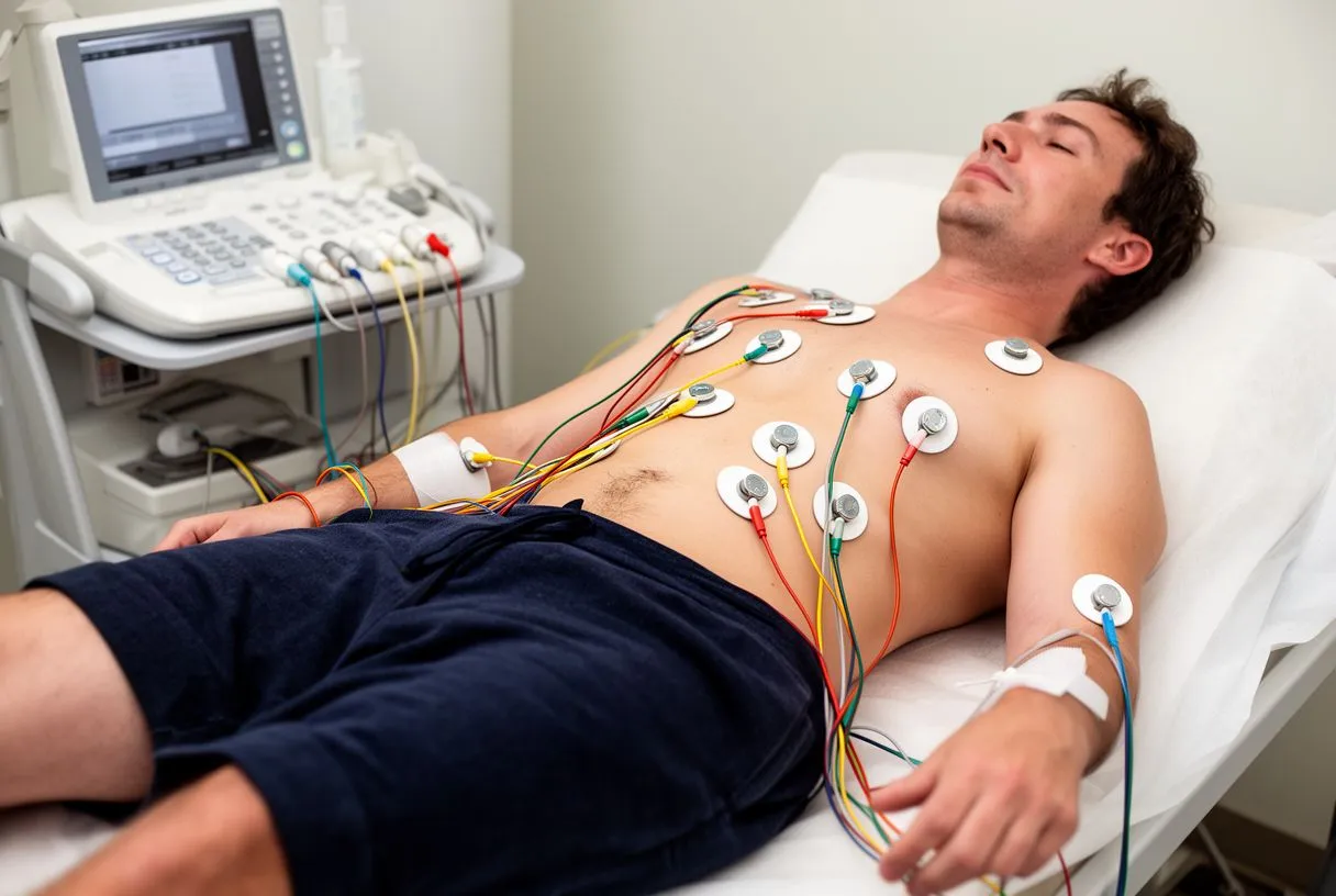
Interpreting the waves on the strip. Each bump and line corresponds to a specific action. The P wave shows the upper chambers squeezing, the QRS complex shows the main pumping chambers firing, and the T wave represents the heart resetting for the next beat. If this resetting phase is disrupted, it can point to an electrolyte imbalance or poor blood flow to the heart muscle. Proper ECG rhythm strip reading requires looking at the timing between these waves.
The importance of signal clarity. Good ECG rhythm strip reading requires clear signals, which is why the technician prepares the skin. Sometimes, initial auscultation with a stethoscope reveals a murmur that prompts the test, but the electrical tracing confirms the diagnosis. Clear signals allow the cardiologist to spot subtle changes, like those seen in an atypical infarction, where classic crushing chest pain is absent but the heart tissue is still starved for oxygen. High-quality tracings ensure no subtle diagnostic clues are missed.
ECG vs EKG Difference Explained for Anxious Patients
Why the two acronyms exist. Many patients worry that ECG and EKG refer to two different tests, but the ECG vs EKG difference is simply a matter of language. ECG stands for electrocardiogram, while EKG comes from the German spelling, Elektrokardiogramm. Doctors adopted EKG to avoid confusion with EEG, a brain wave test, but both terms describe the exact same procedure. Knowing this simple translation eliminates a common source of pre-appointment anxiety for patients.
What this means for your medical chart. When reviewing your 12-lead ECG results, you might see either acronym used interchangeably by nursing staff and physicians. There is no difference in the machine, the placement of the electrodes, or the information gathered. Your cardiac screening ECG test remains identical regardless of which abbreviation the clinic uses on the requisition form. The medical standard for capturing the heart's electrical activity is universally consistent.
- Same Test: Both ECG and EKG refer to the exact same electrocardiogram procedure.
- Same Machine: The 12-lead ECG equipment is used regardless of the acronym.
- Same Results: The 12-lead ECG results provide identical clinical data either way.
Focusing on the results, not the letters. Understanding that the terminology is identical helps reduce unnecessary anxiety before an appointment. The focus should remain on what the tracing shows regarding your heart's health. Whether your doctor orders an ECG before heart surgery or as part of a routine check-up, the goal is always to capture the most accurate electrical snapshot of your heart function. We encourage patients to ask their doctors to clarify any confusing terminology on their charts.
Understanding 12-Lead ECG Electrode Placement
Where the sensors go on the body. Proper 12-lead ECG electrode placement is essential for an accurate reading. The technician places six small pads across the left side of the chest in a specific arc over the heart, and four pads on the arms and legs. This configuration creates the twelve distinct viewpoints needed to map the heart's electrical activity comprehensively. The precise anatomical landmarks guide the technician to ensure every lead captures its designated angle.
How placement affects the reading. If a pad is misplaced by even a few centimeters, it can alter the tracing and mimic a heart attack where none exists. The pads on the limbs act as the baseline, while the chest pads provide the horizontal, cross-sectional view. This precise placement allows doctors to track the progression of an electrical block through the heart tissue. Consistency in electrode positioning is vital for comparing tracings taken on different days.
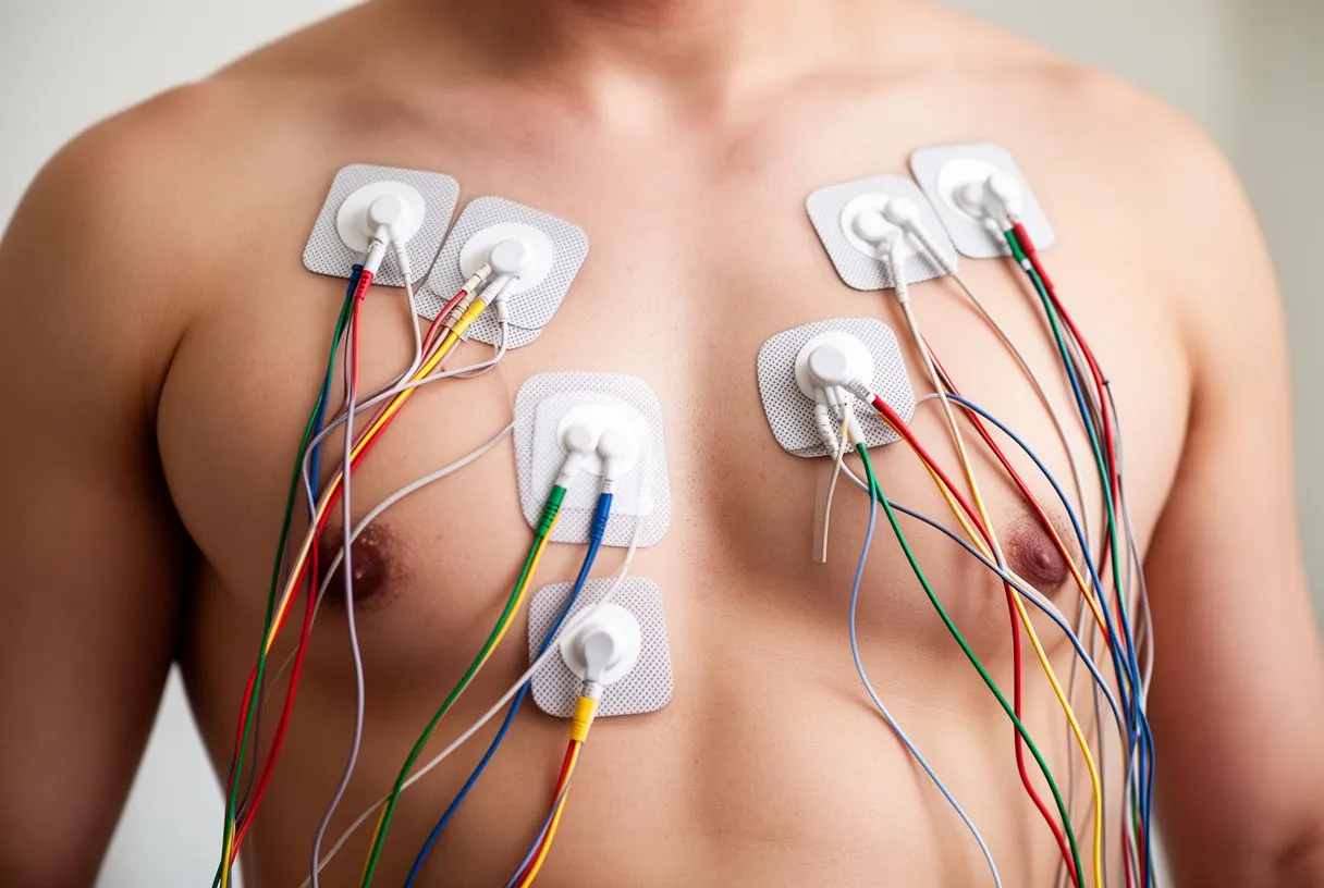
Preparing the skin for accuracy. The skin must be clean and dry, and sometimes a small area is shaved to ensure the pad sticks properly. This preparation helps keep the tiny electrical signals from being blocked by dead skin cells, sweat, or lotions. The entire setup is quick, and once the pads are in place, the actual recording usually takes only a few seconds. Patients must remain completely still and hold their breath briefly to prevent muscle tremors from interfering with the electrical signal.
What ECG Shows About Heart Electrical Activity
Detecting rhythm disturbances. One of the primary things the test reveals is the heart's rhythm. A normal heart beats in a steady, predictable pattern, but conditions like atrial flutter disrupt this flow. The tracing will show distinct sawtooth patterns instead of normal P waves, alerting the cardiologist to a rapid, irregular rhythm in the upper chambers that requires medical management. This detailed electrical view helps differentiate between harmless palpitations and serious arrhythmias.
Evaluating structural size and strain. The electrical impulses travel slower through thickened or enlarged heart muscle. By measuring the height and width of the waves, a specialist can determine if the walls of the heart are thicker than normal, a condition called hypertrophy. This structural insight is invaluable when planning future interventions or determining if high blood pressure has damaged the heart over time. The 12-lead ECG acts as an early warning system for chronic structural strain.
Identifying electrical blocks. Sometimes the electrical signal gets delayed or blocked entirely as it travels from the upper to the lower chambers. The 12-lead ECG shows these delays clearly, categorized as first, second, or third-degree blocks. Depending on the severity, a patient might need a pacemaker to ensure the heart maintains a safe, consistent rhythm. This highlights exactly what ECG shows in terms of conduction pathways and the integrity of the heart's internal wiring.
How to Read ECG Normal vs Abnormal Results
Defining a normal tracing. When comparing ECG normal vs abnormal results, a normal tracing shows a steady rate between 60 and 100 beats per minute, with consistent spacing between beats. The waves are present in the correct order, and the ST segment—the flat line after the main squeeze—is perfectly level. This baseline tracing indicates that the electrical pathways are functioning without immediate distress. A normal result brings peace of mind, confirming the heart's basic electrical system is intact.
Recognizing concerning variations. Abnormal findings can range from harmless extra beats to severe signs of oxygen deprivation. A tracing might show rapid, chaotic electrical activity that suggests ventricular fibrillation, an emergency requiring an automated external defibrillator. For stable but abnormal rhythms, doctors might prescribe medications like aspirin to prevent clots, or reference clinical pathways like the ARISTOTLE trial for stroke prevention. More subtle abnormalities warrant immediate clinical correlation with the patient's symptoms.
Contextualizing the findings for the patient. It is important to remember that an abnormal result does not automatically mean you need surgery. Many variations are benign and related to age, fitness level, or electrolyte shifts. Your cardiac team will review the tracing alongside your medical history, physical exam, and any symptoms like unexplained arm pain to determine if further testing is required. We always evaluate the electrical picture in the context of the whole patient, never in isolation.
- Normal: Steady rate (60-100 bpm), regular rhythm, flat ST segments.
- Abnormal (Ischemia): Depressed ST segments, T wave inversions indicating poor blood flow.
- Abnormal (Emergency): ECG ST segment elevation indicating an active, severe heart attack.
Abnormal ECG Findings and ECG ST Segment Elevation
The significance of ST elevation. ECG ST segment elevation is a critical finding that demands urgent attention. This elevation occurs when a section of the heart muscle is actively dying due to a blocked artery, classically known as a STEMI heart attack. The 12-lead ECG is so precise that it can show exactly which artery is blocked based on which leads display the elevated segment. Recognizing this pattern immediately triggers life-saving protocols in the emergency department.
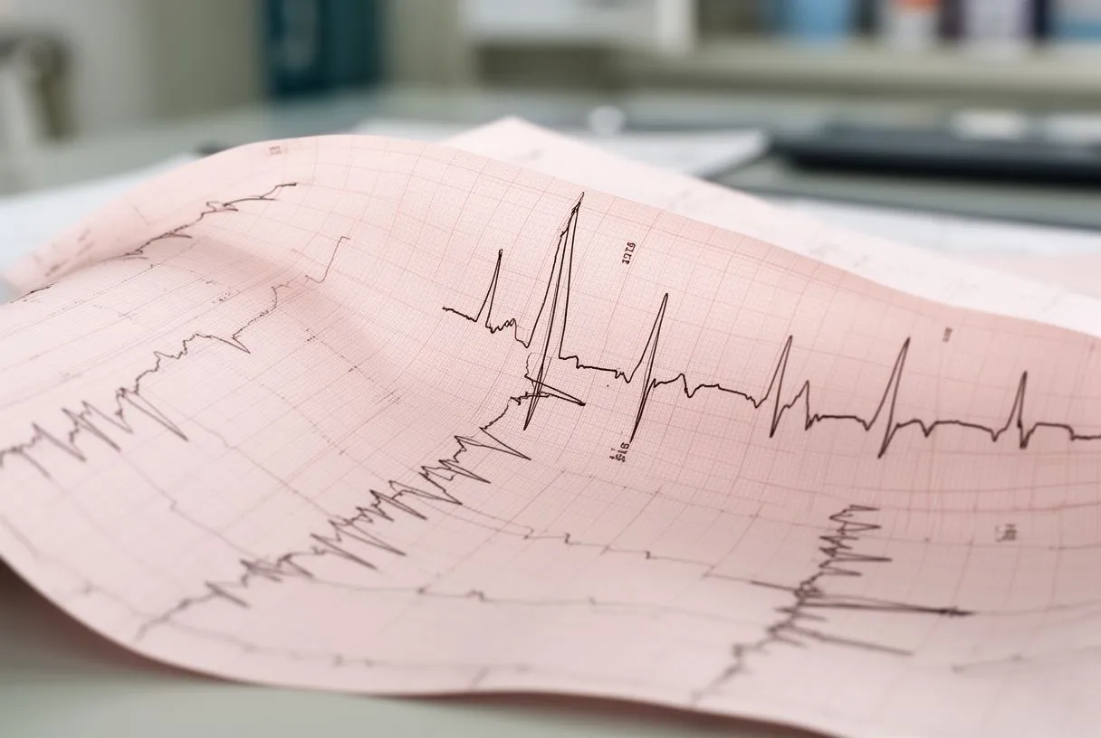
What causes the segment to rise. The ST segment rises because the injured heart muscle lacks adequate blood flow, causing an abnormal electrical current even when the heart is supposed to be resting. This is a true medical emergency. When a patient presents with these findings, guidelines dictate immediate intervention, such as balloon angioplasty, to restore blood flow. In severe cases, maintaining circulation with an arterial graft or supporting the heart with awake ECMO might be necessary while planning definitive surgical repair.
Other abnormal findings to watch for. ST elevation is the most dramatic finding, but other abnormalities carry weight. Depressed ST segments might indicate ischemia, where the heart is starving for oxygen but not yet dying due to a ruptured atheromatous plaque. Pathological Q waves can indicate old scar tissue from a previous, silent heart attack. Each abnormal line tells a story about the heart's history and current crisis, guiding the urgency of the medical response.
Using ECG for Chest Pain Diagnosis and Atypical Infarction
The first step in the emergency room. Using ECG for chest pain diagnosis is standard protocol worldwide. When a patient arrives with chest pressure, the test is administered within minutes to rule out an active heart attack. The tracing helps differentiate between cardiac emergencies, gastrointestinal reflux, or musculoskeletal pain, dictating the entire trajectory of emergency care. A rapid, accurate electrical snapshot prevents dangerous delays in treating acute coronary syndromes.
Catching silent or atypical events. Not all heart attacks present with crushing chest pain. An atypical infarction might present as jaw pain, profound fatigue, or shortness of breath, especially in women and diabetic patients. The 12-lead ECG is crucial here, as it reveals the electrical truth of the heart even when the physical symptoms are misleading or vague. It is the only definitive way to confirm heart muscle injury when clinical symptoms do not follow the classic pattern, which might later require structural interventions like a balloon aortic valvuloplasty or the placement of a balloon-expandable valve if concurrent valvular disease is discovered.
Monitoring evolving conditions. Sometimes a single tracing is not enough. If the initial ECG is normal but suspicion remains high, the doctor will order serial ECGs to watch for evolving changes. An artery might be slowly narrowing, and the electrical pattern might shift over hours. Combining these tracings with blood tests confirms whether heart muscle enzymes are leaking, solidifying the diagnosis. For complex systemic infections affecting the heart, such as those detailed in a Successful treatment of challenging case of infective endocarditis, the ECG remains a vital monitoring tool alongside continuous arterial line monitoring and the careful management of autologous blood if surgery is required.
ECG Before Heart Surgery and Cardiac Screening Tests
Establishing a surgical baseline. An ECG before heart surgery is mandatory to ensure the patient is safe for anesthesia. The anesthesiologist needs to know the exact rhythm and rate to manage medications properly during the operation. This baseline tracing is compared against tracings taken during and after surgery to ensure the heart tolerates the stress of the procedure. Establishing this electrical baseline is a critical safety checkpoint before any surgical intervention involving an arterial cannula.
Assessing risks for complex repairs. For patients undergoing major structural repairs, like fixing an aneurysm in the ascending aorta or preparing a site for an autograft, the preoperative ECG helps assess overall cardiac risk. Surgeons use this information alongside advanced imaging to plan their approach. In highly complex cases, such as those involving connective tissue disorders requiring multi-stage operations as described in the case report Eyes wide shut - unusual two stage repair of pectus excavatum and annuloaortic ectasia in a 37 year old marfan patient: case report, understanding baseline electrical stability is paramount before proceeding.

Routine screening for high-risk patients. A cardiac screening ECG test is often recommended for patients with a strong family history of heart disease or risk factors like high cholesterol, marked by elevated apolipoprotein B levels. Catching electrical abnormalities early can prevent sudden cardiac events. This proactive approach allows doctors to manage conditions with medication long before surgical intervention becomes necessary. Doctors use these screenings to build a comprehensive risk profile for each patient.
Where to Get a 12-Lead ECG in Guadalajara and Mexico
Accessing specialized cardiac care. If you are wondering where to get a 12-lead ECG in Guadalajara or across Mexico, it is a widely available test, usually arranged through a cardiologist, a clinic or a hospital.
Navigating cost and logistics. We understand that international patients have concerns about logistics. A standard test is brief, answering the common question of how long does ECG take—usually less than ten minutes for the actual recording.
Partnering with your cardiac team. Do not ignore symptoms like chest tightness, palpitations, or unexplained fatigue. We encourage you to discuss your specific situation with a qualified cardiac team rather than self-diagnosing based on internet searches. Booking a consultation with Dr Alejandro Molina allows us to review your 12-lead ECG results together, explain exactly what the tracing means, and chart the safest, most effective path forward for your heart health.






