


Epicardium and Epicardial Fat: What It Means for the Heart
Epicardial fat heart tissue can protect or harm your heart.
Most people have never heard of the epicardium, yet this thin outer layer of the heart — and the epicardial fat heart tissue that sits against it — plays a surprising role in cardiac health. If you or a family member has been told about "fat around the heart" on a scan, the words can sound alarming. In most cases they are not. This guide explains what the epicardium is, what the fat around it does, when it matters, and when it deserves a specialist's attention.
What Is the Epicardium and Where Does It Sit?
The epicardium is the outermost layer of the heart wall. It is a thin, smooth membrane that covers the entire surface of the heart, like the skin of an apple. In simple terms, when someone asks about the meaning of epicardium, the answer is: the heart's own outer covering. It is also the inner layer of the pericardium, the double-walled sac that encloses the heart and anchors it inside the chest.
This layer is more than wrapping paper. The epicardium carries the coronary arteries — the vessels that feed the heart muscle with blood — on its surface. Nerves and lymphatic vessels also run through it. During development before birth, the epicardium gives rise to cells that help build parts of the heart itself, a role researchers continue to study for its potential in healing damaged tissue.
Because it sits directly on the beating muscle, the epicardium is exposed to everything happening in and around the heart. Changes in this layer, or in the fat lying on top of it, can offer early clues about conditions elsewhere in the cardiovascular system. That is why cardiologists pay attention to it even though patients rarely hear the word outside a report.
The Layers of the Heart Wall Explained
The heart wall is made of three distinct layers, each with its own job. Understanding them makes the role of the epicardium much clearer.
- Epicardium: the smooth outer covering, rich in fat tissue in adults, which protects and nourishes the surface of the heart.
- Myocardium: the thick muscular middle layer that actually contracts and pumps blood to the whole body.
- Endocardium: the delicate inner lining that contacts the blood itself and covers the heart valves, keeping flow smooth and preventing clots from forming on healthy tissue.
What is the function of the endocardium? Its main job is to create a friction-free surface for blood moving through the chambers and across the valves. A healthy endocardium also releases chemical signals that help the heart muscle relax properly between beats. When this lining is damaged — for example by prolonged high blood pressure — it can contribute to stiffening and poor heart function over time.
What is the function of the fluid in the pericardial sac? Between the epicardium and the outer pericardial sac sits a small amount of lubricating fluid, usually just a few millilitres. This fluid lets the heart glide with every beat, without rubbing against the surrounding structures. Too much fluid, or inflamed surfaces, cause the chest pain and breathing discomfort patients know as pericarditis.
The three layers work together as one organ. Disease rarely respects boundaries — inflammation on the surface can affect the muscle beneath, and muscle disease can distort the lining. That interdependence is a recurring theme throughout this article.
What Is Epicardial Fat and Why Does the Heart Have It?
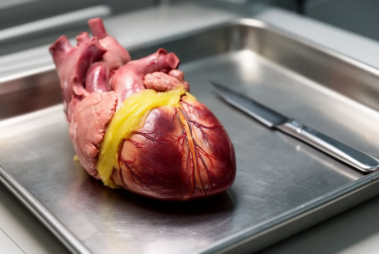
Epicardial adipose tissue is the fat depot that lies directly on the epicardium, between the heart muscle and the pericardial sac. It is not the same as ordinary body fat. It shares the heart's blood supply, wraps around the coronary arteries, and communicates actively with the muscle beneath it by releasing fatty acids and signalling molecules.
In healthy amounts, this fat is genuinely useful. It stores energy for the tireless heart muscle, cushions the heart against the mechanical movement of the lungs and chest wall, and helps regulate local temperature around the vessels. Everyone has some; it is a normal anatomical structure, not a defect.
Why does it grow? Epicardial fat tends to expand alongside overall weight gain, metabolic syndrome, type 2 diabetes and chronic inflammation. It also increases with age. The pattern matters: a modest, evenly distributed layer is normal, while a thickened, inflamed layer is what researchers associate with epicardial fat heart problems down the line.
Epicardial Fat Heart Function: The Helpful Side
When we talk about epicardial fat heart function, we mean the jobs this tissue performs for a healthy heart. Its closest analogue is the fat around the kidneys, which protects and feeds a nearby organ with constant energy demands. The beating heart uses fatty acids as a preferred fuel, and the epicardial depot delivers them locally through small connecting vessels.
Protection and repair. The epicardium and its fat respond to injury. Research summarised in Frontiers describes how epicardial-derived cells can reactivate repair programmes and participate in the heart's response to damage, a crosstalk scientists are still working to understand fully. This is one reason the layer is far more than passive padding.
Local immune role. Epicardial fat contains immune cells and helps orchestrate the local inflammatory response. In balance, this is protective: it clears debris and defends the vessel walls. Out of balance, the same machinery becomes a source of chronic, low-grade inflammation — the pivot point between a healthy depot and a harmful one.
In our practice we explain this balance to patients in plain words: the fat around the heart is neither an enemy nor an ornament. It is a working tissue, and our goal is to understand what state yours is in before deciding anything.
When Epicardial Fat Becomes a Risk for Heart Disease
The same tissue that helps in health can harm in excess. Enlarged epicardial adipose tissue behaves differently: it releases more inflammatory signalling molecules, fewer protective ones, and can influence the coronary arteries it touches from the outside in. This is the core of epicardial fat heart risk as researchers currently understand it.
What changes in an enlarged depot? Studies in obesity and metabolic disease describe fat cells that swell, immune cells that accumulate, and a shift toward chronic inflammation. Because the fat sits directly against the vessel walls, those signals do not need to travel far. Thickened epicardial fat has been linked in the literature to coronary calcium, arterial plaque burden and atrial fibrillation, although the exact cause-and-effect relationships are still being mapped.
Who is most at risk? People with central obesity, insulin resistance or type 2 diabetes, those with sleep apnoea, and patients with established coronary disease tend to have larger depots. Age and genetic factors also contribute. Importantly, epicardial fat can be increased in people who look otherwise well — which is why it comes up as an incidental finding on scans done for other reasons.
It is worth being precise here: an increased amount of fat does not mean disease is present, and no scan finding replaces a full clinical evaluation. In our team every imaging finding is read together with symptoms, history and guideline-based testing before we tell a patient what it means.
Pericardial Fat vs Epicardial Fat: What Is the Difference?
This is one of the most common points of confusion, and the distinction matters. Pericardial fat is the broader term for all fat inside the chest but outside the heart muscle itself. Epicardial fat is the specific portion lying between the myocardium and the visceral pericardium — directly on the heart. Some fat deposits sit between the pericardial layers or outside the sac entirely; those behave differently and carry different implications.
Why the distinction matters. Epicardial fat shares blood flow with the heart and touches the coronary arteries, so it can influence them directly. Fat outside the pericardium is anatomically more distant and is generally considered less metabolically active in relation to the heart. When a radiology report mentions a "cardiac fat pad" or measures "pericardial fat volume", it is worth asking which compartment the measurement refers to.
The epicardial fat heart difference in one sentence: epicardial fat is the inner, metabolically active compartment in contact with the heart and its vessels, while the rest is pericardial fat lying further out. Surgeons and imaging specialists care about this because the inner depot is the one most consistently associated with cardiovascular outcomes in published research.
If a report has left you unsure which fat you were told about, ask. A short clarifying conversation is often all it takes, and a good team will never make you feel the question is too basic.
How an Epicardial Fat Heart Scan Detects Problems
An epicardial fat heart scan is usually not a dedicated test. The fat shows up on imaging already being done for other reasons: a CT coronary calcium study, a CT angiogram, an echocardiogram or a cardiac MRI. On echocardiography it appears as a bright echo-dense space near the right ventricle; on CT it can even be measured precisely as a volume.
What the images actually show. Radiologists look at thickness and distribution — commonly measuring the fat over the right ventricle, in the grooves between chambers, and around the coronary arteries. They also note whether the fat appears to compress structures, encase vessels, or show features suggesting inflammation. Epicardial fat heart health assessment is therefore a descriptive exercise, read against standard reference ranges.
What a finding does and does not mean. An increased measurement is a risk marker, not a diagnosis. It prompts questions: Is there metabolic disease? Are there symptoms? Is there coronary plaque or valve disease that needs attention? In our consultations we take the time to explain what a study actually shows in everyday language, and to say plainly which additional tests are genuinely needed — and which are not.
Imaging technology keeps improving, and researchers are working to make fat quantification a routine part of cardiovascular risk scoring. For now, treat it as one useful signal among many, never as a verdict on its own.
Epicardial Fat and Coronary Disease: What Is the Link?
The relationship between epicardial fat and coronary disease is one of the most studied areas in this field. The coronary arteries run directly within the epicardial fat, so any inflammatory molecules the fat produces reach the vessel walls almost immediately. Autopsy and imaging studies consistently show that patients with more extensive coronary plaque tend to have larger, more inflamed epicardial depots.
From fat to plaque. The proposed chain of events is straightforward: inflamed fat promotes inflammation inside the artery wall, inflammation destabilises plaque, and unstable plaque is what causes heart attacks. The fat also secretes molecules that influence blood clotting and vessel tone. None of this means fat causes every case of coronary disease — cholesterol, blood pressure, smoking and genetics remain the dominant drivers — but it adds a local dimension to a systemic problem.
Why surgeons care. In the operating room, epicardial fat can influence how the surgical field looks and how structures are approached, particularly around the right ventricle and the coronary targets for bypass grafting. Experienced surgical teams account for it when planning conventional, minimally invasive or robotic procedures, and Dr Molina's team plans every operation with the patient's exact anatomy in view.
For patients, the practical takeaway is this: if you have coronary disease, it is reasonable to assume your epicardial fat is part of the picture, and managing the whole metabolic situation — not the fat in isolation — is what changes outcomes.
How to Reduce Epicardial Fat Around the Heart
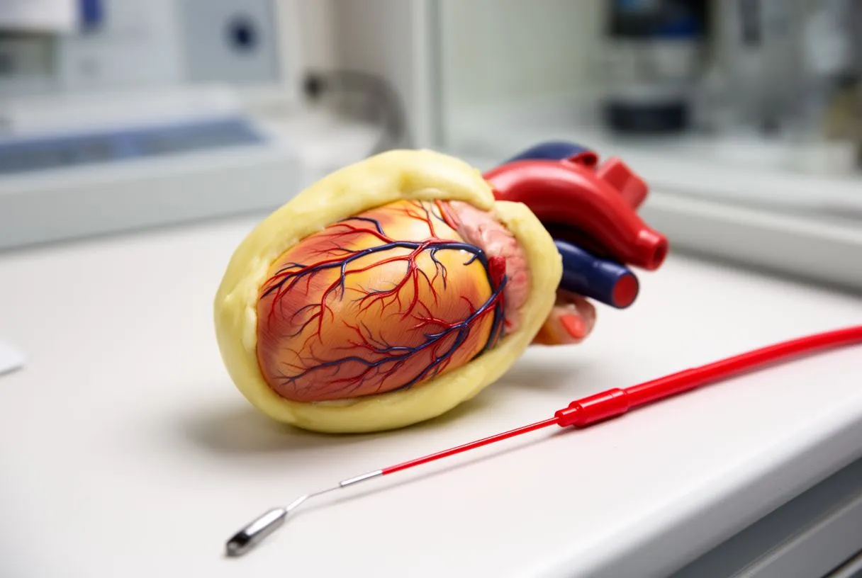
The good news is that the strategies to reduce epicardial fat are the same ones that reduce overall cardiovascular risk. Research shows epicardial fat responds to weight loss, exercise and improved metabolic control, and in some studies it responds faster than fat elsewhere in the body.
- Sustained exercise: regular aerobic activity plus resistance training, consistent with general physical-activity guidelines, is the most consistently supported intervention in the literature.
- Weight and metabolic control: meaningful weight reduction, good diabetes control and treatment of sleep apnoea all correlate with shrinking epicardial depots in published studies.
- Managing the underlying drivers: blood pressure, cholesterol and smoking cessation remain foundational, because epicardial fat rarely shrinks while the metabolic environment stays hostile.
Is medication part of epicardial fat heart treatment? There is no drug approved specifically to shrink epicardial fat. Some medications used for diabetes, obesity or cholesterol have been observed in studies to reduce fat volume as a secondary effect, but decisions belong with your own physicians, based on your whole situation. Beware of anyone selling a supplement or programme claiming to target cardiac fat specifically — no credible evidence supports such claims.
Realistic expectations. Change takes months, not weeks, and the goal is better metabolic health rather than a number on a report. Prevention, in the sense of epicardial fat heart prevention, starts decades earlier: keeping weight, activity and glucose in healthy ranges through adult life is the most reliable strategy known today. There is no situation where medication or surgery is chosen solely because a fat measurement is high.
When to Talk to a Heart Surgeon About Epicardial Fat
Most people who learn about epicardial fat will never need a heart surgeon, and that is worth saying clearly. A surgeon enters the picture when there is disease that surgery can treat — significant coronary blockages, valve disease, an aortic condition or a structural heart problem — and where epicardial fat is an associated finding rather than a reason for surgery on its own.
Situations that genuinely warrant specialist input: chest pain or breathlessness being investigated, a scan showing significant coronary plaque or calcium, a heart murmur or diagnosed valve disease, an aorta measured above guideline thresholds, or a report mentioning a cardiac fat pad alongside other abnormalities. Fat deposition combined with symptoms is what deserves workup — the fat alone, rarely.
What a good evaluation looks like. It is multidisciplinary and guideline-based, with a clear proposal, the alternatives laid out, and the final decision always left with the patient. A second opinion should be treated as a normal part of deciding about heart surgery — if a team resists your wish for one, that itself is information. We actively encourage patients to seek second opinions, and our multidisciplinary team reviews every case against international practice guidelines before any recommendation is made.
Access and first steps. Dr Molina is a heart surgeon trained in cardiac, minimally invasive, robotic and structural surgery, with consulting rooms in Guadalajara, Querétaro and Hermosillo, and the team performs conventional, minimally invasive, robotic and transcatheter procedures including TAVR. The first contact is a free video consultation: we get to know you, talk through your condition, and identify exactly which studies are still needed so no unnecessary ones are ordered. For patients searching for an epicardial fat heart specialist in Guadalajara or elsewhere in Mexico, that conversation is the honest starting point — not a scan purchased in isolation.
One last practical note: bring your imaging reports and questions to any consultation, and expect plain-language answers. If you leave an appointment more confused than you arrived, keep looking for the right team.

The epicardium is the heart's outer covering, and the epicardial fat heart layer against it is a living tissue with real jobs: fuelling, cushioning and protecting the muscle. In healthy amounts it is a friend; when it enlarges with metabolic disease it becomes a messenger worth reading, because it tracks with coronary risk and inflammation. The word itself should never be a source of panic — it should be a prompt for good questions, sound imaging interpreted in context, and honest conversations with a qualified cardiac team. If any of this has struck a chord with your own situation, book a consultation and discuss it with specialists rather than drawing conclusions from a report alone; your heart deserves nothing less.






