

Sinotubular Junction
Explore the sinotubular junction’s role in cardiac anatomy, pathology, dilatation, surgery, and aortic valve function with expert insights.
What is the sinotubular junction?
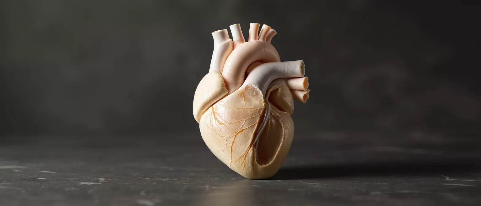
The sinotubular junction is a key structural landmark within the heart, specifically marking the boundary between the aortic root and the ascending aorta. For patients preparing for or recovering from cardiac surgery, understanding this region offers clarity about why certain procedures are necessary and how the heart maintains its remarkable, rhythmic function. It sits just above the aortic valve, acting as a physiological bridge between the heart's pumping chambers and the body's main arterial highway.
When surgeons evaluate the heart, this junction is one of the most carefully measured anatomical zones. It is not merely a passive border but an active, functional segment of the aortic root that plays a crucial part in maintaining unidirectional blood flow. Recognizing its location helps demystify many terms you may hear during consultations, such as aortic root enlargement or valve-sparing operations.
Because the sinotubular junction is intimately connected to the aortic valve's operation, any structural change in this area can have cascading effects on overall cardiac performance. Whether you are researching a known diagnosis or simply trying to better understand the surgical landscape of the heart, grasping the role of this junction is a foundational step in cardiac health literacy.
Sinotubular junction anatomy and structure
The sinotubular junction anatomy defines the upper margin of the aortic sinuses, which are the bulbous, crescent-shaped pockets that house the coronary artery ostia. Structurally, it forms a distinct ridge where the wider, curved sinuses transition into the tubular, straighter shape of the ascending aorta. This structural narrowing creates a localized geometric shift that is vital for the mechanical dynamics of the cardiovascular system.
What structures surround the aortic sinotubular junction? The aortic root contains three sinuses of Valsalva, and the sinotubular ridge forms the ceiling of each. The left and right coronary arteries branch off just below this junction, meaning the structural integrity of the ridge directly impacts the origin of the vessels supplying blood to the heart muscle itself.
Composed of elastic fibers and smooth muscle cells, the tissue at this junction allows for controlled expansion during systole, the phase of the heartbeat when blood is actively pumped forward. This elasticity is essential; it lets the aorta absorb the mechanical stress of pulsatile blood flow. Over time, structural changes in this tissue can lead to pathological expansion, known as aortic root aneurysm repair devices, which are designed to stabilize the vessel.
Sinotubular junction function in the heart
Sinotubular junction function is primarily mechanical, acting as a supportive anchor for the aortic valve leaflets. When the left ventricle contracts, blood surges through the aortic valve, pushing the leaflets open against the walls of the sinuses. The sinotubular ridge helps limit how far the leaflets can open, preventing them from collapsing or hitting the aortic wall too forcefully.
During diastole, when the heart relaxes, the elastic recoil of the sinotubular junction assists in snapping the aortic valve leaflets shut. This prevents blood from leaking back into the left ventricle, ensuring that the blood propelled outward continues moving forward through the systemic circulation. Without this elastic support, the precise, synchronized closure of the aortic valve would be compromised.
How does the sinotubular ridge support valve closure? By maintaining a specific geometric tension on the valve's commissures, the points where the leaflets attach to each other. If the ridge dilates or loses its natural tension, the commissures stretch outward, preventing the leaflets from meeting properly at the center, which leads to valve leakage.
Sinotubular junction diameter and dimensions
Cardiologists and surgeons closely monitor the sinotubular junction diameter during imaging studies. In a healthy adult heart, this diameter typically measures between 28 to 34 millimeters, though individual variations exist based on age, body size, and sex. The critical factor is the relationship between this diameter and the size of the aortic annulus, the ring at the base of the valve.
Normally, the sinotubular junction is slightly narrower than or equal to the annular diameter, maintaining a geometric harmony that ensures proper leaflet coaptation. When dimensions are tracked over time, stable measurements indicate structural health, while a progressive increase in diameter often signals underlying pathology. Patients often become familiar with these dimensions when utilizing advanced cardiac imaging services to monitor heart health.
Understanding these dimensions helps medical teams determine the threshold for surgical intervention. A significantly dilated junction is not just a numerical anomaly; it is a mechanical failure point. By understanding the structural ideal of the aortic root, patients can better comprehend why their surgeon may recommend prophylactic repair before symptoms arise.
Sinotubular junction dilatation and ectasia
Sinotubular junction dilatation occurs when the ridge loses its natural structural tension and expands outward. Ectasia refers to a mild, diffuse enlargement, while aneurysm implies a more pronounced, localized dilation. These conditions often develop slowly due to genetic tissue disorders, longstanding hypertension, or age-related degeneration of the aortic wall's elastic fibers.
What causes sinotubular junction ectasia? The primary drivers are connective tissue disorders like Marfan syndrome or Loeys-Dietz syndrome, but they also include wear and tear from aging and chronic high blood pressure. The elastic tissue framework degrades, causing the aortic root to widen over time.
When the junction dilates, the delicate spatial relationship required for aortic valve closure is disrupted. The distance between the valve's commissures increases, pulling the leaflets apart. Consequently, the valve no longer seals correctly, resulting in aortic regurgitation, a condition where blood flows backward into the heart, ultimately requiring surgical correction.
Sinotubular junction pathology and aneurysm risk
Understanding sinotubular junction pathology is vital for assessing the risk of aortic aneurysm. When the tissue at this junction weakens, the constant force of blood pressure pushes the wall outward, creating a bulge. This pathology is dangerous because as the aneurysm grows, the wall tension increases, raising the risk of a life-threatening dissection or rupture.
What are the warning signs of aortic root pathology? Most patients with isolated sinotubular pathology experience no symptoms until the valve begins to fail or the aneurysm grows significantly. When symptoms do appear, they often include shortness of breath, chest pain, or exercise intolerance, signaling that the heart's efficiency is dropping due to aortic regurgitation.
Because the risk of catastrophic tearing increases as the diameter expands, preemptive monitoring and surgical planning are standard protocols. Identifying the exact location and extent of the pathology allows surgeons to determine whether the native valve can be saved or if a full root replacement is necessary to restore safe cardiac function.
How sinotubular junction changes affect the aortic valve
The aortic valve sinotubular junction is structurally dependent, meaning the valve relies heavily on the junction's geometric stability to function. When the junction expands, it pulls the valve's suspension points outward. This outward tension causes the valve leaflets to stretch and fail to meet at the center during diastole, creating a central leak.
This type of valve failure is purely geometric rather than structural. The leaflets themselves may be perfectly healthy and pliable, but because their supporting frame has dilated, they cannot seal. This distinction is crucial for surgical planning, as it opens the door for valve-sparing root replacement implants that correct the frame without replacing the native leaflets.
Patients facing this specific mechanism of valve leakage often benefit from a highly specialized surgical approach. By restoring the sinotubular junction to its proper diameter, the surgeon can re-establish the tension needed for the native leaflets to close properly, preserving the patient's own tissue and avoiding the need for lifelong blood thinners associated with mechanical valves.
Restoring this geometric tension is the core principle behind valve-sparing operations, making it one of the most effective modern interventions for isolated sinotubular dilation.
Sinotubular junction surgery and repair options
Sinotubular junction surgery focuses on restoring the normal anatomy of the aortic root to support valve function and prevent aneurysm progression. The specific technique chosen depends on the extent of the dilation, the condition of the native valve leaflets, and the patient's overall health profile.
When is aortic root surgery recommended? Surgeons typically recommend intervention when the diameter reaches a critical threshold, often around 5.5 centimeters for general patients, or earlier for those with connective tissue disorders or a family history of aortic dissection.
- Valve-Sparing Root Replacement: The native aortic valve is preserved while the enlarged aortic tissue is replaced with a graft, restoring the correct sinotubular geometry.
- External Ring Annuloplasty: A rigid or flexible ring is placed around the sinotubular junction to bring it back to its correct diameter, supporting leaflet closure.
- Bentall Procedure: When the native valve is diseased, both the aortic root and the valve are replaced, completely resolving the structural defect.
The choice between these procedures requires meticulous preoperative imaging and a highly specialized surgical team. The goal is always to provide a durable repair that eliminates the risk of rupture while optimizing long-term cardiovascular function.
Diagnosing sinotubular junction disorders
Diagnosing sinotubular junction disorders relies on advanced cardiac imaging. Echocardiography is usually the first-line tool, allowing doctors to visualize the aortic root and assess valve function dynamically. It provides real-time insight into blood flow and the presence of aortic regurgitation.
For precise measurement of the sinotubular junction dimensions, a cardiac MRI or CT scan of the chest is often ordered. These cross-sectional imaging modalities offer millimeter-level accuracy, allowing surgeons to track the growth of the aortic root over time and determine the optimal timing for intervention.
Why is accurate measurement of the sinotubular ridge critical? Because surgical decisions are based entirely on these dimensions. A millimeter difference can change the surgical strategy from watchful waiting to immediate operative planning. Consistent, high-resolution imaging ensures patients receive the right intervention at the right time.
Living with sinotubular junction conditions
For patients diagnosed with mild sinotubular junction ectasia or recovering from aortic root repair, lifestyle and consistent medical management are key pillars of long-term health. Managing blood pressure is the most critical factor, as high pressure directly stresses the aortic wall and accelerates dilation over time.
Regular cardiology follow-ups with serial imaging are essential for monitoring the stability of the sinotubular junction. Patients are typically advised to avoid heavy isometric straining and to engage in moderate, heart-healthy exercise, though specific restrictions vary based on the individual's anatomy and surgical history.
Ultimately, understanding the sinotubular junction demystifies a complex region of the heart, transforming an intimidating diagnosis into a manageable condition. With modern imaging, meticulous surgical techniques, and active patient engagement, individuals with sinotubular junction conditions can lead full, active lives with reassuring clinical stability.






