

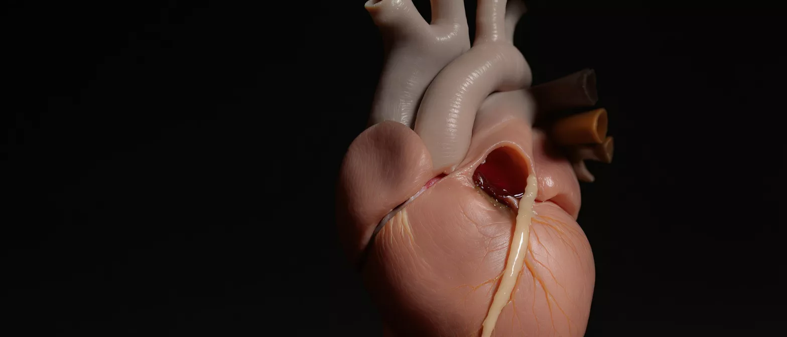
Tricuspid (three-leaflet)
Learn tricuspid (three-leaflet) valve anatomy, function, and related heart conditions.
What is the tricuspid (three-leaflet) valve?
The tricuspid (three-leaflet) valve is one of the four essential valves inside the human heart. It acts as a one-way gate that manages blood flow on the right side of the heart, ensuring that blood moves correctly from the body into the lungs to pick up oxygen. Understanding this valve is a vital part of learning about overall cardiac anatomy.
Why does this valve matter to a heart surgery patient? When you or a loved one is facing cardiac surgery, knowing exactly what the surgeon is repairing brings peace of mind. The tricuspid valve ensures that deoxygenated blood returning from the body flows smoothly from the right atrium to the right ventricle without leaking backward.
A healthy tricuspid valve opens fully to let blood pass through and closes tightly to prevent any backflow. If the valve becomes diseased or damaged, it strains the right side of the heart. Cardiologists and surgeons focus closely on this structure because right-heart function is crucial for long-term circulation and recovery.
Tricuspid valve anatomy and location
Understanding tricuspid valve anatomy helps patients visualize what happens during cardiac surgery. The valve sits between the right atrium (the upper chamber that receives blood from the body) and the right ventricle (the lower chamber that pumps blood to the lungs). It is called "tricuspid" because it consists of three flexible flaps, known as leaflets.
What are the three leaflets of the tricuspid valve? The three leaflets are medically known as the anterior, posterior, and septal leaflets. These thin but strong tissues are anchored to the heart wall by string-like structures called chordae tendineae and papillary muscles. This intricate suspension system keeps the leaflets stable, preventing them from collapsing backward under pressure.
The precise location of the right heart valve means it is uniquely positioned to handle deoxygenated blood. When the right ventricle contracts, the pressure pushes the three leaflets together to seal the opening. This coordinated anatomical design ensures that blood only moves forward toward the lungs, maintaining efficient circulation.
How the tricuspid valve functions in the heart
The primary tricuspid valve function is to act as a highly efficient mechanical door. When the heart muscle relaxes after a beat, the valve opens. This allows the right atrium to empty its entire volume of deoxygenated blood into the right ventricle, preparing the blood for its journey to the lungs.
When the right ventricle contracts to pump blood into the pulmonary artery, the pressure inside the chamber rises instantly. This pressure change forces the three valve leaflets to snap shut. By closing tightly, the valve prevents the blood from washing back up into the atrium.
Because it handles blood returning from the body, the tricuspid valve operates under lower pressure compared to the left side of the heart. However, this does not make it any less important. Any dysfunction in this step-by-step function creates a ripple effect that limits the amount of oxygen-rich blood the heart can eventually send to the rest of the body.
What is tricuspid regurgitation?
Tricuspid regurgitation is a condition where the three leaflets fail to close completely, allowing blood to leak backward into the right atrium. This means the heart has to work twice as hard to pump the same amount of blood forward, leading to stretching and enlargement of the right ventricle over time.
What causes tricuspid valve leaking? The most frequent cause is functional regurgitation, where the right ventricle stretches out due to left-sided heart conditions or high blood pressure in the lungs. As the chamber enlarges, the valve opening stretches, and the leaflets can no longer meet in the center to form a seal.
Patients with this condition may experience fatigue, swelling in the legs and abdomen, or an irregular heartbeat. If the leaking becomes severe, surgeons often recommend intervening to restore proper forward blood flow.
What is tricuspid stenosis?
Tricuspid stenosis is the opposite of leaking. In this condition, the three leaflets become thick, stiff, or fuse together, causing the valve opening to narrow. This restriction physically blocks blood from flowing easily from the right atrium into the right ventricle, creating a dangerous bottleneck for circulation.
What causes the right heart valve to narrow? This narrowing is most commonly caused by rheumatic fever, a complication of untreated strep throat. In rarer cases, it can be caused by congenital heart defects that present at birth, or by carcinoid syndrome, a condition that releases chemicals into the blood that damage cardiac tissue.
When the valve opening shrinks, the right atrium must generate much higher pressure to push blood through the narrowed gap. This eventually leads to right atrial enlargement and fluid buildup in the veins. Surgeons must carefully assess the stiffness of the leaflets to determine the best method to open the valve and relieve the pressure.
Common tricuspid heart conditions and symptoms
Tricuspid heart conditions usually develop gradually. Patients might not notice symptoms until the disease has progressed significantly. The body initially compensates for the reduced cardiac efficiency, but eventually, the strain becomes too great, and clear physical signs begin to surface.
What are the warning signs of tricuspid disease? The most common symptoms include severe fatigue, irregular heartbeats, pulsing veins in the neck, and noticeable swelling in the abdomen, ankles, and legs. This fluid retention occurs because blood backs up into the venous system instead of moving forward into the lungs.
If you or a family member experiences these symptoms, it is crucial to seek a cardiology evaluation. The medical team will determine if the right side of the heart is failing due to a faulty three-leaflet valve. Prioritizing rest and tracking symptoms daily is highly recommended for patient safety.
How is tricuspid valve disease diagnosed?
Diagnosing right heart valve disease requires specialized imaging and careful clinical evaluation. Cardiologists use a combination of physical exams and advanced medical technology to see exactly how the three leaflets are opening, closing, and handling the blood flow.
What tests evaluate the three-leaflet heart valve? An echocardiogram is the gold standard for diagnosing tricuspid issues. This ultrasound creates moving pictures of the heart, allowing the doctor to measure the size of the valve opening and the volume of any backward leaking.
Beyond imaging, doctors may order an electrocardiogram (ECG) to check for irregular heart rhythms, or a chest X-ray to see if the heart muscle is enlarged. Cardiac magnetic resonance imaging (MRI) provides highly detailed pictures of the right ventricle, giving the surgical team a complete roadmap before any operation is scheduled.
Tricuspid valve repair vs replacement surgery
When treating tricuspid (three-leaflet) disease, surgeons have two main options: repairing the patient's native valve or replacing it with a prosthetic device. The decision depends entirely on the specific damage, the underlying cause, and the patient's overall heart health.
When is valve repair preferred over replacement? Surgeons generally prefer to repair the valve whenever anatomically possible. Repair preserves the natural heart tissue, keeps the complex suspension system intact, and removes the need for lifelong blood thinners that are often required with mechanical replacements. This leads to better long-term heart function.
- Valve Repair: Surgeons tighten the stretched valve opening, reshape the leaflets, and reinforce the structure. Best for functional leaking and native tissue preservation.
- Valve Replacement: The damaged native valve is entirely removed and replaced with a biological tissue valve or a mechanical valve. Best for stiff, fused, or irreparably damaged leaflets.
If the valve cannot be saved and replacement is necessary, patients may be fitted with a durable mechanical device or a biological tissue substitute.
Recovery after tricuspid valve surgery
Recovery after right-heart valve treatment is a critical phase that requires patience and careful monitoring. The heart needs time to adjust to its new, improved blood flow dynamics, and the chest muscles and bone require weeks of gentle healing.
What is the timeline for heart valve surgery recovery? Patients typically spend one or two days in the intensive care unit (ICU) followed by several days in a regular hospital room. Over the next six to eight weeks, activity levels slowly increase under strict medical supervision, focusing entirely on rebuilding cardiovascular strength without overexerting the chest.
During this time, swelling in the legs and abdomen usually begins to reduce rapidly as the right side of the heart pumps efficiently once again. Cardiac rehabilitation, a medically supervised exercise program, plays a vital role in safely rebuilding stamina and guiding the patient back to normal daily activities.
Living well after right heart valve treatment
Understanding the tricuspid (three-leaflet) valve and its impact on circulation gives patients a foundation for long-term health. Whether you undergo a repair or a full replacement, successful surgery restores efficient blood flow, instantly reducing the strain on the heart and alleviating uncomfortable symptoms.
Adopting a heart-healthy lifestyle is the final step in the journey. This includes maintaining a balanced diet low in excess sodium, engaging in regular but doctor-approved physical activity, and attending all scheduled follow-up appointments. Consistent monitoring ensures the valve continues to function perfectly over the years.
By understanding the anatomy and function of your heart, you become an active, empowered participant in your own cardiac care. With the right surgical treatment and dedicated recovery, patients can look forward to a full, active, and healthy life after right-heart valve surgery.






