Hemiarch Replacement for Ascending Aortic Aneurysm
Hemiarch replacement aneurysm surgery explained by Guadalajara heart specialists.
What is Hemiarch Replacement for an Aneurysm?
The diagnosis of an enlarged aorta can be alarming, but a hemiarch replacement is a highly effective surgical procedure designed to correct this specific defect. The aorta is the largest artery in your body, carrying blood from your heart to the rest of your system. When the first part of this vessel, known as the ascending aorta, expands like a balloon, it becomes an aneurysm. A hemiarch replacement involves removing the diseased portion of this tube and replacing it with a durable synthetic graft, preventing a life-threatening rupture. Hemiarch replacement for an aneurysm of this kind is planned around the size and shape of your aorta.
Understanding the surgical landscape: During a hemiarch replacement, the surgeon carefully excises the weakened tissue of the front half of the aortic arch. The arch is the curved portion of the aorta that gives rise to the blood vessels supplying the brain. By replacing only the undersection, or hemiarch, the surgeon maintains the natural blood flow to the brain while reinforcing the entire structure against future expansion.
How does the heart keep beating? To perform this delicate work, the surgical team uses a heart-lung machine. This machine temporarily takes over the job of the heart and lungs, allowing the surgeon to work on a still, bloodless field. Blood returning to the right atrium is diverted into the machine. The medical team closely monitors the patient's hemodynamics using an arterial line, which provides real-time blood pressure readings directly from the arteries. According to research aggregated by PubMed, elective repair of the ascending aorta carries excellent long-term survival rates when performed at experienced centers.
What Causes an Ascending Aortic Aneurysm?
Genetic and degenerative factors: An ascending aortic aneurysm rarely develops overnight; the eventual need for a hemiarch replacement is usually the result of long-term structural changes in the blood vessel wall. One primary cause is the slow accumulation of atheromatous plaque, a buildup of cholesterol and inflammatory cells that weakens the vessel. High blood pressure continuously pounds against the aortic walls, accelerating this wear and tear. Over time, the elastic fibers within the aorta break down, causing the vessel to stretch outward.
Connective tissue disorders: Some patients are born with genetic conditions that predispose them to aortic enlargement. Syndromes like Marfan or Loeys-Dietz affect the connective tissues that give the aorta its strength. In these patients, the aorta may enlarge at a younger age, requiring earlier surgical intervention. During evaluation, doctors may check levels of apolipoprotein B, a protein involved in cholesterol transport, to assess cardiovascular risk. Sometimes, coronary blockages are treated with balloon angioplasty before the aneurysm is addressed.
Age and lifestyle contributions: Beyond genetics, aging naturally degrades the aortic tissue. Chronic smoking severely accelerates this damage by inflaming the blood vessels and degrading the structural integrity of the aortic wall. If the aneurysm begins to leak or tear, known as a dissection, it can sometimes present as an atypical infarction, making immediate diagnosis through imaging absolutely critical for survival and long-term recovery.
When is Hemiarch Replacement Indicated?
Surgical size thresholds: Determining the right time for a hemiarch replacement is a critical decision guided by established clinical guidelines. For most patients, elective surgery is recommended when the diameter of the ascending aorta reaches or exceeds 5.5 centimeters. If a patient has a known genetic connective tissue disorder, the threshold may be lowered to 5.0 centimeters to prevent rupture before symptoms occur. These thresholds represent the point where the risk of rupture begins to outweigh the risks of the surgery itself.
Rapid growth and symptom onset: Surgery is also indicated if the aneurysm is growing rapidly, typically defined as an increase of more than 0.5 centimeters per year. Sometimes, patients experience chest pain or back pain as the aneurysm stretches the surrounding tissues. In rare cases, a patient might undergo evaluation for a related issue, such as a study on the Frequency, Severity, and Management of Left Main Disease in Acute Coronary Syndrome in Sulaimani Cardiac Hospital, and the aneurysm is discovered incidentally on a routine echocardiogram.
Emergency presentations: In acute scenarios, such as a Type A aortic dissection, emergency surgery is required immediately. A dissection is a tear in the inner layer of the aorta that allows blood to split the wall. This is a life-threatening emergency where a hemiarch replacement is often performed to remove the diseased segment, restore normal blood flow, and reinforce the aortic wall to prevent further tearing or rupture.
Hemiarch vs Total Arch Replacement: What is the Difference?
Anatomical extent of repair: The primary difference between a hemiarch replacement and a total arch replacement lies in how much of the curved portion of the aorta is replaced. The aortic arch has three major branches that supply blood to the brain and arms. A hemiarch replacement involves replacing only the underside of the arch, preserving those three critical branch vessels. A total arch replacement removes the entire arch and reattaches those three branches to a specialized multi-branched synthetic graft.
When is a total arch necessary? A total arch replacement is reserved for cases where the aneurysm or dissection extends fully across the arch, threatening the branch vessels. This is a more complex surgery requiring longer circulatory arrest times, meaning the body's circulation is temporarily stopped to allow the surgeon to work safely on the blood vessels. The decision depends heavily on precise imaging to map the exact extent of the disease.
Surgical planning and trade-offs: Choosing the right approach requires balancing surgical risk with the need for complete repair. A hemiarch replacement is generally faster and carries a lower risk of neurological complications because the brain's blood supply is less disrupted. However, if the disease extends further, a total arch repair is necessary to ensure the aneurysm does not return in the unrepaired sections.
- Hemiarch: Replaces the underside of the arch, preserving native blood vessels to the brain.
- Total Arch: Replaces the entire arch and reattaches all three head vessels to the new graft.
- Circulatory Arrest: Total arch requires longer stopped-circulation times, increasing complexity.
How is the Hemiarch Surgery Performed?
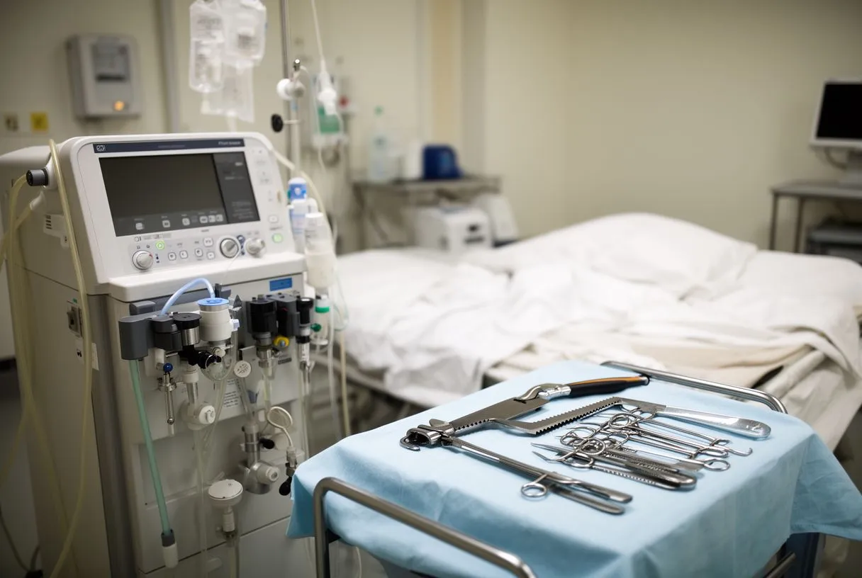
Preparation and access: The hemiarch replacement procedure begins with a median sternotomy, a careful incision down the center of the chest to access the heart. Once inside, the surgical team prepares the patient for cardiopulmonary bypass. An arterial cannula is placed to connect the patient to the heart-lung machine, and venous drainage is established. This setup allows the team to safely stop the heart and drain blood from the surgical field.
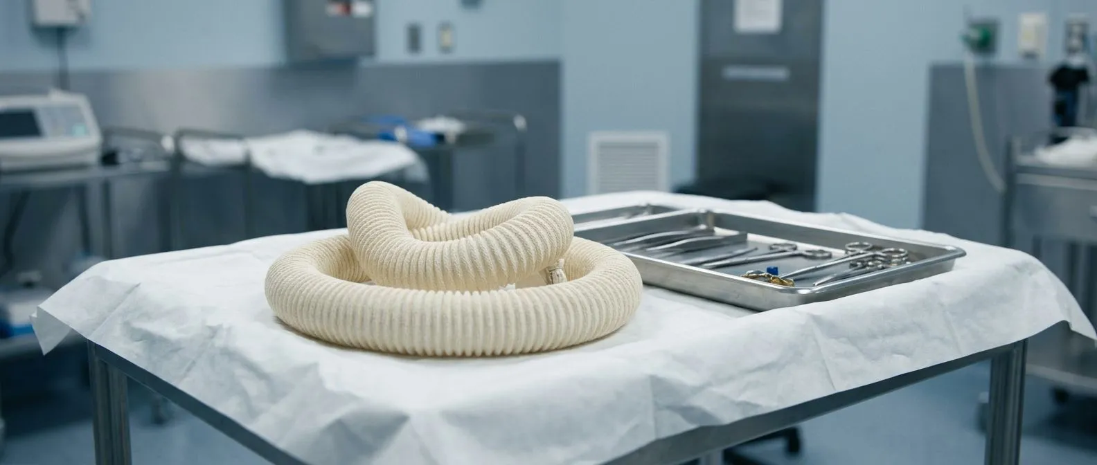
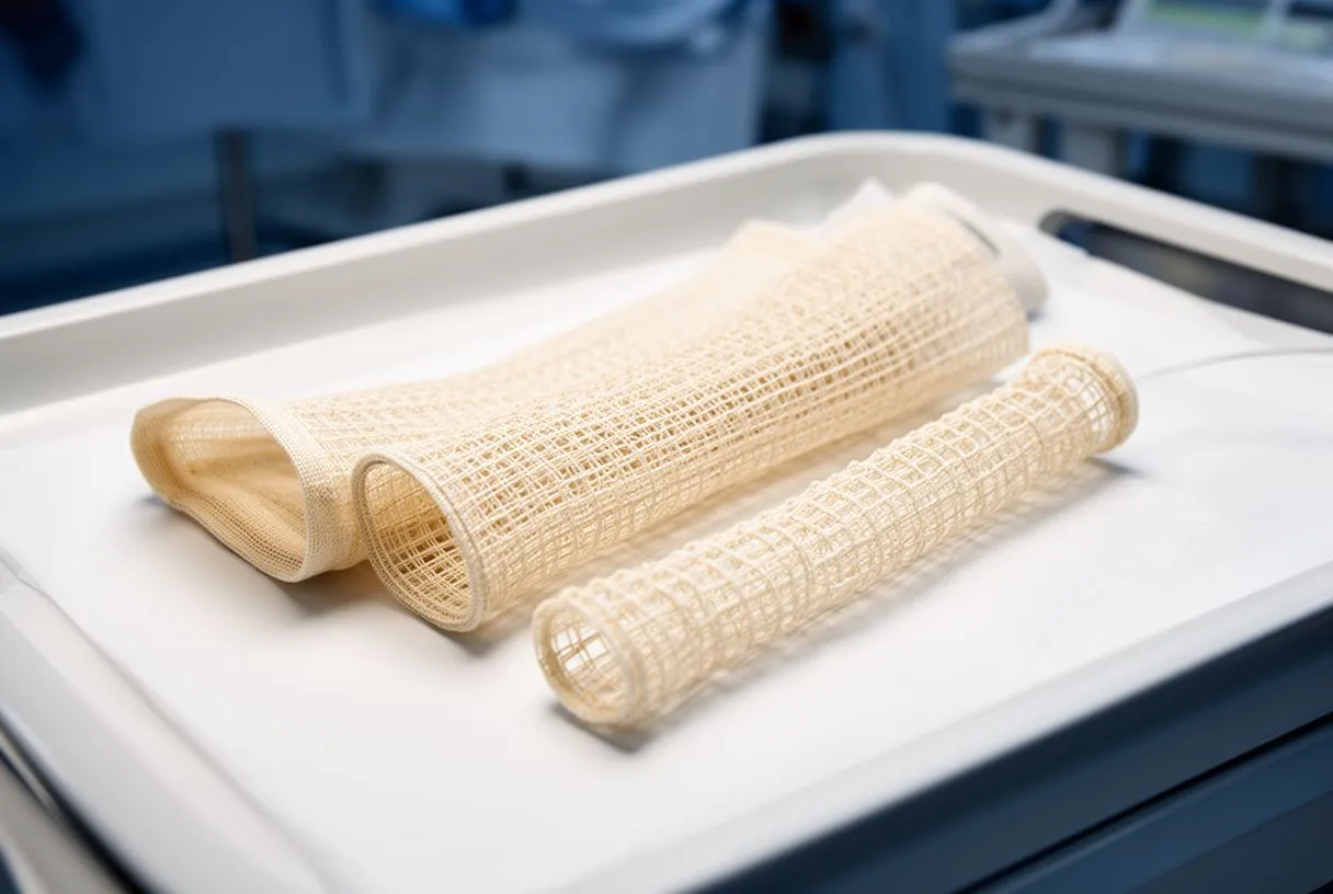
Executing the repair: With the patient on bypass and the body cooled to protect the brain, the surgeon opens the diseased aorta. The damaged tissue is trimmed away. The surgeon then sizes a woven polyester graft to match the patient's anatomy and sews it precisely into place. If the aortic valve is also diseased, the team may use an autograft or a synthetic valve, or perform a balloon-expandable valve replacement if appropriate for the anatomy.
Restoring circulation: Once the graft is securely sutured, the heart is slowly rewarmed and the heart-lung machine is gradually withdrawn. The surgical team meticulously checks for any bleeding. If a patient requires anticoagulation for a mechanical valve, careful management protocols, similar to those studied in the ARISTOTLE trial, are initiated to prevent blood clots while minimizing bleeding risks. Sometimes, an arterial graft is used to reinforce the repair.
What are the Risks and Complications of Hemiarch Surgery?
Common surgical risks: Like all major cardiac procedures, a hemiarch replacement carries inherent risks. Bleeding is a primary concern, requiring meticulous surgical technique and careful management of the patient's clotting system. In some cases, the team may use autologous blood, meaning the patient's own blood is collected and reused during surgery to reduce the need for donor transfusions. Infection and adverse reactions to anesthesia are also standard risks monitored closely in the intensive care unit.
Neurological and cardiac complications: Because the surgery involves the aortic arch near the blood vessels supplying the brain, stroke is a significant concern. Micro-debris or air bubbles can compromise cerebral blood flow. Post-operative arrhythmias are also common; patients may experience atrial flutter, an abnormal heart rhythm that requires medication or temporary pacing. In severe cases, an automated external defibrillator may be used to restore a normal heartbeat.
Long-term monitoring of complications: Understanding the consequences of potential complications is vital for patient education. Research on neurological events, such as the Stroke and its consequences: protocol and pilot data of the observational Berlin Long-term Observation of Vascular Events (BeLOVE) stroke stratum, helps teams anticipate recovery needs. Paralysis due to spinal cord ischemia is rare in arch surgery but requires immediate attention if it occurs.
What to Expect During Hemiarch Replacement Recovery
Immediate post-operative phase: Recovery from a hemiarch replacement begins in the intensive care unit, where the patient is closely monitored for the first 24 to 48 hours. Tubes and monitors, including a temporary arterial line for continuous blood pressure tracking, help the medical team ensure stability. Patients are typically extubated, meaning the breathing tube is removed, within a day. Early mobilization, such as sitting on the edge of the bed, is encouraged to prevent blood clots and promote lung expansion.
Transitioning to the ward: After the ICU, patients move to a regular cardiac ward for several days. The focus shifts to pain management and gradual physical rehabilitation. It is common to experience chest discomfort and even radiating arm pain as the incision heals and nerves recover. The nursing staff will administer medications like aspirin to prevent graft clotting, depending on the specific valve repair used.
Returning home: Most patients spend about a week in the hospital before going home. Complete recovery from a hemiarch replacement takes about six to eight weeks. Patients are advised to avoid heavy lifting and strenuous activity to allow the breastbone to heal. Cardiac rehabilitation is highly recommended to safely rebuild strength under medical supervision.
Can an Aortic Valve Be Spared During Hemiarch Surgery?
The valve-sparing approach: In many cases of ascending aortic aneurysm, the aortic valve itself remains healthy. When possible, surgeons prefer a valve-sparing root replacement alongside the hemiarch replacement. This technique removes the diseased aortic tissue while keeping the patient's own native valve intact. The surgeon then reimplants the valve inside a new synthetic graft. This approach is highly advantageous because it avoids the need for long-term blood thinners.
When replacement is necessary: If the aortic valve is diseased or leaking heavily, it must be replaced during the surgery. Options include mechanical valves, which require lifelong anticoagulation, or biological tissue valves. Sometimes, a balloon aortic valvuloplasty is attempted beforehand to delay surgery, but aneurysm repair will ultimately require definitive surgical management. If a transcatheter approach is used later in life, it is referred to as TAVR, never TAVI.
Assessing valve health: The decision to spare or replace the valve is made using transesophageal echocardiography during the surgery. The surgeon evaluates the leaflets of the valve and the aortic root. If the valve is stretched but structurally sound, the sparing operation offers excellent long-term outcomes. If the leaflets are damaged or calcified, a mechanical or biological valve, or in select cases a balloon-expandable valve, is implanted.
Long-Term Outcomes and Quality of Life After Aortic Repair
Life expectancy and follow-up: Following a successful hemiarch replacement, patients can expect to return to a full and active life. The synthetic graft used is incredibly durable and does not wear out. However, lifelong surveillance is mandatory. Patients undergo annual CT scans or MRIs to monitor the remaining portions of the aorta, ensuring no new aneurysms develop downstream.
Managing residual aortic disease: Even after the ascending aorta is repaired, patients must manage underlying risk factors. High blood pressure must be strictly controlled with medication. Regular cardiology check-ups are essential to listen for any new murmurs via auscultation and to review imaging. In complex cases, advanced support like Awake ECMO might be discussed if severe cardiac failure occurs, though this is rare after elective repairs.
Psychological recovery: Surviving a major cardiac surgery or an aortic dissection is a profound experience. Many patients benefit from psychological support to process the trauma of the diagnosis and surgery. Support groups and cardiac rehabilitation programs offer a community of survivors. With proper care, the long-term quality of life is generally excellent.
- Annual Imaging: Lifelong CT or MRI scans to monitor the remaining aorta.
- Blood Pressure Control: Strict management to protect the repaired and downstream vessels.
- Cardiac Follow-up: Regular visits to detect any valve or graft issues early.

Getting a Second Opinion for Aortic Aneurysm Surgery in Mexico
The value of a specialized team: Facing aortic surgery is a major life event, and seeking a second opinion is a wise step. Aortic surgery requires a highly specialized team with specific expertise in complex aortic repairs. When looking for a surgeon, ask about their volume of aortic cases and their outcomes. Dr Alejandro Molina and the team at Cirugía de Corazón offer expert evaluation for patients in Guadalajara, Querétaro, and Hermosillo.
International patient considerations: For international patients, traveling for cardiac surgery involves logistical planning. They provide clear estimates and work with patients to understand the financial aspects without hidden costs. It is crucial to have transparent discussions about the entire treatment plan before committing to surgery.

Taking the next step: If you have been diagnosed with an ascending aortic aneurysm, do not delay in seeking expert advice. Whether you need monitoring, medical management, or a hemiarch replacement, having a clear understanding of your specific situation is empowering. We encourage you to discuss your case with a qualified cardiac team and book a consultation with Cirugía de Corazón to explore your safest path forward.