

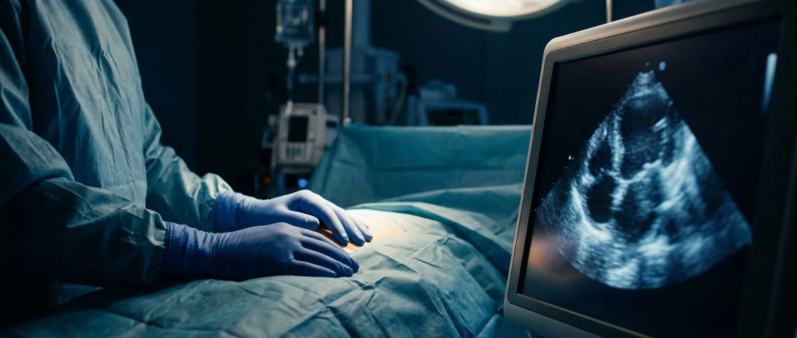
Intraoperative Echocardiography During Heart Surgery
Intraoperative echocardiography heart surgery lets your team see the heart in real time for safer, more precise results.
Intraoperative echocardiography heart surgery teams rely on it for one simple reason: once the chest is open, the surgeon cannot see the heart beating inside. An ultrasound exam performed during the operation gives the whole team a live picture of the heart's chambers, valves and blood flow while the operation is underway. For patients and families, understanding this tool takes away some of the mystery of the operating room and explains why it has become a standard part of modern cardiac care.
What Is Intraoperative Echocardiography During Heart Surgery?
Echocardiography is an ultrasound study of the heart. It uses sound waves, not radiation, to build a moving image of the heart as it beats. When the same study is performed in the operating room, during the operation itself, it is called intraoperative echocardiography.
The idea is straightforward. Before the surgeon makes a single cut inside the heart, the echo confirms the diagnosis. After the repair is finished, while the patient is still on the operating table, the echo checks that the repair actually works. If something is not right, the surgeon can go back in immediately, rather than discovering a problem days later in the intensive care unit.
This makes the study a form of quality control performed in real time. Intraoperative echocardiography during cardiac surgery is now considered standard practice for most valve operations and many coronary and aortic procedures, and textbooks such as StatPearls on NCBI describe it as a core monitoring and diagnostic tool in the operating room.
What Is Transesophageal Echocardiography Heart Surgery Imaging?
Most intraoperative imaging uses transesophageal echocardiography heart surgery teams call TEE for short. "Transesophageal" means the ultrasound probe is placed in the esophagus, the swallowing tube that runs directly behind the heart. Because there is no skin, fat or lung between the probe and the heart, the images are far sharper than those obtained from the chest wall.
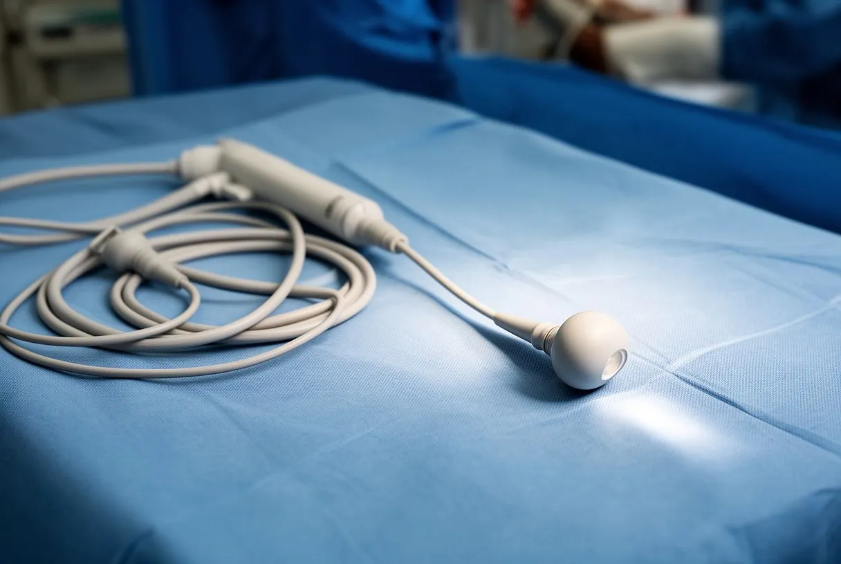
During surgery, the anesthesiologist or a specially trained cardiologist passes a soft, flexible probe about the thickness of a thumb through the mouth and into the esophagus after you are asleep. The probe then stays in place for the whole operation, taking pictures on demand.
You may wonder why the team checks your neck during an echocardiogram. When the probe is being guided into the esophagus, the team gently holds and inspects the neck and watches the swallow to confirm the probe passes smoothly and sits comfortably. It is a safety check, and it takes only seconds.
How Does a TEE Probe Work During Cardiac Surgery?
A TEE probe cardiac surgery teams use looks like a long, flexible endoscope with an ultrasound tip. The tip can flex and rotate, which lets the operator view the heart from many angles without moving the patient. Modern probes produce both two-dimensional images and Doppler echocardiography, which measures the direction and speed of blood flow using sound.
Real time heart imaging surgery means exactly that: the images update with every heartbeat, on a screen beside the operating table. The surgeon, the anesthesiologist and the imaging doctor all watch the same screen and make decisions together.
Heart surgery safety monitoring is the other half of the job. Beyond the operation itself, the TEE watches the heart continuously throughout the case. It can detect sudden changes in pumping function, air or clot entering the circulation, and problems with blood flow, allowing the team to respond within seconds rather than minutes.
When Is an Echocardiogram Recommended Before Surgery?
An echocardiogram is recommended before heart surgery whenever the operation depends on knowing the exact structure of the heart. Valve surgery, repair of congenital defects, aortic procedures and many coronary operations all begin with a preoperative echo to map out the plan.
Does echo detect heart blockage? It detects the consequences of blockage, such as areas of the heart muscle that pump poorly after a heart attack, and it can show valve problems caused by coronary disease. But the coronary arteries themselves are usually assessed with other studies, such as a catheter angiogram or a CT scan, which show the vessel walls directly.
Perioperative echocardiography heart imaging covers the whole journey: before the operation to plan, during the operation to guide and verify, and after the operation to confirm the result. In our practice every patient's imaging is reviewed by the multidisciplinary team against international guidelines before we recommend any procedure, and you receive a clear proposal with the alternatives.
How Does Intraoperative Echo Guide Valve Repair?
Nowhere is echocardiography guided heart surgery more valuable than in valve operations. Before the operation begins, the echo confirms which valve is leaking or narrowed, and how severely. Surgeons know that the preoperative study and what they find in the operating room do not always match, and the echo settles any doubt immediately.
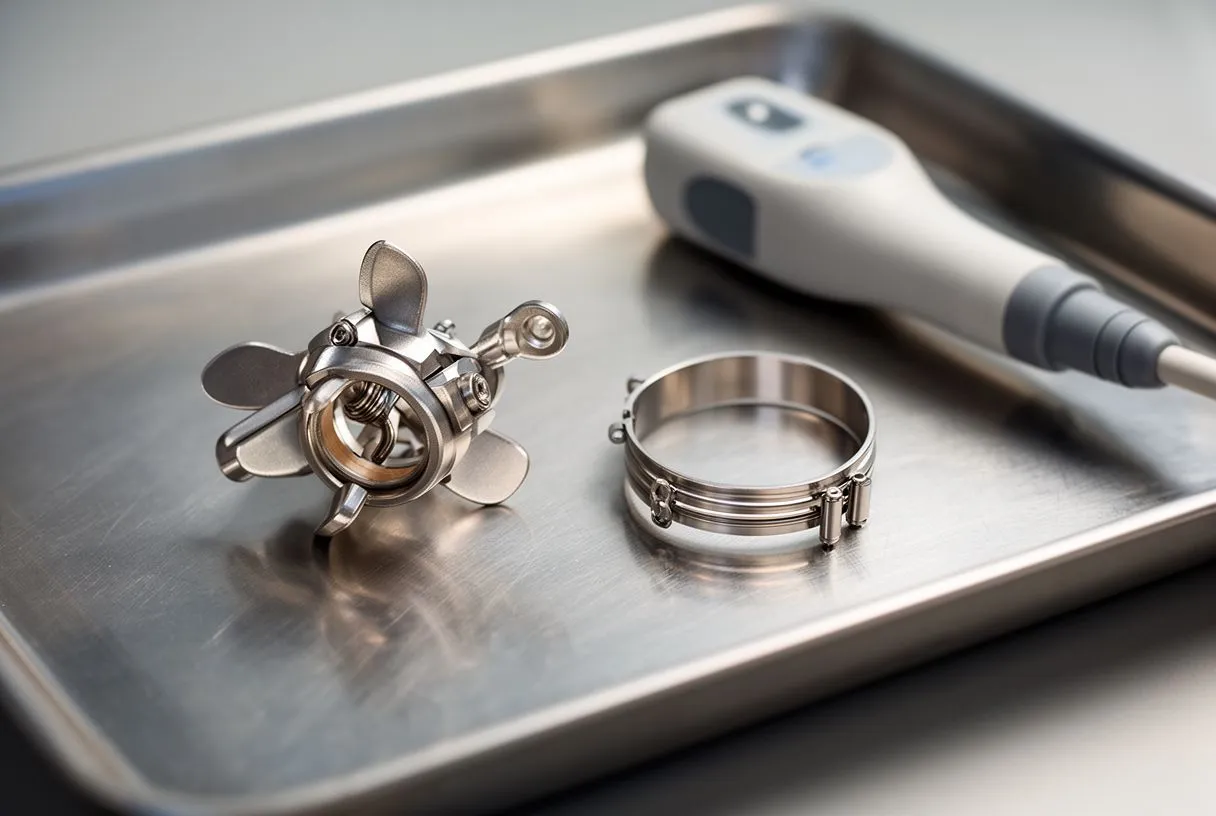
Intraoperative echo for valve repair has changed mitral valve surgery in particular. After the repair is completed and the patient is weaned from the heart-lung machine, the echo shows whether the valve leaks, whether blood flow through it is obstructed, and whether the repair looks durable. If the result is not good enough, the repair is revised before the chest is even closed.
This immediate verification is one of the main reasons valve repair outcomes have improved over the decades. It is also why surgical volume and imaging expertise go hand in hand. The Japanese Association for Thoracic Surgery's annual report of thoracic and cardiovascular surgery in Japan documents the very large number of valve and other cardiac operations performed there each year, in health systems where intraoperative echo is routine.
What Does an Echocardiogram Detect During Open Heart Surgery?
During an echocardiogram during open heart surgery, the study acts as the surgeon's eyes inside the chest. It confirms the anatomy before incision, verifies that all air has been removed from the heart after the heart-lung machine period, and checks the function of the heart muscle as it resumes work.
The study also detects the unexpected. Occasionally the echo reveals a problem nobody suspected, such as a previously unrecognized leak, a blood clot inside a chamber, or a hole between chambers. Finding these while the patient is still on the table means they can be corrected in the same operation.
Finally, the echo measures how the heart is pumping at the end of the operation. This baseline guides the intensive care team in the hours that follow, because they know exactly what the heart looked like when the patient left the operating room.
Are You Awake During a Transesophageal Echocardiogram?
During heart surgery, the answer is simple: you are fully asleep under general anesthesia, so you feel nothing and remember nothing about the probe or the images.
A TEE is also performed outside the operating room, as a diagnostic study. In that setting you are usually given a light sedative and a throat spray, so most patients remain drowsy but responsive. Policies vary by center, and some centers offer deeper sedation on request.
We take the time in consultation to explain, in plain words, what each study shows and exactly what it will feel like, and we answer every question before any decision is made. If a TEE is part of your plan, you should know in advance whether it will happen asleep in the operating room or as a separate, sedated study.

How Uncomfortable Is a TEE Probe and How Long Is Recovery?
For a sedated, outpatient TEE, most patients describe the sensation as unusual rather than painful: a brief gagging feeling as the lubricated probe passes the back of the throat, then remarkably little discomfort once it is in place. The study itself usually takes ten to twenty minutes.
What should you not do before an echocardiogram? For a TEE, the rule that matters most is fasting: no food for roughly six to eight hours and restricted fluids beforehand, exactly as your team instructs, because the probe is passed into the esophagus and stomach. You should also tell the team about any swallowing problems, loose teeth or allergies in advance.
Recovery is short. After a sedated TEE you rest until the sedation wears off, avoid eating and drinking until your gag reflex returns, and may have a mild sore throat for a day. After surgery, the probe is removed while you are still asleep, and most patients never notice it was there. Serious complications, such as perforation of the esophagus, are rare and the team screens for risk factors before passing the probe.
Why Is Echo Essential for Minimally Invasive Heart Surgery?
Minimally invasive heart surgery echo dependence is even greater than in open operations. When the surgeon works through a small incision between the ribs, or through catheters in the groin as in TAVR, a transcatheter aortic valve replacement, there is no direct view of the heart at all. The echo image becomes the primary view.
Heart surgery imaging technology has evolved to match. Three-dimensional TEE now renders the mitral valve from the atrial side almost as a surgeon would see it, and live guidance software overlays catheter positions on the echo image during structural heart procedures.
Pediatric intraoperative echocardiography deserves its own mention. Children with congenital heart defects often have small chests and complex anatomy, and the TEE, or ultrasound probes placed directly on the heart surface, is indispensable for confirming that a repair is complete before the operation ends. Our team performs conventional, minimally invasive, robotic and transcatheter procedures, and we recommend the less invasive route only where the evidence supports it for that specific patient.
How Do You Choose a Heart Surgery Team That Uses Echo Well?
The quality of intraoperative echocardiography depends less on the machine than on the people interpreting it. Look for a team where cardiac surgery, anesthesiology and cardiology work together, and where someone with advanced echo training is present for the whole case, not just called in when trouble appears.
Good teams also use the echo to support honest conversations. Cardiac surgery quality assessment increasingly includes whether the intraoperative findings matched the plan and whether repairs were revised immediately when needed. These questions are reasonable to ask any team you are considering.
Our first contact is a free video consultation: we get to know you, talk through your condition, and tell you exactly which studies are still needed so no unnecessary ones are ordered, and we actively encourage second opinions as a normal part of deciding about heart surgery. The final decision is always yours.
Intraoperative echocardiography heart surgery imaging has quietly become one of the great safety advances in cardiac care. It confirms the diagnosis before the first incision, guides the repair while it happens, verifies the result before the chest is closed, and watches over the heart through every stage in between. For patients, it means problems are caught in the operating room rather than afterward. If you or a family member are facing valve surgery, a TAVR procedure, or another cardiac operation, ask your team how they will use echo during your case, and discuss your own situation with a qualified cardiac team. A consultation is the right place to start, not a self-diagnosis.






