


Left Atrium Enlargement: Causes and Atrial Fibrillation
Left atrium enlargement raises atrial fibrillation risk.
Left atrium enlargement is one of the most common findings on an echocardiogram, and also one of the most misunderstood. Many patients receive the report, read the words "left atrial enlargement," and immediately fear the worst. The reality is more nuanced: the left atrium — the small upper chamber on the left side of the heart — acts as a barometer of what is happening to the rest of the cardiovascular system. In this guide, we explain in plain language what the left atrium does, why it enlarges, how it relates to atrial fibrillation, and what can genuinely be done about it.
What Does Your Left Atrium Do?
The receiving chamber. The left atrium is the upper-left chamber of the heart. It receives oxygen-rich blood returning from the lungs through the pulmonary veins, then passes it down through the mitral valve into the left ventricle — the main pumping chamber that sends blood to the entire body. In simple terms, the left atrium is a waiting room: blood arrives there and waits for the valve below to open.
Although it sounds passive, the left atrium does real work. Near the end of each heartbeat, it contracts and squeezes an extra portion of blood into the left ventricle. This "boost" can account for a meaningful share of the blood the heart pumps with each beat. When the chamber contracts properly, the Left Ventricle fills efficiently and the whole system runs smoothly.
Why function matters. Cardiologists increasingly look at how well the left atrium works, not only how big it is. A chamber that is stiff, scarred, or contracting poorly can affect exercise tolerance and set the stage for rhythm problems. This is why your cardiologist cares about the left atrium even when the rest of the echocardiogram looks normal: it is often the first chamber to show the strain of high blood pressure, valve disease, or a failing ventricle.
What Is Left Atrium Enlargement? The Meaning of Left Atrium Dilation
The basic definition. Left atrium enlargement — sometimes called left atrial dilation — means the upper-left chamber is bigger than it should be. The heart is a muscular pump, and like any chamber working against pressure or volume it was not designed for, the atrium can stretch over time. Some stretching is a healthy adaptation; a chamber that stays stretched for months or years, however, undergoes structural change in its walls.
Enlargement is not a disease in itself. It is a finding — a signal that something upstream or downstream is making the chamber work harder. Commonly, that something is long-standing high blood pressure, a leaking or narrowed mitral valve, or a rhythm disorder. Think of it like a tyre bulging on one side: the bulge tells you where the pressure problem is.
Why the chamber stretches. The left atrium enlarges for two broad reasons. First, it may hold more blood than usual — for example, when the mitral valve leaks and blood flows backwards into the atrium with every beat. Second, it may pump against higher pressure — for example, when the Left Ventricle becomes stiff and resists filling. Over years, either mechanism remodels the chamber, and the walls may develop fibrosis, a type of internal scarring that changes how the atrium conducts electrical signals.
What Does Left Atrial Enlargement on Echocardiogram Mean?
How it is measured. An echocardiogram — an ultrasound scan of the heart — is where enlargement is usually first noticed. The report may describe a linear dimension (the anteroposterior diameter) or, more accurately, a volume. Specialists prefer volume because the atrium does not enlarge evenly in every direction.
To make results comparable between people of different body sizes, the volume is divided by the patient's body surface area. This gives the left atrial volume index, the figure most guidelines rely on. When a report quotes a volume index rather than a single linear measurement, it is using the more modern and reliable standard.
- Normal range: a left atrial volume index up to roughly 34 mL/m² is considered normal for most adults; mild enlargement starts above that threshold.
- Linear measurement: many reports still quote a diameter; a healthy adult atrium is typically under about 4 cm across, but volume is the better guide.
- What it means: enlargement on a scan is a prompt to look for the cause — blood pressure, valve function, sleep apnoea, and rhythm history all deserve attention.
In our practice, when a patient brings an outside echocardiogram, we review the actual images and measurements rather than the one-line conclusion, because technique varies between laboratories and a borderline measurement may not mean what the report suggests. In the consultation we take the time to explain what the studies actually show, in plain words, and to answer every question before any decision is made.

What Causes Left Atrium Enlargement?
High blood pressure. Hypertension is the most common cause worldwide. Chronically elevated pressure makes the Left Ventricle stiffen as its muscle thickens, and a stiff ventricle resists filling. The left atrium pushes against that resistance for years and gradually stretches. This is why enlargement is so frequent in adults over 60, and why controlling blood pressure is the first step in almost every treatment plan.
Mitral valve disease. The mitral valve sits between the left atrium and the Left Ventricle. When it leaks (regurgitation) or narrows (stenosis), the atrium absorbs the consequences. A leaking valve means the atrium receives blood twice — once from the lungs and once as backflow — so it enlarges to accommodate the extra volume. The team treats valve disease with conventional, minimally invasive, robotic, and transcatheter repairs, depending on what the evidence supports for each patient.
Other contributors. Several less obvious factors also matter, and a complete evaluation should consider each of them:
- Sleep apnoea: repeated night-time oxygen drops strain the heart and are strongly associated with atrial enlargement and rhythm problems.
- Obesity and age: both independently enlarge the atrium, even when blood pressure is well controlled.
- Heart failure and aortic valve disease: any condition that raises filling pressures on the left side of the heart will eventually be reflected in the atrium.
- Structural and rare causes: problems affecting the atrium's own structure — from neighbouring anatomy to rare tumours — can also influence its behaviour. Case reports in cardiology literature describe Left Atrium Papillary Fibroelastomas, small benign tumours, as a recognised cause of cerebral emboli, illustrating why unexplained findings on the left side of the heart always merit proper investigation.
Even surgery elsewhere in the chest can play a role. Research has examined how removing the left upper lung lobe alters left atrial haemodynamics, showing that the atrium's filling pattern can change after lung surgery. Frontiers in Physiology published work modelling these haemodynamic shifts, a reminder that the atrium responds to pressure and flow changes from many directions.
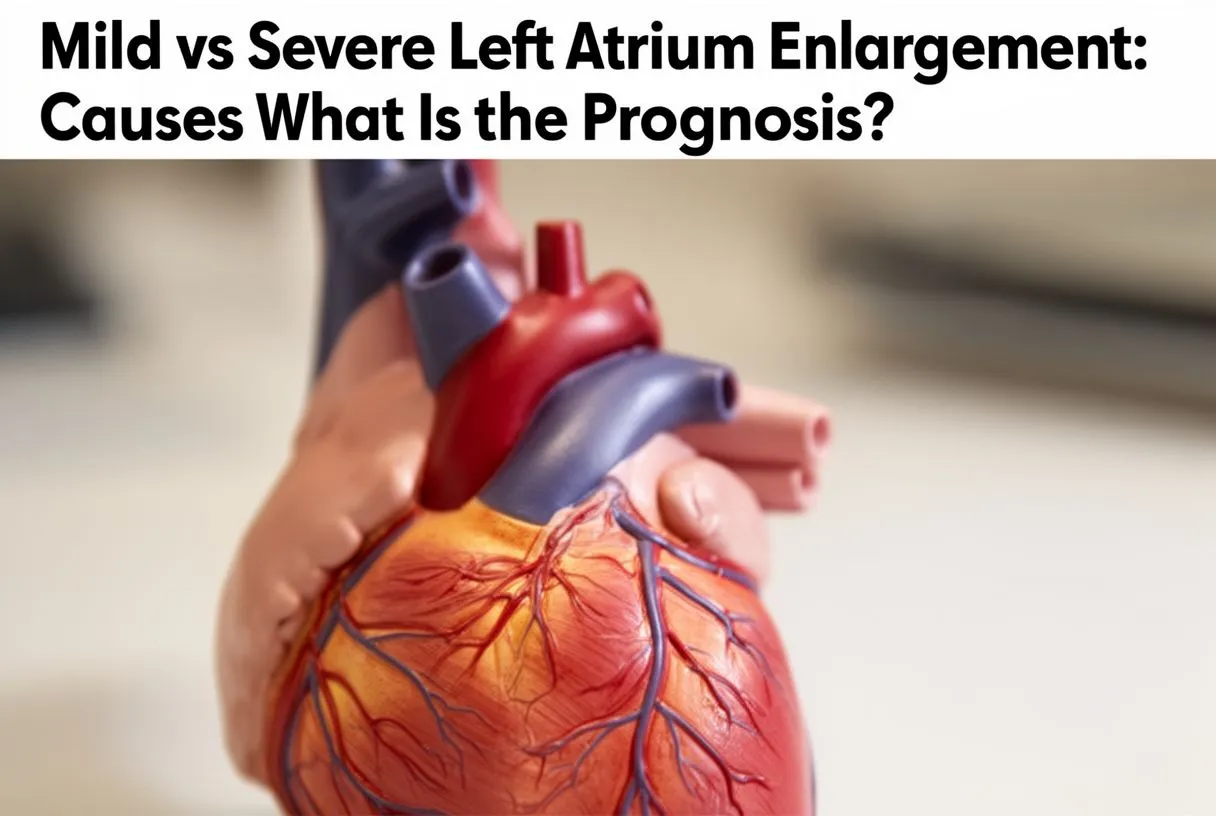
Mild vs Severe Left Atrium Enlargement: What Is the Prognosis?
Mild enlargement. Mild left atrium enlargement is extremely common and, on its own, rarely urgent. It often reflects long-standing blood pressure or simply ageing. The important question is not "how big is it?" but "why is it big, and is it getting bigger?" A stable, mildly enlarged atrium with a normal rhythm and good valve function usually needs monitoring and risk-factor control, not intervention.
Severe enlargement. Severe dilation changes the picture. A markedly stretched atrium loses its orderly contraction, blood flows more slowly inside it, and clots can form — particularly in the Left Atrial Appendage, a small ear-shaped pouch off the atrium where blood stagnates most. This is the mechanism linking severe enlargement to stroke risk, and it is why patients with big atria and irregular rhythms are often offered anticoagulation (blood-thinning) medication.
Prognosis in context. The long-term outlook depends on the underlying cause and on whether atrial fibrillation develops or is controlled. Population studies, indexed in registries such as DOAJ, consistently associate larger atrial volumes with worse cardiovascular outcomes — but association is not destiny. Patients who control blood pressure, treat valve disease early, and manage rhythm problems can live full, active lives. What matters is acting on the finding rather than ignoring it.
Enlarged Left Atrium and Atrial Fibrillation: How Are They Connected?
What is atrial fibrillation? Atrial Fibrillation — often shortened to AF — is the most common sustained rhythm disorder. Instead of beating in an organised way, the atria quiver chaotically at several hundred impulses per minute. The result is an irregular pulse, inefficient filling of the ventricles, and, importantly, sluggish flow inside the left atrium that raises clot and stroke risk.
A two-way street. The relationship between enlargement and AF runs both directions. A stretched, scarred atrium provides the electrical "substrate" — the abnormal tissue pathways — that lets atrial fibrillation start and persist. Once AF takes hold, the atrium stops contracting, enlarges further, and scars more. Each condition feeds the other, which is why cardiologists try to break the cycle early: restoring or maintaining normal rhythm is easier before the atrium has remodeled extensively.
Why size guides treatment. Atrial size is one of the factors cardiologists weigh when choosing between rhythm-control strategies. A modestly enlarged atrium responds well to cardioversion (an electrical reset of the rhythm) or catheter ablation. A severely enlarged, heavily scarred atrium is harder to keep in normal rhythm, so the plan may lean toward rate control and stroke prevention instead. There is no single correct answer — the decision is individualised.
What Are the Symptoms of Left Atrium Enlargement?
Often, there are none. Most people with a mildly enlarged left atrium feel perfectly well, which is why the finding usually appears on a scan done for another reason. This silence is normal and, in itself, reassuring — it does not mean the problem is being missed.
Symptoms that do appear. When enlargement is significant or when it has triggered rhythm or pressure changes, patients may notice breathlessness on exertion, a fluttering or irregular heartbeat, fatigue, reduced exercise tolerance, or swelling of the ankles. Palpitations that start and stop abruptly deserve particular attention, as they may indicate atrial fibrillation. Some patients describe waking at night breathless, a symptom called paroxysmal nocturnal dyspnoea, which suggests pressure is building on the left side of the heart.
Red flags. Seek urgent care for chest pain, fainting, or sudden weakness or numbness on one side of the body — the latter could indicate a stroke and needs emergency assessment. For everything less urgent, a planned evaluation with a cardiac team is the right next step. When patients contact our team with palpitations or an abnormal scan, the first step is always a careful history and a review of existing studies, so we can tell them which tests are genuinely needed and which are not.
Left Atrial Enlargement Treatment: What Are Your Options?
Treat the cause. There is no "pill for an enlarged atrium." Effective left atrial enlargement treatment means treating what is stretching it. For high blood pressure, that means medication optimised to target and lifestyle change. For mitral valve disease, it may mean valve repair or replacement — and modern options range from open surgery through minimally invasive and robotic approaches to transcatheter procedures performed through a small tube in the groin.
Managing atrial fibrillation. When AF is present, treatment has two goals: control the rhythm or the rate, and prevent stroke. Options include medication, electrical cardioversion, and catheter ablation — a procedure where thin wires are threaded into the heart to isolate the areas firing abnormal signals, most of which sit at the junction of the pulmonary veins and the left atrium. Anticoagulation decisions follow validated stroke-risk scores, not atrial size alone.
Advanced and surgical options. For carefully selected patients, surgical approaches can address the atrium directly. During valve surgery, surgeons can perform a Left Atrial Appendage Closure — removing or occluding the appendage where clots form — and some procedures include lesion sets designed to interrupt the electrical circuits of atrial fibrillation. In our multidisciplinary practice, every case is reviewed by the full team against international practice guidelines before a recommendation is made. We recommend the less invasive route only where the evidence supports it for that specific patient, and the patient always receives a clear proposal with the alternatives — the final decision is always theirs.
Can Left Atrium Enlargement Be Reversed?
It depends on the cause and the timeline. The honest answer is: sometimes, partially. When enlargement is driven by something treatable and caught early — a leaking valve repaired promptly, blood pressure brought under control, sleep apnoea treated, significant weight lost — the atrium can shrink back toward normal, and studies of valve repair and AF ablation have documented measurable reductions in atrial size after successful treatment.
When reversal is limited. Long-standing, severe enlargement with fibrosis (internal scarring) is harder to undo. Scar tissue does not turn back into healthy muscle. That said, even when full reversal is not possible, stopping progression is a genuine win: halting further dilation preserves function, lowers clot risk, and improves how the patient feels day to day.
What patients can control. The controllable factors are the ones worth focusing on: blood pressure at or near target, healthy weight, treated sleep apnoea, limited alcohol, and regular moderate exercise. None of these will shrink a scarred atrium, but all of them remove forces that keep stretching it — and together they meaningfully change the long-term trajectory.
When Should You See a Left Atrium Enlargement Cardiologist?
Clear indications. Book a consultation if your echocardiogram mentions left atrial enlargement and no one has explained the cause; if you have palpitations, breathlessness, or an irregular pulse; if you have known valve disease or hypertension that is not at target; or if you have been offered conflicting opinions about what to do next. These are exactly the situations where a specialist review changes the plan.
What a proper evaluation includes. Expect a careful history, a repeat or reviewed echocardiogram with volume measurements, assessment of valve function, screening for sleep apnoea, and — if the rhythm is in question — a monitor or event recorder to catch intermittent atrial fibrillation. For patients considering treatment in Mexico or travelling for surgery, logistics matter too: imaging can usually be arranged close to home and reviewed remotely before any travel.
How we work. Our first contact is a free video consultation: we get to know the patient, talk through the condition, and identify which studies are still needed so no unnecessary tests are ordered; a face-to-face visit at our consulting rooms in Guadalajara, Querétaro or Hermosillo follows only when the patient decides to go ahead. Our team is led by Dr Alejandro Molina, a cardiac surgeon trained in Guadalajara, Montreal and Bordeaux, and we actively encourage second opinions — seeking one is a normal, sensible part of deciding about heart surgery.
Left atrium enlargement is a finding, not a verdict. It tells you where to look: at blood pressure, at the mitral valve, at the rhythm, at sleep and weight. Caught early, its causes are among the most treatable conditions in cardiology, and the connection between an enlarged left atrium and atrial fibrillation is precisely why early action pays off. If your scan mentions enlargement and you still do not understand why it happened or what to do about it, that uncertainty is itself a reason to speak with a qualified cardiac team. Discuss your own situation with a specialist rather than self-diagnosing — and if you would like a calm, thorough explanation of your own results, book a consultation with our team.






