Pericardial Effusion: Symptoms, Causes and Drainage
Pericardial effusion treatment from Guadalajara heart specialists: causes, symptoms, pericardiocentesis, and surgical drainage options explained.
Pericardial effusion treatment usually begins with a simple question: why has fluid collected around the heart, and is it pressing on it? The pericardial sac is a thin, double-layered membrane that wraps around the heart like a loose fitting bag. A small amount of lubricating fluid sits between its layers, which lets the heart beat and move freely. When more fluid gathers than the sac can accommodate, the heart has less room to fill with blood, and that is when symptoms appear and decisions about drainage become real. This guide walks through the causes, the warning signs, and every drainage option, so you can arrive at a consultation understanding what your own scan is showing.
What Is Pericardial Effusion and Why Fluid Builds Around the Heart
Why fluid builds around the heart. The pericardial sac normally holds only a few millilitres of fluid, enough to reduce friction between the heart and its lining. An effusion is simply an excess: inflammation, injury, or another condition causes the sac to produce fluid faster than the body absorbs it. The reasons range from a passing viral infection to kidney failure, cancer, or a complication after cardiac surgery.
When it matters. A modest effusion, discovered by chance on a scan, often needs no drainage at all. The sac is elastic and can stretch gradually, so fluid that accumulates slowly may reach a considerable volume without squeezing the heart. Fluid that accumulates quickly, however, can trap the heart even in small amounts, because the sac cannot stretch fast enough.
How it is detected. An echocardiogram, the ultrasound scan of the heart, is the standard test. It shows the size of the effusion, its location, and whether it is pressing on the chambers. Doctors also grade effusions as small, moderate, or large based on the distance between the sac layers measured on that scan. The same imaging principles are used even before birth, and fetal echocardiography guidelines describe how effusions are identified and followed during pregnancy Diagnosis and Treatment of Fetal Cardiac Disease.
What Are the Common Causes of Pericardial Effusion
Inflammation and infection. The most frequent causes of pericardial effusion are inflammatory. Viral infections can inflame the pericardium, the sac's lining, which then produces extra fluid. Bacterial infections, tuberculosis in particular in some regions, and autoimmune conditions such as lupus or rheumatoid arthritis can do the same. Uremia, the toxin buildup in advanced kidney disease, is another well-recognised trigger.
Medical and surgical causes. Cancer that involves the chest, especially lung, breast, or lymphoma, can seed the pericardium and cause fluid to accumulate. Injury to the sac during procedures, catheter wire perforation during pacemaker implantation, or trauma after a road accident can all fill the sac with blood. Hypothyroidism, an underactive thyroid, is a quieter cause that is easily missed and easily treated.
What the fluid itself tells us. The fluid can be drawn and analysed. Watery, straw-coloured fluid suggests inflammation or heart failure. Bloody fluid raises suspicion of tumour, trauma, or a complication after a procedure. Cloudy or purulent fluid points to infection, which changes the plan entirely because pus around the heart is an emergency. Part of every assessment is deciding whether the fluid needs to be sampled, not just drained.
What Are the Symptoms of Fluid Around the Heart
The quiet effusion. Many people have no symptoms at all, especially when the effusion is small or developed slowly. It may be found incidentally on a chest X-ray or ultrasound done for another reason. In these cases the signs you need pericardial drainage may simply not be present yet, and careful observation is the right response.
Pressure symptoms. When the effusion grows, people typically describe a heavy or aching discomfort behind the breastbone. Lying flat makes it worse, and sitting up and leaning forward eases it. Shortness of breath on exertion is common, because the crowded heart cannot pump efficiently. Some people notice a fullness in the chest that they struggle to put into words.
Warning signs that demand urgent attention. Certain symptoms mean the heart is being squeezed now, not just crowded. Seek emergency care immediately for any of the following:
- Sudden, severe breathlessness with fainting or near-fainting
- Sharp chest pain that worsens sharply when lying down and eases when sitting forward
- Pale, clammy skin with a racing or oddly quiet heartbeat
- Confusion or extreme restlessness in someone known to have an effusion
These symptoms suggest cardiac tamponade, the compression emergency described below. They are the clearest signs that fluid around the heart treatment cannot wait for a scheduled appointment.
Pericardial Effusion vs Pleural Effusion: How They Differ
Two different spaces. People often confuse pericardial effusion vs pleural effusion because both involve fluid in the chest. A pericardial effusion collects inside the sac that directly encloses the heart. A pleural effusion collects in the pleural space, the thin gap between the lung and the chest wall, around one lung rather than the heart itself. The two can coexist, but they arise from different problems and are drained differently.
Different consequences. Pleural fluid compresses the lung and causes breathlessness and cough. Pericardial fluid compresses the heart and threatens the circulation itself, which is why a relatively small pericardial collection can be far more dangerous than a larger pleural one. On imaging, the distinction is clear: pericardial fluid hugs the heart, while pleural fluid sits behind and beside the lung.
Different drainage routes. Pleural fluid is usually drained through the back or side of the chest with a chest tube. Pericardial fluid is drained from just below the breastbone or through a small window in the sac. Knowing which space holds the fluid determines the entire procedure, which is why the echocardiogram matters so much before any needle is placed.
Types of Pericarditis and How Long It Takes to Clear Up
The main types. Pericarditis, the inflammation of the pericardium, comes in several forms, and each has a different timeline. Acute pericarditis appears suddenly, often after a viral illness, with sharp chest pain and sometimes a friction rub the doctor can hear. Recurrent pericarditis returns after periods of calm. Constrictive pericarditis is the chronic form in which the sac becomes stiff and scarred, restricting the heart permanently. Effusive-constrictive disease combines fluid with tightening of the sac.
Recovery timelines. How long pericarditis takes to clear up depends on the type. Most acute viral cases settle within a few weeks on anti-inflammatory medication, often colchicine added to reduce recurrence. Recurrent cases can smoulder for months and need longer courses of medication. Constrictive pericarditis may not clear with medicine at all and can eventually require surgical removal of the scarred sac.
Why the diagnosis matters. The type of pericarditis shapes the pericardial effusion treatment plan. Inflammation without much fluid is treated with medication and monitoring. Inflammation with a large effusion may need drainage. A sac that has scarred stiff needs surgical assessment. In our consultation rooms in Guadalajara, Querétaro and Hermosillo, our multidisciplinary team reviews every one of these possibilities against international guidelines before recommending anything, and the patient receives a clear proposal with the alternatives. In the consultation we take the time to explain what the studies actually show, in plain words, and to answer every question before any decision.
How Cardiac Tamponade Is Treated
What tamponade is. Cardiac tamponade happens when fluid pressure inside the pericardial sac exceeds the pressure inside the heart chambers, so the heart cannot fill. Blood pressure falls, the neck veins bulge, heart sounds soften, and the person can deteriorate quickly. It is one of the true emergencies in cardiology, and it is the reason any new effusion with symptoms is taken seriously.
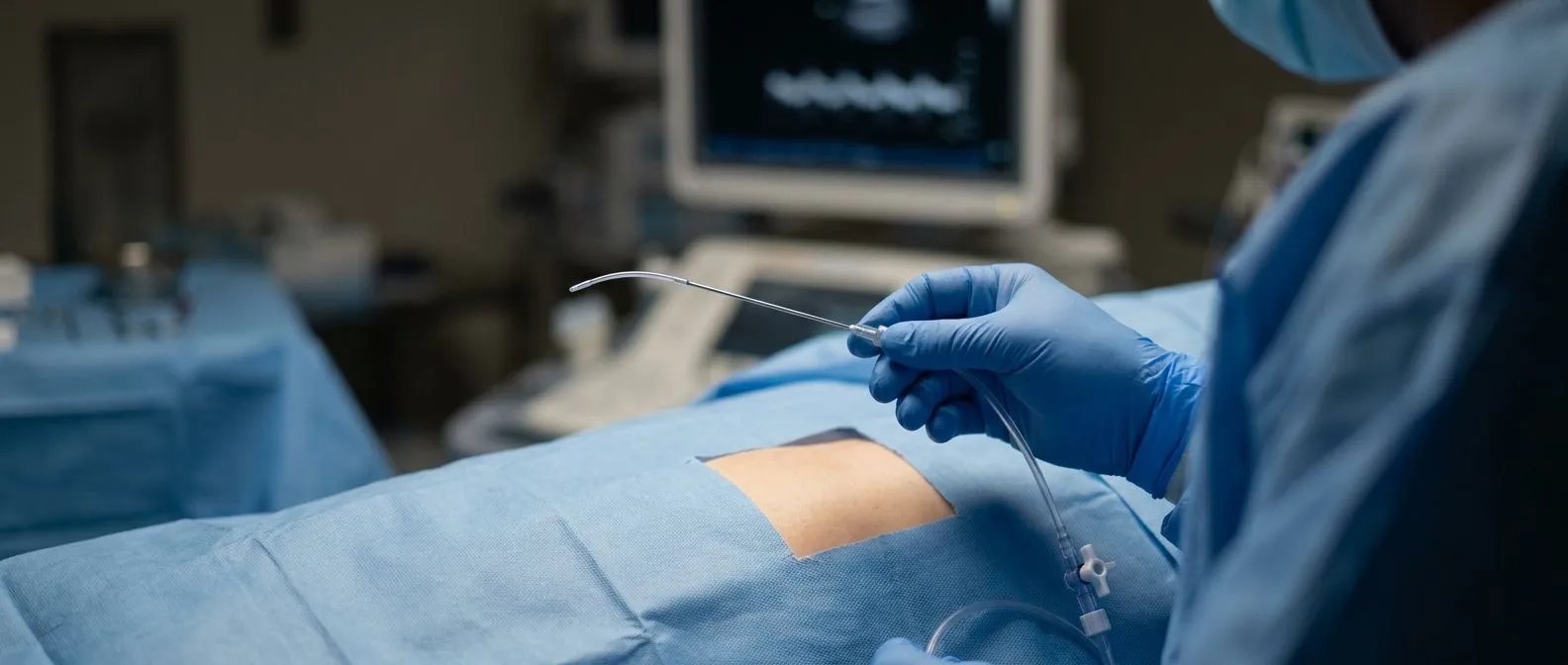
The immediate treatment. How cardiac tamponade is treated comes down to one thing: removing the fluid, urgently. Fluids and medicines given through a drip can buy minutes by temporarily supporting blood pressure, but they do not solve the problem. The definitive step is pericardiocentesis, a needle inserted under echo guidance from just below the breastbone into the sac, with the fluid drawn out. Emergency drainage in a catheterisation laboratory or at the bedside can transform a critical situation within minutes.
Preventing it from developing. Cardiac tamponade prevention, in the planned rather than emergency setting, rests on surveillance. Patients with a known moderate or large effusion are followed with repeat echocardiograms, and drainage is scheduled before the fluid reaches a tipping point rather than after. Our team reviews every case of significant effusion together before recommending drainage, precisely so that a scheduled, controlled procedure replaces a midnight emergency whenever possible. Johns Hopkins Medicine describes this compression of the heart as a condition requiring immediate decompression of the sac Johns Hopkins Medicine.
Pericardial Drainage Procedure: Pericardiocentesis Explained
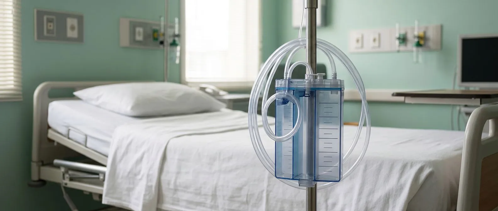
What the procedure involves. The pericardial drainage procedure known as pericardiocentesis is usually performed under local anaesthetic with sedation. The patient lies flat, an echocardiogram maps the safest path through the chest, and a needle is advanced from just below the breastbone into the sac. Once fluid is confirmed, a thin flexible catheter is exchanged for the needle and left in place, usually for one to three days, connected to a drainage bag.
How to drain fluid from the heart sac safely. The echo guidance is what makes this procedure safe. The needle is steered along a path that avoids the lung, the liver, and the coronary arteries, and the operator can watch the needle tip reach the fluid in real time. Removing the fluid is often staged rather than all at once, because rapid decompression can cause sudden shifts in blood pressure. We perform these drainages with the patient fully informed beforehand, and we sample the fluid for analysis whenever the cause of the effusion is not already clear.
Pericardiocentesis recovery. Pericardiocentesis recovery is usually short. Most patients stay in hospital for one to three days while the catheter drains and the team watches for re-accumulation. Once the drainage slows to a trickle, the catheter is removed and a follow-up echocardiogram confirms the sac is empty. Soreness at the entry site is common for a few days. Most people return to normal light activity within a week, though the underlying cause still needs treatment to stop the fluid returning.
Why a window is created. When fluid returns after a needle drainage, or when the effusion is loculated, meaning it sits in pockets the needle cannot reach, surgery offers a more durable answer. Pericardial window surgery, sometimes called a pericardial fenestration, creates a small opening in the sac. Fluid drains from the sac into the chest cavity, where the body reabsorbs it naturally, and the opening prevents pressure from building again.
Minimally invasive pericardial drainage. The modern approach is minimally invasive pericardial drainage using video-assisted thoracoscopic surgery, often called VATS. Three small incisions, a tiny camera, and instruments passed between the ribs allow the surgeon to open a generous window in the sac and, where useful, take a biopsy of the pericardium at the same time. Alternatively, a subxiphoid window is created through a small incision just below the breastbone, which can even be done under local anaesthetic in frail patients. Our team performs both conventional and minimally invasive approaches, and we recommend the less invasive route only where the evidence supports it for that particular patient.
Pericardial window surgery cost. Pericardial window surgery cost varies with the technique, the hospital, and whether a biopsy and fluid analysis are included, so no single figure is honest for every case. For international patients considering care in Mexico, the practical advice is to request a written proposal that itemises the surgeon's fee, hospital costs, anaesthesia, imaging, and pathology. Every patient we see receives a clear proposal with the alternatives, and the final decision is always theirs. Anyone planning treatment abroad should also confirm what their insurance or travel coverage will and will not include before travelling.
Pericardial Window Surgery: Cost and What to Expect
Why a window is created. When fluid returns after a needle drainage, or when the effusion is loculated, meaning it sits in pockets the needle cannot reach, surgery offers a more durable answer. Pericardial window surgery, sometimes called a pericardial fenestration, creates a small opening in the sac. Fluid drains from the sac into the chest cavity, where the body reabsorbs it naturally, and the opening prevents pressure from building again.
Minimally invasive pericardial drainage. The modern approach is minimally invasive pericardial drainage using video-assisted thoracoscopic surgery, often called VATS. Three small incisions, a tiny camera, and instruments passed between the ribs allow the surgeon to open a generous window in the sac and, where useful, take a biopsy of the pericardium at the same time. Alternatively, a subxiphoid window is created through a small incision just below the breastbone, which can even be done under local anaesthetic in frail patients. Our team performs both conventional and minimally invasive approaches, and we recommend the less invasive route only where the evidence supports it for that particular patient.
Pericardial window surgery cost. Pericardial window surgery cost varies with the technique, the hospital, and whether a biopsy and fluid analysis are included, so no single figure is honest for every case. For international patients considering care in Mexico, the practical advice is to request a written proposal that itemises the surgeon's fee, hospital costs, anaesthesia, imaging, and pathology. Every patient we see receives a clear written proposal with the alternatives and their costs, and the final decision is always theirs. Anyone planning treatment abroad should also confirm what their insurance or travel coverage will and will not include before travelling.
Pericardial Effusion After Heart Surgery and Chronic Management
Effusion after cardiac operations. Pericardial effusion after heart surgery is common in the first weeks, because the sac has been opened and becomes inflamed as it heals. Most post-operative effusions are small and disappear on their own. Larger ones, or ones appearing later, can cause low blood pressure and breathlessness and may need drainage. Anyone who has had valve surgery, coronary bypass, or any cardiac operation should mention new chest discomfort or breathlessness to their surgical team rather than assume it is normal healing.
Chronic pericardial effusion management. Chronic pericardial effusion management, for fluid that persists or keeps returning without a fixable cause, follows a stepwise path. First, treat the underlying driver: thyroid replacement for hypothyroidism, dialysis optimisation for kidney disease, chemotherapy or targeted therapy for malignancy, anti-inflammatory medication for recurrent pericarditis. Second, if fluid persists despite medical treatment and causes symptoms or shows signs of threatening the heart, drainage is reconsidered, often as a window rather than a repeated needle.
Living with an ongoing effusion. People with a stable, moderate effusion can live full lives under surveillance. The rhythm of care is usually a clinic visit and echocardiogram every few months, adjusting as the fluid behaves. The honest picture is that some chronic effusions never fully resolve but remain harmless, while others declare themselves and need intervention. Follow-up scans are compared over time, so that a quiet effusion is left in peace and a changing one is acted on promptly.
Recurrence Prevention and When to Seek a Cardiology Consultation
Stopping the fluid from coming back. Recurrence prevention after pericardial drainage depends mostly on treating the cause, not on the drainage itself. For inflammatory effusions, continuing anti-inflammatory medication and colchicine for the full prescribed course lowers the chance of return. For effusions caused by cancer or kidney disease, controlling that disease is the real prevention. Skipping medication early because the fluid is gone is the most common self-inflicted setback we see.
- Complete the medication course: anti-inflammatory treatment often needs to continue for weeks after the fluid clears
- Keep the surveillance scans: each echocardiogram catches re-accumulation before symptoms do
- Use the fluid analysis: knowing the cause, infection, malignancy, or inflammation, directs the treatment that prevents recurrence
Pericardial effusion life expectancy. Pericardial effusion life expectancy is governed far more by its cause than by the fluid itself. An effusion from a viral pericarditis that is treated fully usually leaves no trace. An effusion from advanced cancer reflects the outlook of that cancer. The reassuring news is that when drainage is performed before tamponade develops, and the underlying condition is managed, most people return to their previous life and activity.
Natural approaches and honest limits. Some readers search for pericardial effusion natural treatment, and the honest answer is that no home remedy can drain fluid from around the heart. Rest, sensible activity limits, and general health measures support recovery, but the medical treatment of the cause and, when needed, drainage are the interventions with evidence behind them. Anyone tempted to manage a diagnosed effusion without medical follow-up should understand that the main risk, tamponade, gives little warning.
Taking the next step. If you or someone close to you has been told there is fluid around the heart, a cardiology consultation is the sensible next move, not self-diagnosis. Dr Molina trained in cardiothoracic surgery at the Universidad de Guadalajara, in cardiac, minimally invasive, robotic and structural surgery at the University of Montreal, and in structural cardiology and TAVR at CHU Bordeaux, and he performed the first robotic mitral valve repair in the Ángeles Health System and the first robotic heart surgery in Guadalajara. The first contact with our team is a free video consultation: we get to know the patient, talk through the condition, and say which studies are still needed so no unnecessary ones are ordered; a face-to-face visit at the consulting rooms follows when the patient decides to go ahead. We encourage second opinions and treat seeking one as a normal part of deciding about heart care. Bring your scans, ask your questions, and make the decision with a team that has explained every option in plain words.