

Pulmonary Veins and AFib: Why Ablation Targets Them
Pulmonary veins afib ablation stops irregular rhythm by isolating the triggers in your pulmonary veins.
When cardiologists explain a pulmonary veins afib ablation, they are describing one of the most studied procedures in modern heart rhythm care. The pulmonary veins are four vessels that carry oxygen-rich blood from your lungs back to the left atrium, the upper-left chamber of your heart. Decades of research have shown that these veins are where most episodes of atrial fibrillation (AFib) begin, which is exactly why ablation targets them. If you or a family member has been told that AFib starts "in the veins of the lungs," this guide explains the anatomy, the procedure, the recovery, and the decisions in plain language.
What Are the Pulmonary Veins and What Do They Do?
What are pulmonary veins? They are the only veins in your body that carry oxygen-rich blood. Blood picks up oxygen in the lungs, then travels through the pulmonary veins into the left atrium. From there, it passes through the mitral valve into the left ventricle and is pumped to the rest of the body. There are normally four: two from the right lung and two from the left lung.
Where do the pulmonary veins deliver blood to? Always to the left atrium — never directly to the ventricles or the aorta. This is the key point for understanding AFib treatment. The left atrium wraps around the openings of these veins, so the muscular sleeves that extend from the atrium into the vein walls are in constant electrical contact with the heart's conduction system.
How do they differ from the pulmonary artery? The pulmonary artery carries oxygen-poor blood from the right ventricle to the lungs, while the pulmonary veins return freshly oxygenated blood in the opposite direction. One vessel sends blood away for oxygen pickup; the others deliver it back for circulation. Their names confuse many patients because "pulmonary" simply means "related to the lungs."
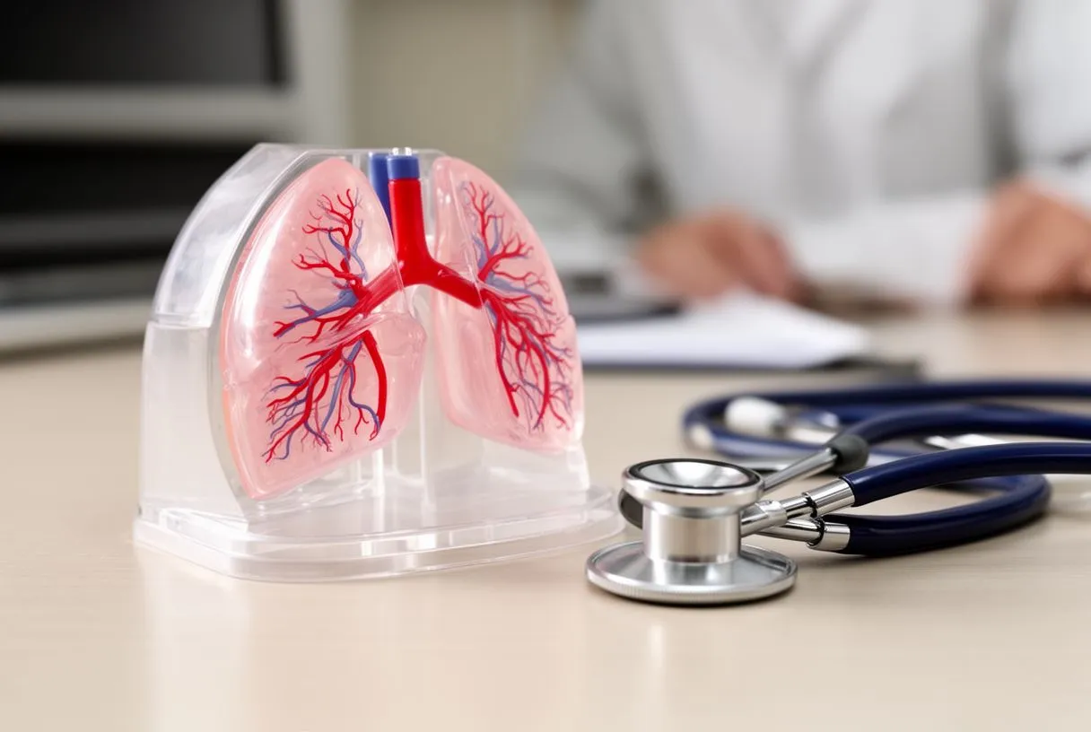
Why are there four pulmonary veins? It is the usual human pattern, but anatomy varies more than most patients expect. Some people have a fifth vein, or two veins from one side can join into a common trunk before reaching the atrium. Rarely, all four veins drain abnormally into other structures — a condition called total anomalous pulmonary venous drainage — which is usually repaired in infancy.
Their position matters too. The veins sit just behind the heart, close to the esophagus and the descending aorta. A case reported in the Brazilian Journal of Cardiovascular Surgery described aortic aneurysms compressing the left atrium and pulmonary veins, which shows how snugly these structures fit together in the chest (Brazilian Journal of Cardiovascular Surgery). In our consultations we often show patients a simple diagram of this anatomy first, because once you see where the veins sit, the whole treatment logic makes sense.
Why Do Pulmonary Veins Cause AFib?
The trigger zone lives inside the veins. The muscular tissue that lines the pulmonary veins can fire electrical signals on its own. These rapid, chaotic bursts escape the vein, hit the left atrium, and set off atrial fibrillation — a fast, irregular rhythm that replaces the heart's normal beat. This is why pulmonary veins cause AFib in the large majority of patients, especially the intermittent type called paroxysmal AFib.
Muscle sleeves are the culprit. Unlike most veins, which are thin-walled and passive, the pulmonary veins carry extensions of atrial muscle in their walls. These sleeves can behave like small electrical pacemakers. When they fire faster than the sinus node — the heart's natural pacemaker — the atrium is overwhelmed and fibrillates.
Anatomy and other conditions play a role. Vein diameter, the shape of the junction with the atrium, and the number of veins all influence how arrhythmogenic the region is. Conditions that enlarge or pressurize the left atrium, such as mitral valve disease or pulmonary hypertension, make AFib more likely. A published case of Lutembacher's syndrome — a rare combination of atrial septal defect and mitral stenosis — also described partial anomalies of the pulmonary veins, a reminder that congenital variations do occur (European Journal of Case Reports in Internal Medicine). Every case is different, which is why imaging of your own vein anatomy comes before any recommendation.
What Is Pulmonary Vein Isolation Ablation?
Pulmonary vein isolation ablation is a procedure that creates a ring of scar tissue around the opening of each vein. The goal is simple to state: trap the erratic electrical signals inside the veins so they can no longer reach the left atrium. The veins keep carrying blood normally — only their electrical connection to the atrium is interrupted.
Why isolation works. Scar tissue does not conduct electricity. Once a continuous barrier of scar surrounds each vein opening, the muscle sleeves inside the vein become electrically silent to the atrium. If the veins were the source of the AFib — and in most patients they are — the fibrillation stops or becomes far less frequent.
It is a treatment, not a cure-all. Isolation addresses the dominant trigger, but AFib also involves changes in the atrium itself, especially in long-standing cases. Some patients need additional ablation of atrial tissue beyond the veins. This is decided during the procedure and explained beforehand, so the plan matches the type and duration of your arrhythmia.
Catheter Ablation Pulmonary Veins: What Are the Procedure Steps?
Preparation and anesthesia. The procedure is performed under deep sedation or general anesthesia. Your team reviews your echocardiogram, CT or MRI of the left atrium, and rhythm recordings beforehand. Blood thinners are usually adjusted in the days before, and the femoral vein in the groin is the standard access point.
The main pulmonary veins afib ablation procedure steps follow a consistent sequence:
- Access: thin tubes (sheaths) are placed in the femoral vein and guided to the right atrium.
- Transseptal puncture: the catheter crosses the wall between the two atria to reach the left atrium.
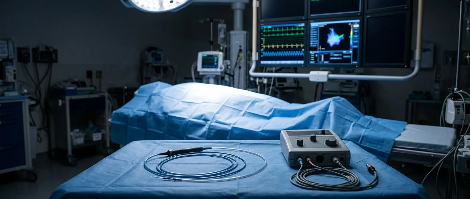
- Mapping: a 3D electroanatomic map of the left atrium and each pulmonary vein is built on screen.
- Ablation: energy is applied point by point in a circular pattern around each vein opening.
- Verification: electrical testing confirms every vein is completely isolated before the catheters are removed.
Duration and monitoring. A typical catheter ablation of the pulmonary veins takes between two and four hours. Patients usually go home the next morning. At our practice, the first contact is a free video consultation where we review which studies you already have and which are genuinely still needed, so nothing unnecessary is ordered before the procedure.
Radiofrequency Ablation Pulmonary Veins or Cryoablation: Which Is Better?
Radiofrequency ablation pulmonary veins uses controlled heat from a flexible catheter tip. The catheter makes point-by-point contact around each vein opening, and each application creates a small lesion. This technique offers precise control and is the traditional standard, performed for more than two decades worldwide.
Cryoablation uses extreme cold instead. A balloon catheter is inflated at each vein opening, freezing a continuous ring of tissue in one application per vein. The result — electrically isolated veins — is the same. Major trials, including work published by JAMA, have compared the two techniques and found similar effectiveness for first-time ablation of paroxysmal AFib.
How we choose for you. The best technique depends on your vein anatomy, how many veins need isolation, and whether additional ablation is likely. When we review a case, our multidisciplinary team weighs the options against international practice guidelines and presents you with a clear proposal — including the alternatives — and the final decision is always yours. We encourage second opinions and see them as a normal part of deciding about heart surgery.
Who Are Candidates for Pulmonary Veins AFib Ablation?
Typical pulmonary veins afib ablation candidates share a few features. They have symptomatic AFib — palpitations, breathlessness, fatigue — despite medication, or they have paroxysmal AFib that keeps returning. Many are otherwise healthy patients who want to reduce their reliance on antiarrhythmic drugs or have not tolerated them well.
Factors that strengthen the case:
- Recurrent episodes despite at least one antiarrhythmic medication
- Early-stage AFib, especially paroxysmal AFib of recent onset
- Reduced heart function where rhythm control may help the ventricle recover
- Intolerance of medications or unwillingness to take them long term
Factors that require extra caution. Long-standing, persistent AFib, a very enlarged left atrium, or severe valve disease change the plan. In those cases, AFib ablation may still be appropriate — sometimes combined with a surgical ablation performed during another cardiac operation — but the expectations must be honest. In our consultations we take the time to explain what the studies actually show, in plain words, and to answer every question before any decision is made.
Pulmonary Veins AFib Ablation vs Medication: Which Should You Choose?
Medication remains the first step for many patients. Antiarrhythmic drugs and rate-control medications can keep AFib manageable, and anticoagulants protect against stroke regardless of which strategy you choose. For some patients, medication alone is enough for years.
Ablation offers a different trade-off. Comparing pulmonary veins afib ablation vs medication, the procedure addresses the triggers directly rather than suppressing symptoms with drugs. Trials have shown ablation reduces AFib recurrence more effectively than antiarrhythmic drugs in patients who fail medication. The trade-offs are the upfront procedure risks, the recovery period, and the possibility of a repeat procedure.
Neither choice is permanent or exclusive. Some patients start with medication and move to ablation later; others ablate early to avoid drug dependence. What matters is matching the strategy to your AFib type, your symptoms, and your overall heart condition — a conversation worth having with a team that performs both approaches rather than only one.
What Is the Pulmonary Veins AFib Ablation Success Rate?
Published results are encouraging, but honest. For paroxysmal AFib, the pulmonary veins afib ablation success rate in published series is generally around 70 to 80 percent freedom from arrhythmia after one procedure, with better results after a second procedure. Persistent AFib responds somewhat less well, because the atrium itself has more advanced changes.
What "success" means varies. Some patients are completely free of AFib; others have far fewer, shorter episodes that no longer cause symptoms. Most studies count freedom from any atrial arrhythmia after a blanking period of three months, which is when the healing tissue stabilizes. Ask your team to define success the way they measure it.
Improvements continue. Better mapping systems, contact-force catheters, and the cryoballoon have all improved outcomes over time. The team also performs robotic and minimally invasive procedures, and we recommend the less invasive route only where the evidence supports it for that specific patient — not as a default for everyone.
What Is the Pulmonary Veins AFib Ablation Recovery Time?
The first days. Most patients spend one night in the hospital. The groin puncture site is checked for bleeding, and mild soreness is normal. You will be told to avoid heavy lifting and strenuous exercise for a few days to a week.
The pulmonary veins afib ablation recovery time at home is typically one to two weeks for most daily activities. Office work usually resumes within days. Energetic exercise waits longer, generally two to four weeks, following your team's instructions.
Healing and the blanking period. For about three months the ablated tissue is inflamed and irritable, so palpitations and even brief AFib episodes during this window do not mean the procedure failed. Many patients feel worse before they feel better. Blood thinners continue during this period, and a follow-up rhythm monitor confirms how the veins are healing. Follow-up visits are the time to review these monitor readings with your doctor, so the recovery is guided by real data rather than guesswork.
What Are the Risks of AFib Ablation?
Pulmonary veins afib ablation risks are real but uncommon. The most serious complication is cardiac tamponade — fluid collecting around the heart — which occurs in roughly one percent of cases and is treated promptly during the procedure. Other risks include bleeding at the groin site, injury to the phrenic nerve (which controls the diaphragm), stroke, and rare damage to the esophagus or to the pulmonary veins themselves.
How risks are minimized. Careful intracardiac ultrasound guidance, esophageal temperature monitoring, continuous anticoagulation, and verification of every vein's isolation all reduce risk. Patients with atrial fibrillation also carry a baseline risk of stroke and pulmonary embolism, which is why anticoagulation continues after the procedure until the team decides otherwise.
Costs in Mexico and practical questions. Ask for a written, itemized estimate that covers the mapping system, the ablation catheters, the anesthesia, and any planned overnight stay. For patients traveling from abroad, confirm which pre-procedure studies you can complete at home and which must be done locally.
Choosing your team matters as much as the technique. Look for a team that does high volumes of ablation, explains alternatives honestly, and offers a realistic picture of recovery. Dr Molina trained in cardiothoracic surgery at the Universidad de Guadalajara, in cardiac, minimally invasive, robotic and structural surgery at the University of Montreal, and in structural cardiology and TAVR at CHU Bordeaux — and the team reviews every case together against international practice guidelines before recommending anything. In Guadalajara, Querétaro and Hermosillo we receive many patients who arrive with a second opinion in hand, and we welcome it.

The takeaway. The pulmonary veins are the starting point of most AFib, and isolating them is the most direct way to stop the arrhythmia at its source. The procedure is well established, the recovery is measured in weeks, and the decision deserves a careful conversation with a team that knows your anatomy. If AFib is affecting your life, book a consultation with a qualified cardiac team — including ours if you are in Guadalajara, Querétaro or Hermosillo — rather than trying to self-diagnose. Pulmonary veins afib ablation is a proven option, but the right plan is the one built around your heart, not around a headline.






