


Right Atrium Enlargement: Causes and What It Means
Right atrium enlargement signals underlying heart conditions.
If your recent echocardiogram report mentioned right atrium enlargement, you are not alone in wondering what it means. The right atrium is one of the heart's four chambers, and when it grows larger than normal, it is usually a signal that something else is asking it to work harder. This guide walks you through what the right atrium does, why right atrium enlargement happens, which symptoms matter, how doctors measure it, and what treatment paths exist — in plain language, without alarm.
What Is the Right Atrium and What Does It Do?
The heart has four chambers: two upper chambers called atria and two lower chambers called ventricles. The right atrium is the upper chamber on the right side of the heart. It receives oxygen-poor blood returning from the whole body through two large veins, the superior vena cava and the inferior vena cava, and then passes that blood into the right ventricle, which pumps it to the lungs to pick up fresh oxygen.
What does the right atrium do in daily life? It works as a receiving chamber and a gentle booster pump. Between heartbeats it relaxes and fills with venous blood; just before the ventricle contracts, the right atrium squeezes to top it off. It also acts as a reservoir, smoothing out the pulses of blood arriving from the veins so the heart's rhythm stays efficient.
Because the right atrium sits at the entry point of all systemic venous return, it is sensitive to pressure changes anywhere in the right heart chambers. Raised pressure in the lungs, leaky valves, or lung disease all show up here first. That is why doctors pay close attention to its size — a change in right atrium size often points to a condition worth investigating rather than being a disease in itself.
What Is Right Atrium Enlargement?
Right atrium enlargement — sometimes called right atrial dilation — means the right upper chamber of the heart is bigger than the normal range for your body size. It is a finding, not a diagnosis on its own. The enlargement reflects that the chamber has been stretching, usually because pressure or volume inside it has been higher than usual for months or years.
Is it serious? It depends entirely on the cause. Mild enlargement found incidentally in an otherwise well person may simply be monitored. Significant enlargement, especially with symptoms or other right heart chamber enlargement, deserves a full cardiac workup. Cardiovascular disease remains the leading cause of death worldwide according to the Global Burden of Disease Study, so new heart findings should never be ignored — but neither should they cause panic before the cause is known.
Think of the right atrium like a balloon that has been inflated against resistance for a long time. The muscle wall can stretch and, over years, weaken. Identifying enlargement early gives your cardiac team the chance to treat the underlying driver — a valve, the lungs, or the heart rhythm — before the chamber loses its ability to function well.
What Causes the Right Atrium to Enlarge?
So what causes right atrium to enlarge? The most common drivers fall into a few clear groups, and understanding them helps make sense of the tests your cardiologist will order.
- Valve problems: Tricuspid regurgitation — leakage of the valve between the right atrium and the right ventricle — lets blood flow backwards, volume-loading the atrium until it dilates. Tricuspid regurgitation is one of the most frequent causes of right atrium enlargement seen in clinic.
- Lung-related pressure: Chronic lung disease such as COPD or long-standing sleep apnoea raises pressure in the pulmonary arteries. The right ventricle then pushes harder, and back-pressure reaches the right atrium, producing right heart chamber enlargement over time.
Congenital heart disease and structural causes. Some people are born with openings or abnormalities involving the right atrium, such as an atrial septal defect — a hole in the wall between the two atria — which shunts extra blood into the right side and gradually enlarges it. Congenital heart disease affecting the right atrium can go undetected until adulthood, which is why enlargement found later in life sometimes prompts a search for these conditions.
Rhythm and other contributors. Atrial fibrillation, an irregular rhythm that most often starts in the atria, both causes and is caused by atrial stretching. Thyroid disease, anaemia, kidney failure, and heart failure on the left side can also feed back into the right atrium. Identifying the specific driver in each patient is the essential first step, because treatment follows the cause, not the size measurement.
Right Atrium Enlargement Symptoms to Watch For
Right atrium enlargement symptoms are often subtle at first, and many people have none at all — the finding is discovered on a scan done for another reason. When symptoms do appear, they usually reflect the underlying condition rather than the enlarged chamber itself.
Common warning signs. Breathlessness during everyday activity, swelling of the ankles or legs, a sensation of fluttering or irregular heartbeat, and fatigue that limits what you can do are the patterns we see most. Some people feel fullness or discomfort in the upper right abdomen, where an enlarged liver can signal that back-pressure has built up in the venous system.
Enlarged right atrium heart failure — right-sided heart failure — develops when the right ventricle can no longer keep up with the load. Fluid then accumulates in the legs, abdomen, and sometimes the neck veins. These signs warrant prompt assessment. They are manageable, and treatment is far more effective when it begins before the chamber damage becomes fixed, so report new swelling or breathlessness rather than waiting to see if it settles.
Right Atrium Enlargement Echocardiogram: How It Is Measured
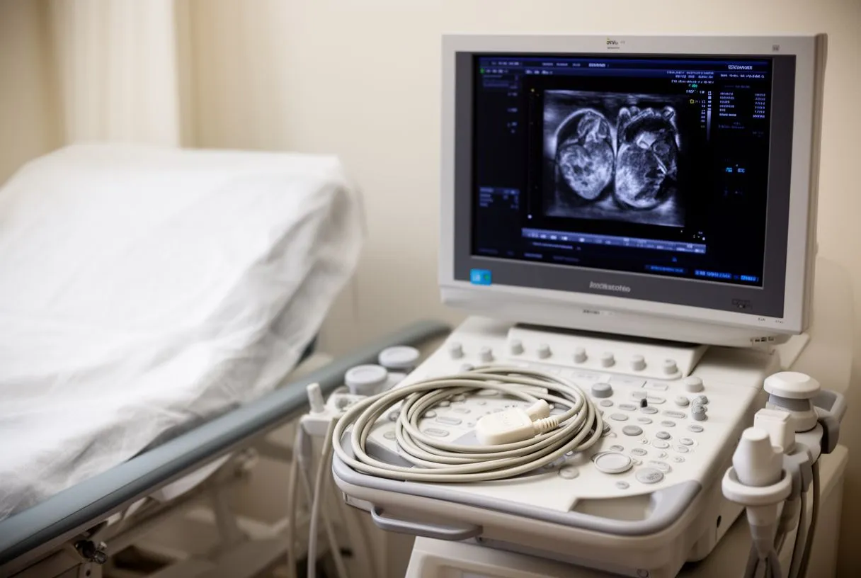
The echocardiogram — an ultrasound scan of the heart — is the standard tool for detecting and grading right atrium enlargement. It is painless, takes under an hour, and gives your cardiologist a moving picture of all four chambers and valves.
How to measure the right atrium on echo? Sonographers measure the chamber's diameter and calculate its volume, then index the volume to your body surface area so it is compared fairly against your size. Modern guidelines define mild, moderate, and severe enlargement by these indexed volumes. The scan also estimates pressures in the pulmonary arteries and shows how the tricuspid valve is holding up — context that matters as much as the size number itself.
What else might be needed? A transoesophageal echo, where a small probe is passed down the gullet, gives sharper images of the atria and any holes or masses. Cardiac MRI and CT offer precise anatomy when planning intervention. In our practice, when we review an echo showing right atrium enlargement, we take the time to explain in plain words what the images actually show and which additional studies are genuinely needed — so no unnecessary ones are ordered.
Right Atrial Enlargement vs Left Atrial Enlargement
These two findings sound similar but point to different problems. The right atrium handles blood returning from the body, so its enlargement usually reflects lung disease, tricuspid valve leakage, or right-side pressure. The left atrium handles oxygen-rich blood returning from the lungs, so its enlargement usually reflects mitral valve disease, high blood pressure, or left ventricular stiffness.
Why the distinction matters. Right atrial enlargement vs left atrial enlargement changes which tests come next and which treatments make sense. A left atrial problem raises the risk of stroke, because clots can form in stagnant blood there and travel to the brain — left atrial masses and thrombi are recognised causes of cerebral emboli, as documented in case reports of left atrium papillary fibroelastomas causing cerebral emboli. Right atrial enlargement instead raises questions about the lungs and right-side pressures.
Sometimes both atria are enlarged. That pattern often appears in long-standing atrial fibrillation or combined valve disease. It does not mean the situation is worse by default, but it does mean the cardiac team needs the full picture — valves, rhythm, lung pressures, and ventricular function — before recommending anything. Every case we review is assessed by the multidisciplinary team against international practice guidelines before any recommendation is made.
Right Atrial Enlargement ECG Findings
An electrocardiogram (ECG) is often the first test to hint at right atrium enlargement, sometimes before an echo is ever done. It records the heart's electrical activity through electrodes on the chest and limbs, and the atria leave recognizable fingerprints on the tracing.
Right atrial enlargement ECG findings. The classic sign is a tall, peaked P wave — the small bump that represents atrial contraction — particularly in the leads that look at the right side of the heart, often called P pulmonale because of its link with lung disease. There may also be signs of right ventricular strain or of atrial fibrillation, which frequently accompanies an enlarged right atrium.
An ECG alone cannot confirm enlargement; it screens and suggests. That is why it usually leads to an echocardiogram rather than replacing one. If your doctor mentioned ECG changes and sent you for further imaging, that sequence is exactly right — the ECG flagged the possibility, and the echo settles the question and looks for the cause behind it.
Right Atrium Enlargement and Pulmonary Hypertension
Pulmonary hypertension means high blood pressure in the arteries of the lungs. It is one of the most important causes of right atrium enlargement, and the connection works like a traffic jam: the right ventricle must push against narrowed, stiff lung vessels, pressure backs up into the right atrium, and the chamber gradually dilates under the strain.
How the two conditions feed each other. As the right ventricle tires, the right atrium stretches further, and the stretched atrium becomes prone to atrial fibrillation — which then makes right-heart function worse. Recognizing this cycle early matters, because treatments for pulmonary hypertension work best before the right side of the heart has remodelled permanently.
Because chronic lung disease right heart enlargement is so common, anyone with significant right atrium enlargement and a history of smoking, COPD, or severe sleep apnoea should have lung pressures formally assessed. Echo gives an estimate; occasionally a right heart catheterisation — a thin tube passed into the heart to measure pressures directly — is needed for an exact answer before treatment decisions are made. It is a well-established procedure and usually a day-case one.

What Are the Treatment Options for Right Atrial Enlargement?
There is no single "right atrium enlargement treatment" — therapy always targets the cause. Once your team knows what is driving the dilation, options usually fall into these groups.
- Medication: Diuretics to relieve fluid overload, drugs to lower pulmonary pressures, anticoagulants if atrial fibrillation is present, and treatment of the underlying lung or systemic condition.
- Procedure-based options: Closing an atrial septal defect with a device, repairing or replacing a leaking tricuspid valve, ablation for rhythm problems, or transcatheter approaches where the anatomy allows — TAVR for the aortic valve is one example of how catheter-based treatment has transformed valve care.
When is right atrium enlargement surgery considered? Direct surgery on the right atrium itself is rare; instead, surgery addresses the cause — severe tricuspid regurgitation, congenital defects, or valve disease needing repair. In our team, we perform conventional, minimally invasive, robotic and transcatheter procedures, and we recommend the less invasive route only where the evidence supports it for that specific patient. Dr Molina performed the first robotic mitral valve repair in the Ángeles Health System and the first robotic heart surgery in Guadalajara, so minimally invasive options are part of our daily practice — but never a default.
Every case is reviewed by our multidisciplinary team against international practice guidelines before a recommendation is given. You receive a clear proposal with the alternatives, and the final decision is always yours. We encourage second opinions and treat seeking one as a normal, sensible part of deciding about heart surgery — a good specialist will never be offended by it.
Enlarged Right Atrium Life Expectancy and When to See a Cardiologist
Questions about enlarged right atrium life expectancy have no single answer, and anyone who gives you a number without knowing your cause, pressures, and ventricular function is guessing. The outlook depends on what is driving the enlargement and how treatable it is: a leaky valve repaired in time, a congenital defect closed, or lung pressures controlled can all change the trajectory substantially. Right atrial dilation prognosis is therefore a prognosis about the underlying disease, not about the chamber measurement alone.
When to see a cardiologist about right atrium enlargement? Seek assessment promptly if you have new breathlessness, leg or abdominal swelling, palpitations, or fainting episodes. Even without symptoms, a newly reported enlarged right atrium on any scan deserves at least one proper evaluation — cause identified, pressures estimated, and a monitoring plan agreed. Mild, stable enlargement with a benign cause may simply be watched; that too is a legitimate outcome of a consultation.
For patients in Mexico and internationally looking for a right atrium enlargement specialist, our consulting rooms in Guadalajara, Querétaro and Hermosillo offer a multidisciplinary team of cardiac surgeons led by Dr Alejandro Molina, whose training spans cardiothoracic surgery at the Universidad de Guadalajara, cardiac and minimally invasive surgery at the University of Montreal, and structural cardiology and TAVR at CHU Bordeaux, with team experience also at Jena, Bad Berka and Buffalo. The first contact is a free video consultation: we get to know you, talk through your condition, and say which studies are still needed so none are unnecessary.

Right atrium enlargement is a signal, not a sentence. It tells your cardiac team to look for a cause — a valve, the lungs, a rhythm, or a hole you were born with — and in most cases that cause can be treated or managed with medication. The key steps are simple: get the finding properly measured, find the driver, and follow a plan built for your situation. If a recent report has left you with questions, the most useful next move is a conversation with a qualified cardiac team rather than self-diagnosis. We are here to help you understand your own heart before any decision is made.






