Sending Your Imaging on Disc for a Second Opinion
Get an imaging on disc second opinion from heart surgeons in Guadalajara.
When a cardiologist hands you a CD or USB after a scan, the natural next question is what to do with it — especially if heart surgery has been mentioned. An imaging on disc second opinion means a heart specialist reviews the actual pictures from your echocardiogram, CT, MRI or catheterization, not just the written report. In our practice, that review is often the turning point: patients arrive with a recommendation for open-heart surgery and leave with a clearer picture of all their options, sometimes including a less invasive one. This guide walks through why your images live on a disc, how to send them safely, and what a cardiac imaging review actually involves from the moment you request it.
Why Is My Imaging on a Disc?
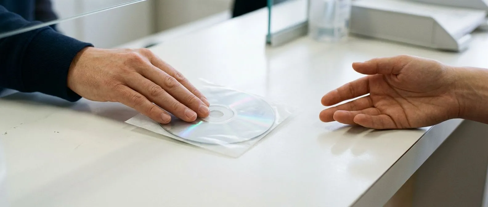
Hospitals and imaging centers store medical images in a system called PACS — Picture Archiving and Communication System — which keeps scans in a hospital's internal network. When you finish a scan, the easiest way for the facility to give you a copy is to burn the study onto a CD or DVD with a small viewing program attached. That is why your MRI is digital yet still arrives in a plastic sleeve: the disc is a portable, low-tech bridge between two computer systems that do not talk to each other.
The disc contains more than pictures. A proper imaging disc holds the original DICOM files — the standard digital format for medical images — which include every slice of the scan, the measurements the radiologist took, and sometimes the raw data behind them. This matters enormously for a second opinion. A printed image or a screenshot shows a single frozen moment; the DICOM data lets a specialist scroll through the entire study and re-measure anything that matters.
The report alone is not the whole story. A written radiology report summarizes findings in words, but words compress detail. When our team reviews a case, we always ask for the disc itself rather than a faxed report, because two readings of the same images can differ — and in heart disease, a few millimeters on a measurement can change the recommendation entirely. We take the time in consultation to explain what the studies actually show, in plain words, before any decision is discussed.
What Does an Imaging on Disc Second Opinion Mean?
An imaging on disc second opinion is a specialist's independent reading of the scans you already have. It answers a different question than the report does. The original radiologist describes what is visible; the reviewing surgeon or cardiologist asks what those findings mean for your treatment — whether surgery is needed, which approach fits, and whether anything was missed.
It applies to every kind of heart study. An echocardiogram CD shows valve function and pumping strength. A cardiac CT disc shows the coronary arteries and the aorta in detail. A cardiac MRI study shows heart muscle health and scarring. A catheterization recording shows blockages from the inside. Each one can be sent on disc and reviewed remotely, which is exactly how modern practices handle patients who live in another city or country.
Reviewing images is standard practice, not an exception. Guidelines from the European Society of Cardiology emphasize that treatment decisions should rest on confirmed findings, and complex valve and coronary cases benefit from multidisciplinary review before surgery is recommended. In our team, every case is reviewed by the multidisciplinary group against international practice guidelines before a recommendation is given — the patient then receives a clear proposal with the alternatives, and the final decision is always theirs.
Can You Request Your MRI Scan Images?
Yes — and you should. The images from your scan belong to you. In the United States, HIPAA gives you the right to your complete medical record, including the imaging itself, not just the report. Mexican law recognizes similar rights of access to your clinical file. Facilities can charge a reasonable copying fee, but they cannot refuse.
Ask for the DICOM files, not just the viewer CD. When you call the imaging center, say exactly this: "I would like the full study on disc, in DICOM format, including all series." If they offer a USB instead of a CD, accept it — it carries the same data and travels better. If you had the scan done at a large hospital, the medical records or radiology department is the place to request it, and it typically takes a few days to prepare.
Some facilities now use online patient portals where you can download images yourself, but a disc remains the most universal format across borders — which matters when your reviewing surgeon is in another country. Keep the disc in its sleeve, away from heat, and note the date of the study on the label.
How Do I Send Imaging for a Second Opinion?
There are three practical routes, and the best one depends on your timeline and comfort with technology. Whatever route you choose, the disc should reach the reviewing team with the accompanying report if you have it, and with a short note about your diagnosis and current symptoms.
- Email or secure upload: many practices accept DICOM files transferred through a secure link or cloud service. This is the fastest option and avoids shipping delays entirely.
- Courier shipping: send the original disc by a tracked courier service in a padded envelope. Never put a bare disc in a standard letter — sorting machines crack them.
- Bring it yourself: if you are already planning a consultation visit, carry the disc with you. It avoids any transfer loss and lets the specialist review it with you in the room.
What to include in the package or upload. Add the imaging disc, the radiology report, any recent lab results, your medication list, and a brief summary of what your current doctor has recommended. In our practice, the first contact is a free video consultation: we get to know the patient, talk through the condition, and say which studies are still needed so no unnecessary ones are ordered. A face-to-face visit at the consulting rooms follows only when the patient decides to go ahead.
Which Studies Are Worth Sending for a Heart Imaging Review?
Not every disc in your drawer needs to travel. The studies that matter are the ones most recent and most relevant to the decision at hand. Sending everything from the past ten years slows the review without adding value.
Prioritize by decision relevance. If surgery has been proposed for a valve, send the most recent echocardiogram and any CT or catheterization done for that problem. If the question is coronary blockages, the catheterization recording and the CT angiogram are central. If you want to send cardiac MRI for review, include the full study — the sequences matter, not just the summary images. When families are unsure which discs to send, a short message listing what they have is usually enough for the reviewing team to say which ones to include.
Age of the study matters as much as the type. The heart changes with time, and so does disease. An echocardiogram from three years ago cannot tell a surgeon what your mitral valve looks like today. As a general rule, imaging done within the last six to twelve months is most useful, and anything showing a rapidly changing condition should be recent. If your most recent study is old, the review will likely include a recommendation about which new study is worth doing — and which is not.
What Does a Heart Surgeon See on Your Disc?
When a cardiac surgeon opens your disc, they are not just re-reading the radiologist's report. They are looking at the anatomy from the point of view of someone who would operate on it. That changes what gets noticed.
Anatomy that drives the surgical approach. On a CT of the chest, a surgeon checks the condition of the aorta, the calcification of the ascending vessels, the size of the access routes, and whether a minimally invasive or robotic approach is feasible. On an echocardiogram, they examine the valve leaflets themselves — whether a mitral valve can be repaired rather than replaced, for instance, depends on details visible only when someone scrolls through the full study. Our work covers valve disease, coronary disease, the aorta and structural heart conditions, and we perform conventional, minimally invasive, robotic and transcatheter procedures — recommending the less invasive route only where the evidence supports it for that specific patient.
Consistency between the report and the images. A second reader also checks whether the written report matches what the images actually show. Discrepancies are uncommon but real, and catching one can change everything. Dr Molina trained in cardiothoracic surgery at the Universidad de Guadalajara, in cardiac, minimally invasive, robotic and structural surgery at the University of Montreal, and in structural cardiology and TAVR at CHU Bordeaux — a background that makes reading another center's imaging a routine part of the work.
When Is a Disc Not Enough? Asking for Newer Studies
Most discs are enough for a meaningful opinion. Sometimes they are not, and a good reviewing team will say so plainly rather than guess.
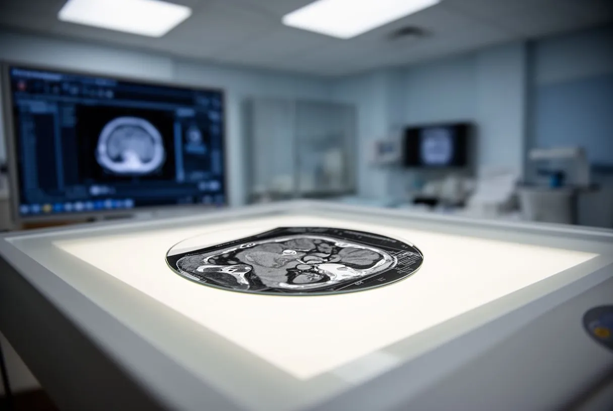
When the images are incomplete or too old. If a study was cut short, if key sequences were not acquired, or if the disc contains only summary screenshots instead of full DICOM data, a proper review is limited. Similarly, if your condition has changed since the scan — new symptoms, a new murmur, a different exercise tolerance — old images describe a heart that no longer exists. The honest answer in those cases is that one focused new study will do more than another reading of the old one.
When a specific question needs a specific test. Deciding between surgery and a transcatheter approach, for example, often requires a dedicated CT with specific measurements of the valve and access vessels. Surveillance imaging after a previous repair follows its own schedule and protocol. Rather than ordering everything, our team tells patients which studies are still needed so no unnecessary ones are ordered — the goal is a complete enough picture to decide safely, nothing more.
Why Ask for a Second Opinion Before Heart Surgery?
Heart surgery is a major decision with real risks and real alternatives, and there are five strong reasons to ask for a second opinion before high-risk surgery proceeds. None of them implies your first doctor is wrong. They simply reflect that complex decisions deserve more than one expert view.
Confirmation of the diagnosis and the approach. The same images can support different recommendations — open repair versus a minimally invasive route, surgery versus a catheter-based option, surgery now versus monitoring with medication. A second reading confirms which of these genuinely fits your anatomy. For aortic aneurysms, for example, guideline thresholds such as a diameter above 5.5 cm generally drive the timing of surgery, and confirming the measurement independently is worth doing before scheduling an operation.
Access to approaches your first center may not offer. Not every hospital performs robotic or transcatheter procedures, so the options you were given may reflect the local menu rather than the full range. A team that performs all of them can compare honestly. Dr Molina performed the first robotic mitral valve repair in the Ángeles Health System and the first robotic heart surgery in Guadalajara, and the team encourages second opinions and treats seeking one as a normal part of deciding about heart surgery — not as a slight against anyone.
For international patients and families in other Mexican cities, the logistics matter as much as the medicine. A well-run imaging review is designed to minimize travel, not require it upfront.
Start remotely, travel only if you proceed. The typical sequence is: send the imaging on disc or by secure upload, have a video consultation where the findings are explained, and only then decide whether a face-to-face visit is worthwhile. Our practice operates this way deliberately — the video consultation is free, and the in-person visit at the consulting rooms in Guadalajara, Querétaro or Hermosillo happens when the patient decides to move forward. Families avoid booking flights for an appointment that a disc review might resolve.
What the review typically covers. A complete second opinion includes the imaging interpretation, the surgical or procedural recommendation with alternatives, an honest discussion of risks and expected recovery, and guidance on which additional tests — if any — would sharpen the decision. Cost discussions belong in this conversation too: an honest team will outline what a proposed procedure involves so families can plan, and will never pressure a decision during a first video call. Patients sometimes arrive with imaging of uncertain quality; rather than repeat everything, the team identifies precisely which single study would resolve the open question.
How Do I Get Started With an Imaging on Disc Second Opinion?
Getting started is simpler than most patients expect, and it begins with a message rather than a plane ticket.
A short, practical first step. Gather your imaging discs, note the dates of each study, and write down in a few sentences what your current doctor has recommended and what worries you most. That summary, plus the discs, is everything a reviewing team needs to begin. You do not need to translate medical reports yourself or organize them perfectly — the reviewing team will tell you what is missing.
What happens next, in order. The typical path looks like this:
- Review of your imaging: the team reads the disc directly, not just the report, and flags anything that needs clarification.
- Video consultation: a free conversation where the findings are explained in plain words and every question is answered before any decision.
- A clear proposal: you receive the recommendation, the alternatives, and the reasoning — and the final decision is always yours.
A second opinion based on your own images is one of the most useful things you can do before committing to heart surgery — it costs little, risks nothing, and replaces uncertainty with understanding. If you have imaging sitting on a disc and a recommendation you are not fully sure about, discuss your situation with a qualified cardiac team rather than deciding alone. Our multidisciplinary team led by Dr Alejandro Molina reviews imaging from patients across Mexico and abroad, and we welcome your questions. Book a consultation — start with the free video call — and take the decision about your heart with clear eyes.