


The Arterial Intima and Plaque Formation
Learn how arterial intima plaque forms in the artery wall, what tests detect it early, and which treatments can slow or reverse buildup.
When a cardiologist mentions arterial intima plaque, many patients picture a simple blockage — something that gradually narrows a pipe. The reality is far more complex, and far more treatable. The intima is the innermost living layer of your artery wall, a single cell thick in some places, and it is where the entire story of atherosclerosis begins. Understanding what happens in this microscopic layer — how cholesterol enters, how inflammation takes hold, how plaques grow and sometimes rupture — gives you the knowledge to make informed decisions about prevention, screening, and treatment. This guide walks through the biology in plain language, explains what tests reveal about your arterial health, and describes what modern cardiac care can offer patients at every stage of this common condition.
What Is the Arterial Intima and Why Does Plaque Form There?
The innermost arterial layer: The intima is the thinnest lining of your artery wall, sitting directly in contact with flowing blood. It consists of a single layer of endothelial cells — a delicate barrier that should remain smooth and non-stick under healthy conditions. When this surface is intact, blood flows freely; when it becomes damaged, the stage is set for arterial intima plaque to develop.
Beneath the endothelium sits a thin layer of connective tissue and occasional smooth muscle cells. Together with the media (the muscular middle layer) and the adventitia (the outer connective tissue), the intima forms part of the artery's three-layer architecture. The ascending aorta, coronary arteries, and carotid arteries face the greatest mechanical stress — pulsatile pressure, shear forces, and turbulent flow — making them especially vulnerable to injury and intima plaque formation.
Plaque forms in the intima because this is where circulating lipids, inflammatory cells, and cellular debris first encounter the artery wall. When low-density lipoprotein particles carrying Apolipoprotein B penetrate a damaged endothelium, they lodge within the intimal space, triggering an inflammatory cascade that — over years or decades — can culminate in clinically significant intima plaque narrowing or rupture.
How Does Endothelial Dysfunction Lead to Intima Plaque Buildup?
The endothelium as a dynamic organ: Endothelial cells are not passive wallpaper. They regulate vascular tone, control clotting, manage inflammation, and decide what passes through the arterial wall. When these cells function well, they release nitric oxide, which keeps vessels relaxed and inhibits the processes that drive intima plaque. Endothelial dysfunction — the earliest detectable sign of arterial disease — represents the loss of these protective functions before any visible intima plaque has formed.
Dysfunction begins when risk factors such as high blood pressure, elevated cholesterol, smoking, or diabetes overwhelm the endothelium's capacity to maintain homeostasis. The cells become permeable, allowing LDL cholesterol to slip between tight junctions and accumulate in the intima. Research compiled by Europe PMC documents how this early permeability change precedes measurable intima plaque thickening by years, meaning the disease process is well underway long before a patient feels any symptoms.

Once the endothelium loses its barrier function, a self-reinforcing cycle takes hold. Oxidized LDL particles in the intima attract monocytes — white blood cells that transform into macrophages and engulf the lipid, becoming foam cells. These foam cells accumulate, release inflammatory cytokines, and signal smooth muscle cells from the media to migrate into the intima, where they proliferate and deposit collagen. The result is a growing, evolving intima plaque lesion — the beginning of true atherosclerotic intima thickening.
What Are the Main Causes of Arterial Intima Plaque?
Modifiable risk factors: The major drivers of intima plaque are conditions that either damage the endothelium directly or increase the concentration of harmful lipids in circulation. Hypertension subjects the arterial wall to mechanical stress that erodes endothelial integrity. Elevated LDL cholesterol provides the raw material — the Apolipoprotein B–containing particles — that infiltrates the intima and seeds intima plaque. Smoking introduces toxins that impair endothelial repair and accelerate oxidation. Diabetes and insulin resistance create a pro-inflammatory environment that promotes both intima plaque initiation and progression.
- High blood pressure: sustained pressure damages endothelial cells and increases intimal permeability
- Elevated LDL cholesterol: Apolipoprotein B particles penetrate the intima and become trapped in the intima plaque
- Tobacco exposure: chemicals impair nitric oxide production and promote oxidation of intima plaque components
- Diabetes and metabolic syndrome: chronic inflammation and glycation accelerate intima plaque growth
- Family history: genetic predisposition affects lipid metabolism and inflammatory response in the intima
Non-modifiable and emerging factors: Age is an independent contributor — arterial stiffening and cumulative endothelial exposure increase intima plaque likelihood over decades. Male sex, postmenopausal status, and certain genetic conditions such as familial hypercholesterolemia raise baseline risk substantially. Emerging evidence also points to chronic stress, sleep apnea, and periodontal disease as contributors that amplify inflammation through systemic pathways, reminding us that intima plaque is a multifactorial disease rather than a single-cause condition.
How Does Atheromatous Plaque Develop in the Intima Layer?
From fatty streak to complex lesion: The earliest visible manifestation of arterial disease is the fatty streak — a yellowish band of lipid-laden macrophages within the intima that can appear even in adolescence. Not every fatty streak progresses to a clinically meaningful intima plaque, but in the presence of ongoing risk factors, it can evolve into a fibrous plaque and eventually into a complex atheroma. This progression reflects a dynamic balance between lipid accumulation, inflammation, and the body's attempts at repair.
A mature atheromatous plaque has a characteristic architecture: a lipid-rich core covered by a fibrous cap made of collagen and smooth muscle cells. The stability of this cap determines whether the intima plaque remains silent or becomes dangerous. Thin-cap fibroatheromas — intima plaque with caps thinner than 65 micrometers — are especially prone to rupture. When a plaque ruptures, the thrombogenic core is exposed to blood, triggering platelet aggregation and clot formation. Techniques for managing vascular access in high-risk patients, such as those described in a report on achieving hemostasis after transcatheter aortic valve replacement in a patient with an aortobifemoral bypass graft, illustrate how arterial pathology complicates even unrelated cardiac procedures.
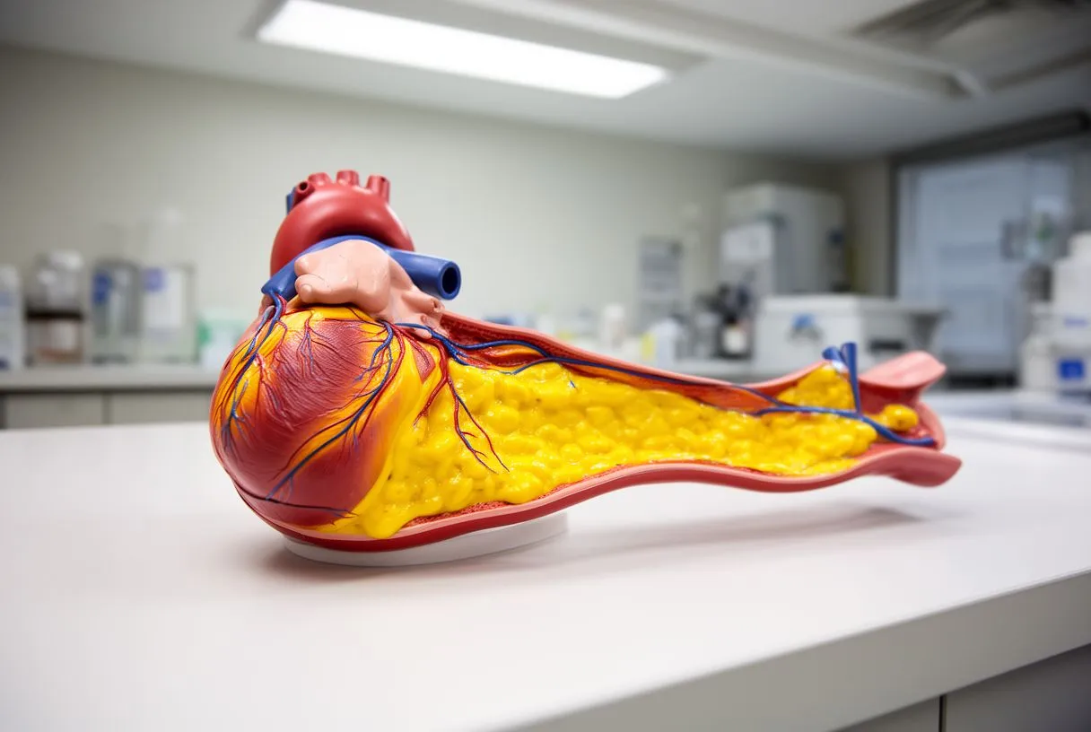
Plaque progression is not linear. Intima plaque lesions can grow slowly for decades, remain stable indefinitely, or undergo periods of rapid expansion following silent ruptures that heal with scar tissue — each healing cycle adding bulk to the plaque. This episodic growth pattern explains why a coronary artery that looked unremarkable on an angiogram a year earlier can suddenly show a critical stenosis. Understanding this biology helps patients grasp why prevention of intima plaque must be sustained rather than episodic.
What Is Intima Media Thickness Testing for Early Plaque Detection?
Ultrasound measurement of the arterial wall: Carotid intima-media thickness (CIMT) is a non-invasive ultrasound test that measures the combined width of the intima and media layers in the carotid artery. Because the carotid is accessible and mirrors the atherosclerotic burden elsewhere in the body, CIMT serves as a surrogate marker for arterial health. An abnormally thick intima-media complex suggests subclinical atherosclerosis — including intima plaque — even when no symptoms are present, allowing earlier intervention.
The test involves placing an ultrasound probe on the neck to capture high-resolution images of the far wall of the common carotid artery. Measurements above approximately 0.9 mm, or the presence of focal intima plaque, generally indicate increased cardiovascular risk. CIMT does not visualize coronary arteries directly, but studies have shown correlation between carotid findings and coronary intima plaque burden. It is particularly useful for patients with intermediate risk — those for whom the decision to start statin therapy or intensify prevention is not otherwise clear-cut.
While CIMT is a valuable screening tool for intima plaque, it has limitations. It does not distinguish between intimal thickening from atherosclerosis and medial thickening from aging or hypertension. It also does not assess intima plaque composition — whether a lesion is stable or vulnerable to rupture. For this reason, CIMT is best used as one data point among several, combined with traditional risk calculators, lipid panels, and — when appropriate — coronary calcium scoring. A cardiac team can help interpret results in context rather than in isolation.
Can Intima Plaque Buildup Be Reversed or Treated?
Plaque regression is possible but limited: Complete elimination of established intima plaque is rare, but evidence shows that aggressive lipid lowering can reduce intima plaque volume and stabilize remaining lesions. High-intensity statin therapy, by lowering circulating LDL and reducing inflammation within the intima plaque itself, can shrink lipid cores and thicken fibrous caps — making plaques less likely to rupture. The goal of treatment is not necessarily to erase intima plaque but to convert dangerous, rupture-prone lesions into stable, quiescent ones.
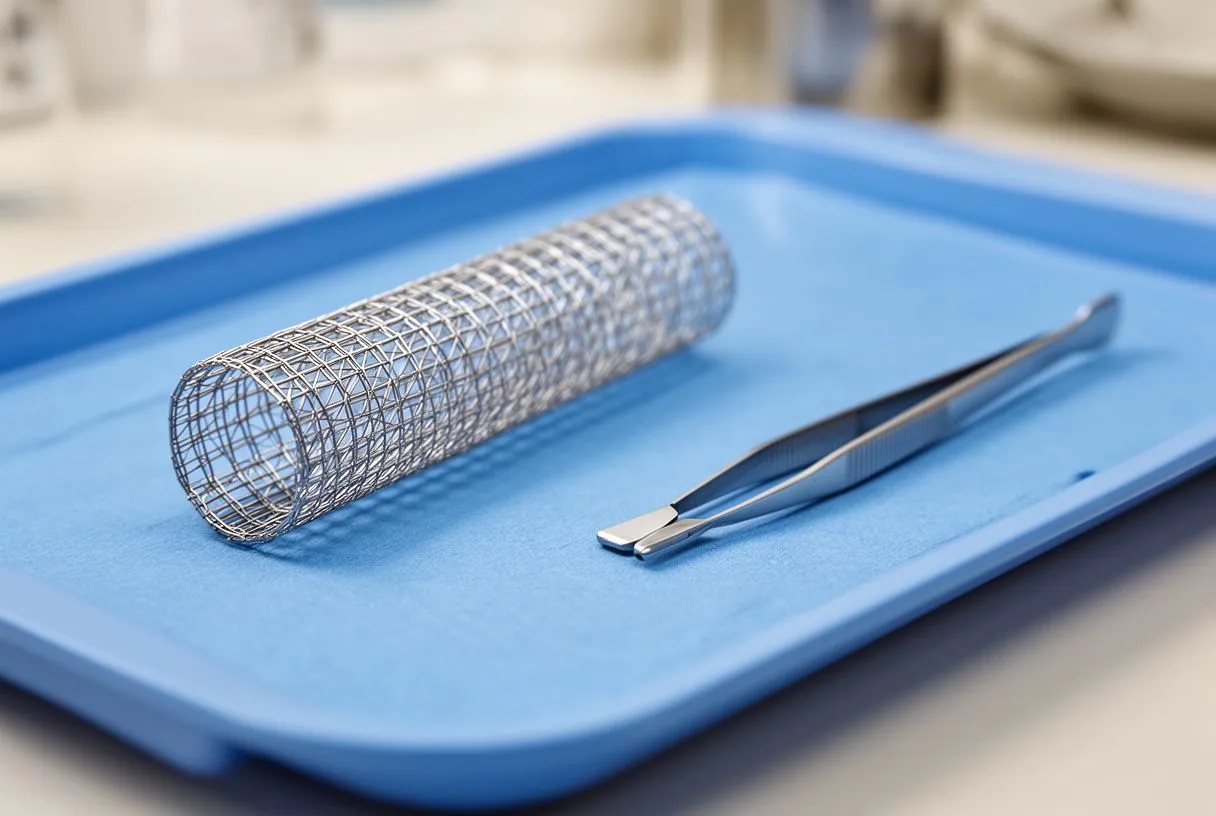
Medications form the backbone of intima plaque management. Statins reduce cholesterol synthesis and have anti-inflammatory effects. Ezetimibe blocks intestinal cholesterol absorption. PCSK9 inhibitors dramatically lower LDL in patients who cannot reach targets with statins alone. Aspirin reduces the risk of thrombus formation on existing intima plaque, though its use must be individualized based on bleeding risk — considerations evaluated in large trials such as ARISTOTLE for related anticoagulation contexts. Blood pressure medications, particularly ACE inhibitors and angiotensin receptor blockers, help reduce mechanical stress on the arterial wall.
Lifestyle changes are equally essential and synergistic with medication for managing intima plaque. A Mediterranean-style diet rich in vegetables, olive oil, nuts, and fish provides anti-inflammatory benefits. Regular aerobic exercise improves endothelial function and lipid profiles. Smoking cessation is perhaps the single most impactful intervention — tobacco cessation halts ongoing endothelial damage and allows partial recovery of vascular function. When these measures are sustained over years, imaging studies have documented measurable reductions in intima plaque burden, offering patients tangible evidence that their efforts matter.
What Happens When Arterial Intima Plaque Ruptures?
The anatomy of an intima plaque rupture: When the fibrous cap overlying a lipid-rich plaque tears, the thrombogenic interior is exposed to circulating blood. Platelets adhere to the exposed collagen and tissue factor, activating the coagulation cascade. Within minutes, a thrombus can form — partially or completely occluding the artery. In the coronary circulation, this produces an acute coronary syndrome: unstable angina if the occlusion is partial, or a heart attack if it is complete.
Not every intima plaque rupture causes a catastrophic event. Many ruptures are clinically silent — the thrombus is small, the body's fibrinolytic system dissolves it, and the intima plaque heals with scar tissue that increases its bulk. This silent rupture-and-repair cycle is a major mechanism of intima plaque growth. A case report on coronary arterial disease after electroconvulsive therapy highlights how acute physiological stress can unmask or exacerbate underlying intima plaque pathology in unexpected clinical settings.
- Chest pressure or tightness: classically central, may radiate to the arm or jaw during an intima plaque event
- Sudden shortness of breath: may occur with or without chest discomfort from intima plaque rupture
- Cold sweat, nausea, or lightheadedness: autonomic response to cardiac ischemia from intima plaque occlusion
- Symptoms at rest: chest pain occurring without exertion suggests intima plaque instability
Atypical presentations are common and important to recognize. Women, older adults, and people with diabetes may experience atypical infarction — symptoms such as profound fatigue, indigestion-like discomfort, or even arm pain without classic chest pressure. Because these presentations are easily mistaken for non-cardiac problems, delays in seeking care are frequent and dangerous. Any sudden, new, or worsening chest symptoms deserve immediate evaluation — calling emergency services is always appropriate when intima plaque complications are suspected.
How Is Coronary Plaque Formation Diagnosed?
From functional to anatomical testing: The diagnostic pathway for coronary plaque begins with assessing whether the heart's blood supply is adequate under stress. Auscultation — listening to the heart with a stethoscope — may reveal murmurs or extra sounds suggesting valve disease, but it cannot detect intima plaque directly. Exercise stress tests evaluate whether the heart develops ischemia under exertion, indicating functionally significant narrowing. These functional tests tell us if intima plaque is causing problems but not where or how much intima plaque exists.

Coronary angiography has long been the gold standard for visualizing coronary arteries. A catheter is threaded — typically via an arterial line in the wrist or groin — to the coronary ostia, and contrast dye is injected while X-ray images are captured. This reveals the location and severity of luminal narrowings. However, angiography shows only the artery's interior contour; it cannot assess the intima plaque within the wall or determine whether a intima plaque is stable or vulnerable. Intravascular ultrasound and optical coherence tomography — catheter-based imaging tools inserted during angiography — provide detailed cross-sectional views of the arterial wall, revealing intima plaque composition and cap thickness.
Non-invasive coronary CT angiography has emerged as a powerful alternative for selected patients. A CT scan with contrast can visualize coronary intima plaque, assess its composition (calcified versus non-calcified), and measure the coronary calcium score — a marker of overall plaque burden. Calcium scoring is particularly useful for risk stratification in asymptomatic patients. For patients facing surgery, understanding the extent and nature of coronary intima plaque is essential — it informs decisions about whether to proceed with balloon angioplasty or whether surgical revascularization using an arterial graft is more appropriate.
What Are the Best Prevention Strategies for Intima Layer Disease?
A tiered approach to vascular protection: Prevention of intima plaque operates on two levels: preventing intima plaque formation in healthy arteries and slowing progression in arteries that already have intima plaque. The fundamentals are the same — control blood pressure, manage lipids, avoid tobacco, maintain healthy weight, and stay physically active — but the intensity differs based on individual risk. For someone with no risk factors, lifestyle measures may suffice. For someone with established intima plaque or multiple risk factors, medication becomes essential.
Diet plays a central role in preventing intima plaque. The evidence consistently supports Mediterranean and DASH dietary patterns, which emphasize vegetables, whole grains, legumes, nuts, olive oil, and fish while limiting processed foods, refined sugars, and red meat. These patterns improve lipid profiles, reduce blood pressure, and provide anti-inflammatory compounds. Physical activity — at least 150 minutes of moderate aerobic exercise weekly — improves endothelial function and reduces intima plaque risk. The benefits of exercise on the arterial wall are measurable: regular aerobic activity increases nitric oxide bioavailability and reduces intima-media thickness over time.
Medical management should not be viewed as an alternative to lifestyle change but as a complement for intima plaque prevention. Statin therapy for patients at elevated risk is supported by decades of evidence demonstrating reduced cardiovascular events. Blood pressure control to targets appropriate for individual circumstances reduces mechanical stress on the arterial wall. For patients with diabetes, glycemic control reduces glycation-related arterial damage. Regular follow-up with a cardiac team ensures that intima plaque prevention strategies are tailored, monitored, and adjusted as circumstances change.
When Should You Seek Cardiac Evaluation for Plaque Concerns?
Knowing when to act: Many patients with intima plaque feel entirely well until a complication occurs — this is the fundamental challenge of coronary artery disease. However, certain situations warrant proactive evaluation even without symptoms. A strong family history of early coronary disease (male relatives under 55, female relatives under 65), known familial hypercholesterolemia, or personal history of conditions that accelerate intima plaque — diabetes, chronic kidney disease, inflammatory conditions — all justify screening discussions with a cardiologist.
Symptoms, even subtle ones, should never be dismissed. Exertional chest discomfort that resolves with rest — classic angina — is a clear signal that intima plaque may be limiting blood flow. New shortness of breath, particularly with activity, may represent the earliest sign that coronary intima plaque is significant. For patients already diagnosed with intima plaque, changes in symptom pattern — chest pain occurring at lower exertion levels, lasting longer, or happening at rest — indicate intima plaque instability and require urgent assessment.
Understanding the arterial intima and how intima plaque forms within it transforms a frightening diagnosis into a manageable condition. The intima is where the disease begins, but it is also where prevention and treatment have their greatest impact. Whether you have been told you have early intima plaque changes, are recovering from a cardiac event, or simply want to understand your risk, the most important step is the same: have a thorough conversation with a qualified cardiac team. Dr Alejandro Molina and the specialists at Cirugía de Corazón offer comprehensive cardiac evaluation in Guadalajara, Querétaro, and Hermosillo — book a consultation to discuss your individual situation, review your risk factors, and build a prevention or treatment plan grounded in current evidence and tailored to your needs. Do not wait for symptoms to make the first appointment.






