


The Four Cardiac Chambers and How They Work Together
Four cardiac chambers work as a precise team to circulate blood through your heart and body.
If you or someone in your family has been told there is a problem with one of the four cardiac chambers, the first few days can feel like a crash course in a language you never chose to learn. This guide walks through the heart's chambers in plain words: what each one does, how they work together as a single pump, how doctors measure them, and what happens when one of them needs repair. By the end, you should be able to read your own test reports with far less confusion — and ask better questions at your next consultation.
What Are the Four Cardiac Chambers?
The four cardiac chambers are the four hollow spaces inside the heart where blood gathers and is pumped onward. Think of the heart as a two-story house with two rooms on each floor. The upper rooms, called atria, receive blood coming back to the heart. The lower rooms, called ventricles, pump blood out to the body and lungs. A muscular wall called the septum divides the heart into right and left sides, so the four chambers sit in a neat two-by-two arrangement.
Doctors use another word for heart chambers in reports and conversation: "cavities," or simply "the atria and ventricles." Whatever the wording, the idea is the same. Each chamber is a distinct space with its own size, its own muscle wall, and its own job in the circulation.
What are the four cardiac chambers doing all day? They fill with blood, then contract to move that blood forward — roughly one hundred thousand times a day, without you ever noticing. The elegance of the design is that each chamber handles a specific leg of the journey, and the valves between them keep blood moving in one direction only.
Why Does the Heart Have Four Chambers?
The double-circulation design. Mammals and birds use two separate circuits: one circuit sends blood to the lungs to pick up oxygen, and the other sends oxygen-rich blood to the rest of the body. Four chambers make this possible. The right side of the heart handles the low-pressure lung circuit, and the left side handles the high-pressure body circuit. Mixing the two jobs in one or two chambers would mean oxygen-poor blood and oxygen-rich blood blend together — exactly what happens in some congenital conditions, which is why those defects matter so much.
Pressure without leakage. A four-chamber heart can generate very different pressures on each side. The left ventricle must push blood to your toes, brain and organs, so its muscle is the thickest in the heart. The right ventricle only needs to reach the nearby lungs. Separating these tasks means each pump is built for its own workload — no chamber wastes effort, and no circuit is under-supplied.
Another way to answer why the heart has four chambers is to look at what goes wrong without them. When a defect lets blood cross from the left side to the right side, or a valve leaks so a chamber keeps filling and refilling, the whole circulation slowly gets overloaded. The four-chamber design is what keeps oxygen delivery steady and pressures balanced across a lifetime.
What Are the Four Heart Chambers' Names?
The four heart chambers names are simple enough to memorize: the right atrium, right ventricle, left atrium, and left ventricle. "Atrium" is the singular of "atria," the two upper chambers. "Ventricle" refers to each of the two lower chambers. Doctors usually abbreviate these as RA, RV, LA and LV on reports and screen displays.
- Right atrium (RA): receives oxygen-poor blood returning from the whole body through two large veins, the superior and inferior vena cava.
- Right ventricle (RV): pumps that blood through the pulmonary valve into the lungs, where it picks up oxygen and releases carbon dioxide.
- Left atrium (LA): receives freshly oxygenated blood returning from the lungs through the pulmonary veins.
- Left ventricle (LV): the main pump, sending oxygen-rich blood through the aortic valve into the aorta and out to the entire body.
Once you know the four heart chambers names, the rest of cardiology vocabulary becomes easier. Most diagnoses — valve disease, heart failure, enlargement, congenital defects — are described by naming which chamber is involved and what is happening to it. When your cardiologist says "moderate enlargement of the left atrium," you now know exactly which room in the house is being discussed.
Atria and Ventricles: What Each One Does
Atria are the receiving chambers. Their job is mostly to collect returning blood and give it a final push into the ventricles below. They contract gently, just before the ventricles contract, topping them up for the next big beat. The atria also stretch comfortably to hold extra volume when demand rises, which is why they are sometimes called the "primer pumps" of the heart.
Ventricles are the pumping chambers. The right ventricle and left ventricle function as the engines of the circulation. Their thick muscular walls squeeze inward with each beat, and their shape — a spiral arrangement of muscle fibers — lets them twist slightly as they contract, wringing blood out efficiently rather than just squashing it.
The atria and ventricles work as a team in a carefully timed sequence. The heart's natural pacemaker, the sinoatrial node, fires an electrical impulse that makes the atria contract first. A brief pause at the atrioventricular node lets the ventricles fill completely, then the impulse spreads through the ventricular muscle and the main pumping squeeze follows. When that timing goes wrong — as in atrial fibrillation, where the atria quiver instead of squeezing — the ventricles still work, but the heart loses efficiency and stroke risk rises.
How Do the Four Chambers of the Heart Work Together?
How do the four chambers of the heart work together in a single beat? In exactly the same sequence every time. Blood enters the right atrium, drops into the right ventricle, is pumped to the lungs, returns to the left atrium, drops into the left ventricle, and is pumped out to the body. Four chambers, four valves, one continuous loop that never stops.
Coordination depends on two things: timing and valves. The electrical system sets the rhythm, making sure the atria squeeze before the ventricles. The four heart valves — tricuspid, pulmonary, mitral and aortic — open and close at precise moments so blood flows forward and never backward. A heart attack, a rhythm problem, or a damaged valve can each break the coordination in a different way, and that is often what produces the symptoms that bring patients to a specialist.
In our team's consultations we take the time to show patients their own chamber measurements and explain, in plain words, what each number means for how the heart is working as a whole — because understanding the sequence makes the treatment choice far easier to discuss.
Which Heart Chamber Is Most Important?
Which heart chamber is most important? The honest answer: all four, but the left ventricle usually gets the most attention. It is the strongest chamber and the one whose pumping performance — measured as "ejection fraction" on echo tests — best predicts how well the heart handles everyday life. Most heart failure treatments focus on protecting or strengthening the left ventricle.
That said, every chamber's health matters. The left ventricle cannot pump well if the right ventricle is failing, if the atria are in a disorganized rhythm, or if a valve is leaking badly. Surgeons and cardiologists therefore never look at one chamber in isolation; they assess how the four work as a system. The same condition can affect different chambers differently in different patients, which is why individualized review matters so much.
For our team, every case is reviewed by the multidisciplinary group against international practice guidelines before any recommendation is made — the patient receives a clear proposal with the alternatives, and the final decision is always theirs. The question is never "which chamber matters most" in the abstract, but "what does this particular heart need."
Four Cardiac Chambers Blood Flow: The Path Step by Step
Four cardiac chambers blood flow follows a fixed route you can trace with your finger. Here is the full path, in order, so you can follow along on any diagram of the heart:
- Oxygen-poor blood returns from the body to the right atrium through the vena cava veins.
- The right atrium contracts and sends blood through the tricuspid valve into the right ventricle.
- The right ventricle pumps blood through the pulmonary valve and arteries into the lungs.
- In the lungs, blood releases carbon dioxide and absorbs oxygen.
- Oxygen-rich blood returns to the left atrium through the pulmonary veins.
- The left atrium sends blood through the mitral valve into the left ventricle.
- The left ventricle pumps blood through the aortic valve into the aorta and onward to the whole body.
Notice that the four cardiac chambers blood flow pattern never mixes the two circuits. The right side only ever handles oxygen-poor blood, and the left side only ever handles oxygen-rich blood. When something disrupts the sequence — a narrowed valve, a hole in the septum between chambers, or weakened muscle — the pattern shows up on testing long before symptoms become severe, which is why early measurement matters.
Echocardiogram Four-Chamber View and Other Tests
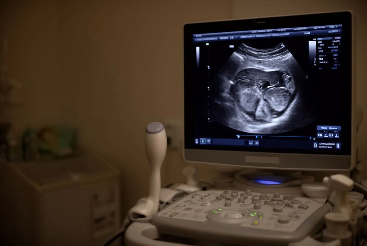
The echocardiogram four-chamber view. An echocardiogram is an ultrasound of the heart. In the standard "four-chamber view," the sonographer angles the probe so all four chambers appear on screen at once, like looking at a two-story house cut open from the front. From this single view, the team can compare chamber sizes side to side, watch the valves open and close, and estimate how strongly the ventricles squeeze.
How chambers are measured. International guidelines give normal size ranges for each chamber, and reports will show whether yours falls inside or outside them. The Recommendations for Cardiac Chamber Quantification by Echocardiography in Adults: An Update from the American Society of Echocardiography and the European Association of Cardiovascular Imaging set the reference values doctors worldwide use to decide whether a chamber is enlarged, and when enlargement signals that a valve problem or heart failure needs closer follow-up.
Beyond echo. When questions remain, other tests fill in the picture. A cardiac MRI of the four chambers gives very precise measurements of volumes and muscle health, and is often used before planning surgery. Cardiac catheterization, where thin tubes are threaded into the heart, measures pressures inside each chamber directly. In our practice, we use these studies to confirm exactly what the studies actually show before recommending surgery, and we tell patients which ones are still needed so no unnecessary ones are ordered.

When Chambers Change: Septal Defects and Enlargement
Atrial ventricular septal defects. A septal defect is a hole in the wall (septum) separating the chambers. An atrial septal defect sits between the two atria; a ventricular septal defect sits between the two ventricles. Either one lets blood flow from the higher-pressure left side to the lower-pressure right side, overloading the right chambers and the lungs over time. Some small defects cause no symptoms for decades; others need closure in childhood or adulthood.
Chamber enlargement from valve disease. When a valve leaks or narrows, the chamber behind it adapts by enlarging. A leaking mitral valve, for example, lets blood flow backward into the left atrium with every beat, and the left atrium slowly stretches. A narrowed aortic valve makes the left ventricle pump harder against resistance, thickening its muscle. These changes are measurable and follow known thresholds — this is where guideline numbers like an aortic diameter above 5.5 cm trigger a surgical recommendation.
Heart failure chamber enlargement treatment. In heart failure, one or more ventricles enlarge and pump less effectively. Treatment ranges from medications that reduce the heart's workload, to device therapy, to valve repair or replacement when a valve is the underlying cause. Atrial ventricular septal defect surgery and valve surgery both aim at the same goal: stop the overload before the chamber muscle changes become permanent. Every case is reviewed by our multidisciplinary team against international practice guidelines, and we recommend the less invasive route only where the evidence supports it for that patient.
Minimally Invasive Heart Chamber Repair: What to Expect
Not every chamber problem needs a full open-heart operation. Today there are several routes to repair or replace what is failing, and the right one depends on the specific valve or defect, the patient's overall health, and the evidence for each approach.
- Transcatheter procedures (TAVR): a replacement aortic valve is delivered through a catheter, usually from an artery in the groin, without opening the chest — often the lowest-risk option for suitable patients.
- Minimally invasive surgery: small incisions between the ribs instead of a full sternotomy, used for some valve repairs and septal defect closures.
- Robotic surgery: instruments guided by the surgeon through tiny ports, allowing very precise work inside the heart with less tissue trauma.
- Conventional open surgery: still the best-proven route for many complex cases, and sometimes simply the right choice.
Choosing among these options is a judgment that weighs the patient's anatomy, age, other conditions, and what the imaging shows about each chamber. Dr Molina trained in cardiac, minimally invasive, robotic and structural surgery at the University of Montreal, and in structural cardiology and TAVR at CHU Bordeaux, and performed the first robotic mitral valve repair in the Ángeles Health System and the first robotic heart surgery in Guadalajara. Our work spans valve disease, coronary disease, the aorta and structural heart conditions, performed conventionally, minimally invasively, robotically and transcatheter — always with the least invasive option that the evidence supports for that patient.
After any chamber or valve procedure, recovery follows predictable milestones: hospital stay, a graded Return to Work over weeks depending on the approach used, and follow-up imaging to confirm the repaired chamber is behaving normally. We encourage patients to seek second opinions and treat that as a normal, sensible part of deciding about heart surgery — no good team should mind. If a question about one of your cardiac chambers has brought you this far, the next step is a conversation with a qualified cardiac team. The first contact with our practice is a free video consultation: we get to know you, talk through your condition, and identify which studies are still needed. A face-to-face visit at our consulting rooms in Guadalajara, Querétaro or Hermosillo follows when you decide to go ahead.

In closing, the four cardiac chambers — right atrium, right ventricle, left atrium, and left ventricle — form a coordinated pump that keeps blood moving through two circuits without ever mixing them. Understanding how the atria and ventricles divide their work helps you read your own reports, understand why a valve or a septal defect can enlarge a chamber, and weigh treatment options with confidence. Nothing here replaces an individual assessment: discuss your own situation with a qualified cardiac team, ask every question you have, and never self-diagnose based on an article — however thorough it may be.






