

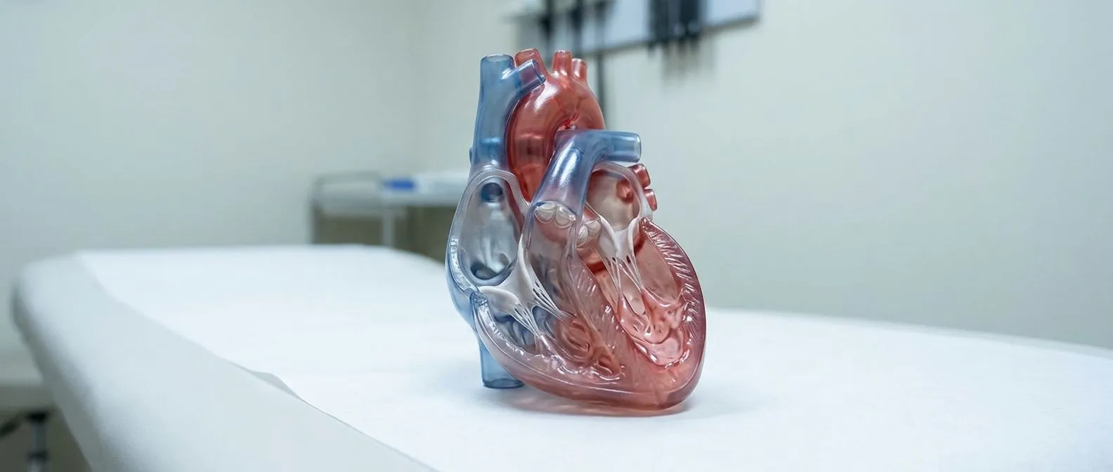
Ventricle Problems: Enlarged and Weak Heart Chambers
Heart ventricle problems can enlarge or weaken heart chambers.
What Are Heart Ventricle Problems?
When a patient first hears they have heart ventricle problems, the immediate concern is how it will affect daily life and longevity. These conditions occur when the lower chambers of the heart, responsible for pumping blood to the lungs and the rest of the body, become enlarged, thick, or weak. It is a daunting diagnosis, but understanding the mechanics helps demystify the journey ahead and allows families to regain a sense of control over their health decisions.
Anatomical changes in the heart: The ventricles are muscular chambers. If they lose their squeezing power or stretch too much, the heart cannot circulate blood efficiently. Initial assessment often begins with simple auscultation, where your physician listens for murmurs or irregular rhythms through a stethoscope. According to the National Center for Biotechnology Information, structural changes in these chambers are often a downstream effect of other cardiovascular stressors rather than a standalone disease.
Why early intervention matters: Waiting until severe symptoms appear limits your options. By understanding the early signs, patients and families can engage in proactive care. We approach these conditions not as an immediate emergency, but as a chronic management process where lifestyle, medication, and sometimes surgery play distinct roles in preserving heart health.
How Heart Ventricle Pump Function Works
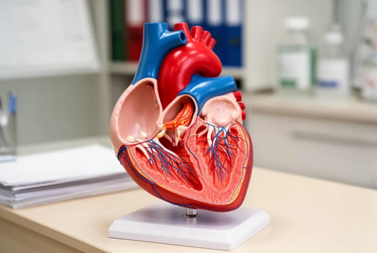
The mechanics of blood flow: To grasp why ventricular dysfunction causes symptoms, it helps to visualize the cardiac cycle. Blood returns from the body into the right atrium, flows into the right ventricle, and is pumped to the lungs to gather oxygen. The oxygen-rich blood then enters the left atrium and fills the left ventricle, which is the most muscular chamber, designed to push blood out to your entire body.
Pressure and volume dynamics: The left ventricle must generate significant pressure to push blood through the aortic valve and into the ascending aorta. When the muscle is weak, it cannot generate this pressure. When it is stiff, it cannot relax to fill properly. Both scenarios lead to fluid backing up, which is the root cause of most heart failure symptoms.
The impact of structural failure: If the heart pump function declines, the body compensates by retaining fluid and making the heart beat faster. Over time, this compensation fails. The heart remodels itself, stretching into an enlarged heart ventricle that contracts poorly, turning a mechanical advantage into a liability that ultimately limits your exercise tolerance and energy levels.
Causes of Left Ventricle Dysfunction
Ischemic heart disease: The most common cause of a weak left ventricle is ischemia, or lack of blood flow to the heart muscle itself. This usually happens when an atheromatous plaque builds up in the coronary arteries, restricting oxygen delivery. High levels of Apolipoprotein B in the blood often drive this plaque formation, making lipid management a cornerstone of long-term prevention.
Electrocardiogram clues: Sometimes, an atypical infarction—a heart attack that does not present with classic crushing chest pain—silently damages the heart muscle. Research such as the Ischemic changes in lead aVR is associated with left ventricular thrombus or high-grade spontaneous echocontrast in patients with acute anterior myocardial infarction highlights how specific electrical patterns on an ECG can warn physicians about severe complications like blood clots forming in the weakened ventricle.
Non-ischemic causes: Dysfunction is not always caused by blocked arteries. Viral infections, severe valve disease, genetic conditions, and chronic high blood pressure can all exhaust the heart muscle. Identifying the exact cause dictates the treatment plan, as repairing a valve will help a ventricle recover far better than simply treating the symptoms of failure.
- Ischemic damage: Reduced blood flow from coronary artery disease weakening the muscle.
- Genetic cardiomyopathies: Inherited conditions that directly alter heart muscle structure.
- Valvular stress: Longstanding leaks or tightness forcing the chamber to overwork continuously.
- Myocarditis: Viral infections causing inflammation and temporary or permanent weakness.
Symptoms of an Enlarged Heart Ventricle
Recognizing early warning signs: Heart failure symptoms often begin subtly. Patients might notice they tire more easily when climbing stairs or carrying groceries. Shortness of breath, especially when lying flat, is a classic sign that the left ventricle is failing to clear fluid from the lungs. Some individuals experience vague discomfort or referred pain, such as arm pain, without realizing the heart is the source of the distress.
Progression of fluid overload: As the right ventricle becomes involved, fluid builds up in the body rather than the lungs. This leads to swelling in the ankles, legs, and abdomen. Patients often complain of waking up in the middle of the night gasping for air, a condition known as paroxysmal nocturnal dyspnea, which signals that the heart is struggling to handle nighttime fluid shifts.
Systemic effects of poor perfusion: When the heart cannot pump effectively, the brain and muscles receive less oxygen. This causes dizziness, confusion, and extreme fatigue. These heart failure symptoms are your body’s alarm system, indicating that the cardiac output has dropped below the threshold needed to sustain normal daily activities without uncomfortable shortness of breath.
How Heart Chamber Enlargement Is Diagnosed
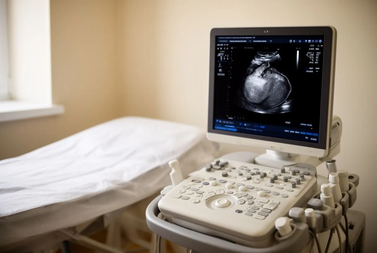
The role of echocardiography: The echocardiogram ventricle assessment is the gold standard for visualizing heart chamber enlargement. Using sound waves, this painless test shows the exact size of the ventricles, how thick the walls are, and critically, how well they squeeze. It allows your cardiologist to calculate the ejection fraction, a percentage representing how much blood the left ventricle pumps out with each beat.
Blood tests and risk ratios: Beyond imaging, blood tests help us understand the metabolic strain on the heart. A study on the Prognostic role of the serum uric acid-to-serum creatinine ratio in patients with st-elevation myocardial infarction and multivessel coronary artery disease demonstrates how simple blood markers can predict outcomes in patients with severe coronary artery blockages and ventricular compromise.
Invasive monitoring techniques: In acute settings, we might use an arterial line to continuously monitor blood pressure, or place an arterial cannula for precise blood gas analysis. These tools provide real-time data on how well the heart is perfusing the body, guiding medication choices in the intensive care unit before any definitive surgical intervention is planned.
Understanding Dilated Cardiomyopathy and Weak Chambers
The remodeling process: Dilated cardiomyopathy is a specific condition where the heart ventricle stretches and becomes thin, floppy, and weak. Unlike hypertrophy, where the muscle thickens, dilation means the chamber volume increases while the muscle mass stays the same, resulting in a weak, inefficient pump. This stretching can also pull apart the mitral valve leaflets, causing leakage.
Blood conservation in surgery: When patients with this condition require surgery, managing their blood volume is critical. Techniques involving autologous blood salvage, where blood lost during surgery is collected, cleaned, and returned to the patient, minimize the need for donor transfusions. If structural repair is needed, using an autograft—living tissue taken from the patient’s own body—reduces the risk of rejection.
Managing the progression: While we cannot always reverse the stretching, we can halt its progression with medications like beta-blockers and ACE inhibitors. The goal is to lower the pressure inside the ventricle, giving the muscle a chance to heal and shrink back toward its normal size, improving the overall heart pump function over months of careful medical management.
Ventricle Arrhythmia Treatment Options
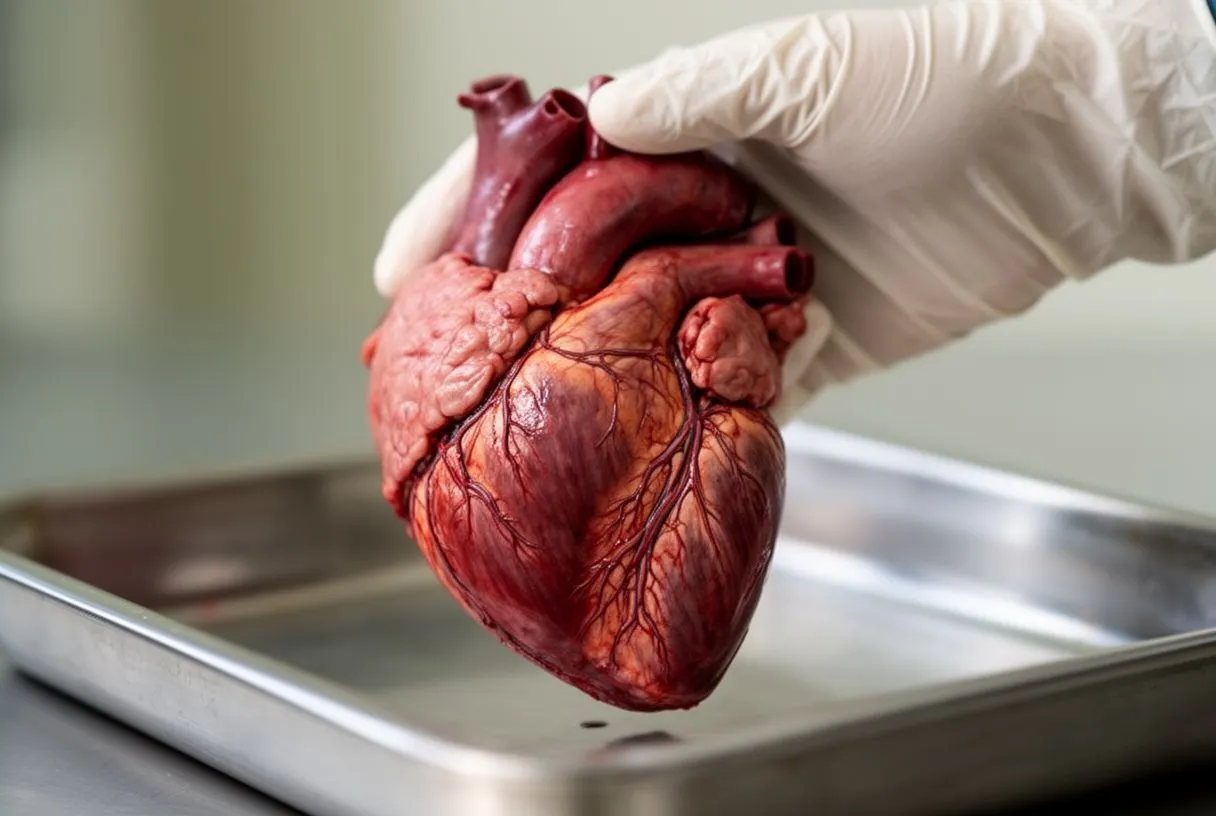
Electrical instability: When ventricles are enlarged or scarred, the electrical pathways become disrupted, leading to dangerous rhythms. While atrial flutter is an upper chamber issue, ventricular tachycardia originates in the lower chambers and can be life-threatening. Ventricular arrhythmia treatment focuses on stabilizing these electrical circuits to prevent sudden cardiac arrest.
Medication and anticoagulation: Physicians often prescribe medications to control the heart rate and rhythm. Depending on the specific arrhythmia, blood thinners may be used; clinical trials like ARISTOTLE guide the use of novel anticoagulants. For patients with coronary artery disease, a daily regimen of aspirin remains a foundational therapy to prevent further ischemic damage that could trigger arrhythmias.
Device therapy: For patients at high risk of lethal rhythms, an implantable cardioverter-defibrillator (ICD) is inserted under the skin. This device monitors the heart continuously and delivers a shock if a fatal rhythm is detected. In emergencies outside the hospital, an automated external defibrillator can be used by bystanders to restore a normal heartbeat before paramedics arrive.
Congenital Ventricle Defects and Repair
Holes in the heart: Some patients are born with congenital ventricle defects, such as a ventricular septal defect (VSD), a hole between the two lower chambers. This allows oxygen-rich blood to mix with oxygen-poor blood, forcing the heart to work harder. Ventricular septal defect repair involves closing this hole, typically with a synthetic patch, to restore normal circulation.
Catheter-based interventions: Not all congenital issues require open surgery. Some structural problems can be addressed using balloon angioplasty to widen narrowed vessels. In older adults with congenital valve issues, balloon aortic valvuloplasty might be used to stretch a stiff valve, or a balloon-expandable valve might be deployed via transcatheter aortic valve replacement (TAVR) to restore unobstructed flow.
Supporting fragile patients: When treating complex congenital defects, the heart may need temporary support to heal. Awake ECMO (extracorporeal membrane oxygenation) can be used to bypass the heart and lungs, allowing the ventricles to rest while still maintaining oxygenation in a patient who is conscious and breathing spontaneously during their recovery period.

Ventricle Surgery in Guadalajara for Advanced Cases
When surgery becomes necessary: When medical therapy fails to control symptoms, ventricle surgery in Guadalajara offers advanced solutions. Procedures may involve coronary artery bypass to restore blood flow, using an arterial graft taken from the chest or arm to bypass blockages. During these operations, an arterial cannula is carefully placed in the aorta to connect the patient to the heart-lung machine.
Evaluating surgical risk: Before proceeding, our cardiac team calculates your risk using tools like EuroSCORE II. We evaluate whether the benefits of surgery outweigh the risks for your specific situation. This ensures we only recommend the lowest risk pathway for your anatomical needs, carefully explaining the trade-offs between medical management and surgical intervention.
- Pre-op assessment: Comprehensive imaging and risk scoring to plan the safest approach.
- Travel planning: Assistance with flights and local recovery housing for international patients.
- Post-op monitoring: Intensive care followed by structured, supervised cardiac rehabilitation.
Recovery and Quality of Life After Treatment
The immediate postoperative phase: Recovering from ventricular surgery requires patience. The first few weeks are focused on wound healing and gradually increasing physical activity. Patients will work with cardiac rehabilitation specialists to safely rebuild their stamina. It is entirely normal to feel fatigued during this time, as the heart muscle itself is recovering from the trauma of surgery.
Long-term maintenance: Treatment does not end when you leave the hospital. Managing heart ventricle problems is a lifelong commitment to medication adherence, dietary changes, and regular follow-up echocardiograms. By monitoring your heart pump function over time, your cardiologist can adjust medications to ensure the ventricles continue to heal and maintain their shape.
Returning to daily life: Most patients see a significant improvement in their symptoms and quality of life after treatment. The shortness of breath and fatigue that limited them before surgery gradually subside. We encourage you to discuss your specific situation with a qualified cardiac team to understand your options and to book a consultation rather than relying on self-diagnosis.






