

Balloon Aortic Valvuloplasty: When It Helps
Learn balloon aortic valvuloplasty indications from Guadalajara heart specialists.
What Is Balloon Aortic Valvuloplasty and How Does It Work?
Balloon aortic valvuloplasty is a minimally invasive cardiac procedure designed to widen a narrowed aortic valve without open surgery. The aortic valve sits at the exit of the left ventricle — the heart's main pumping chamber — and opens to allow blood flow into the ascending aorta, the body's largest artery. When the valve stiffens and narrows, a condition called aortic stenosis, the heart must work harder with every beat to push blood through the restricted opening. Over time this extra strain thickens the heart muscle, reduces exercise tolerance, and can lead to breathlessness, chest tightness, and fainting episodes.
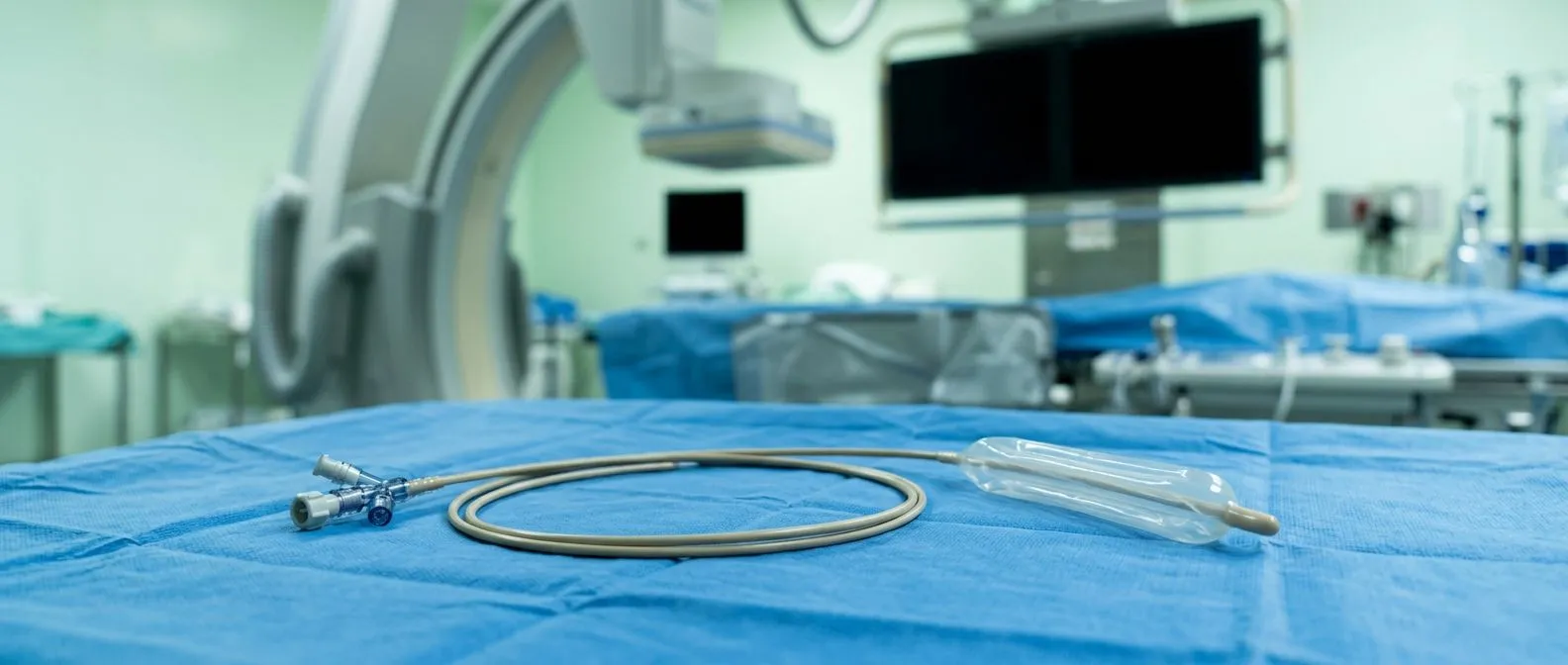
The procedure works by threading a thin, flexible catheter through a small puncture in the groin or wrist, advancing it under X-ray guidance up to the narrowed valve. At the tip of the catheter sits a deflated balloon; once positioned within the valve's fused leaflets, the cardiologist inflates the balloon to crack open the calcium deposits that are holding the valve stiff. This mechanical separation stretches the valve opening and improves blood flow immediately. The technique borrows principles from balloon angioplasty used in coronary arteries, but the target here is the valve itself rather than a blood vessel. Patients are typically sedated but not under general anaesthesia, and the entire procedure usually takes under two hours.
What the valve looks like before and after: Before the procedure, the three leaflets of the aortic valve — the thin flaps that open and close like swinging doors — are fused together by calcium, leaving only a pinhole for blood to pass. After balloon inflation, the leaflets separate along their natural lines of fusion, widening the orifice. The improvement in valve area is measurable and often translates into noticeable relief of symptoms within days. However, the valve remains structurally abnormal; the calcium is redistributed, not removed. This is why balloon aortic valvuloplasty is understood as a temporary measure rather than a permanent fix, and its role must be weighed carefully against definitive valve replacement options such as TAVR or surgical valve replacement.
Who Needs Balloon Valvuloplasty for Severe Aortic Stenosis?
Not every patient with aortic stenosis needs balloon aortic valvuloplasty. The decision hinges on a combination of symptom severity, valve anatomy, overall health, and whether the patient can tolerate a more definitive operation. Severe aortic stenosis is generally defined by a valve area below 1.0 cm², a mean pressure gradient above 40 mmHg, or a peak jet velocity exceeding 4.0 m/s on echocardiography. Once symptoms appear — classically the triad of chest pain, exertional shortness of breath, and syncope — the prognosis without intervention worsens significantly. According to the American Heart Association, symptomatic severe aortic stenosis carries a high mortality risk when left untreated, making timely intervention essential (American Heart Association).
Symptoms that prompt evaluation: Patients often first notice a decline in stamina — stairs that were manageable a year ago now cause breathlessness. Some describe a heavy, squeezing sensation in the chest during exertion that eases with rest. Others experience dizziness or outright fainting when walking uphill or carrying groceries. A careful history and physical examination, including auscultation (listening with a stethoscope) to detect the characteristic harsh, diamond-shaped systolic murmur, guide the cardiologist toward echocardiographic confirmation. It is worth noting that atypical presentations exist; an atypical infarction pattern, where the patient feels minimal chest pain despite significant cardiac stress, can delay diagnosis especially in older adults or those with diabetes.
Patients who are not ideal candidates for valve replacement: Some individuals have medical conditions that make open-heart surgery or even transcatheter valve replacement too risky in the near term. These may include severe frailty, advanced lung disease, kidney failure requiring dialysis, or a combination of factors that pushes their surgical risk score — often calculated using EuroSCORE II — into a prohibitive range. For these patients, balloon aortic valvuloplasty can serve as a palliative option, buying months of improved comfort and mobility. The procedure is also considered for patients who need time to recover from another acute illness before facing a larger operation, a concept explored in more detail in the section on bridge-to-surgery strategies below.
Balloon Aortic Valvuloplasty vs TAVR: Which Is Right for You?
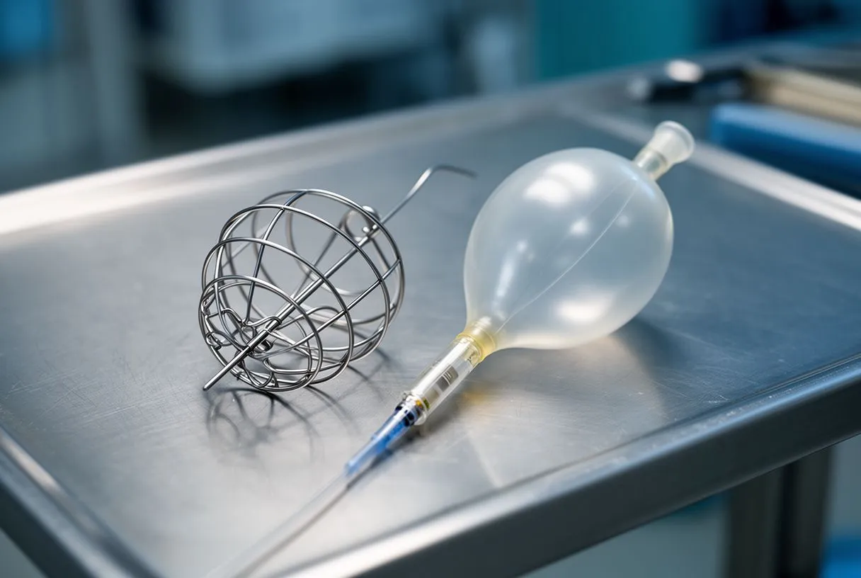
Patients diagnosed with severe aortic stenosis often hear two terms during consultations: balloon aortic valvuloplasty and TAVR (transcatheter aortic valve replacement). While both are catheter-based and avoid opening the chest, they differ fundamentally in what they achieve. Balloon valvuloplasty stretches the existing valve but leaves the calcified leaflets in place; TAVR compresses the old valve out of function and implants a new balloon-expandable valve within the old one, providing a permanent solution. Understanding this distinction is central to making an informed treatment decision.
- Duration of benefit: Balloon valvuloplasty typically provides symptom relief for 6–12 months before restenosis occurs; TAVR offers durable valve function measured in years.
- Procedural complexity: Both use catheter access, but TAVR involves positioning a prosthetic valve — a more technically demanding step that requires a larger delivery sheath and a specialised heart team.
- Resource requirements: TAVR demands a hybrid operating theatre, general or deep sedation anaesthesia, and a multi-disciplinary team; valvuloplasty can often be performed in a standard cardiac catheterisation laboratory under moderate sedation.
- Clinical evidence: Long-term data from the PARTNER trial programme demonstrate superior survival and symptom relief with TAVR compared to medical therapy alone in inoperable patients (European Heart Journal).
When valvuloplasty is preferred despite TAVR availability: There are clinical scenarios where the cardiology team may recommend balloon valvuloplasty over TAVR. In children and young adults with congenital valve narrowing, the anatomy may not accommodate a prosthetic valve, and stretching the native valve can delay the need for surgical replacement for years. In adults with moderate stenosis who require urgent non-cardiac surgery — for example, cancer resection or hip fracture repair — a quick valvuloplasty can reduce cardiac risk during the other operation without committing the patient to a permanent implant. And in patients whose expected survival is limited by another terminal illness, the shorter procedure and lower complexity of valvuloplasty may align better with goals of care focused on comfort rather than longevity.
Balloon Aortic Valvuloplasty Indications in High-Risk Patients
High-risk patients represent the largest group for whom balloon aortic valvuloplasty indications are carefully evaluated. These individuals often carry multiple comorbidities — chronic obstructive pulmonary disease, diabetes with end-organ damage, prior stroke, or advanced kidney disease — that collectively push their operative risk beyond what most cardiac surgeons would accept for elective valve replacement. The EuroSCORE II and STS (Society of Thoracic Surgeons) risk calculators are tools the heart team uses to estimate mortality probability; when the predicted risk exceeds roughly 8–10 percent, alternatives to open surgery become the primary discussion.
Frailty and functional status as decision factors: Beyond numerical risk scores, clinicians assess frailty through measures such as grip strength, gait speed, and the ability to perform activities of daily living independently. A patient who cannot walk five metres without stopping or who has unintentionally lost significant weight over the preceding six months may not tolerate the physiological stress of cardiopulmonary bypass, which is required for surgical valve replacement. In these situations, even TAVR — which avoids bypass but still involves a large catheter and rapid ventricular pacing — may be considered too demanding. Balloon aortic valvuloplasty, with its shorter procedural time and lower hemodynamic burden, becomes a reasonable middle path. The arterial line monitoring and vascular access considerations are similar to a standard catheterisation, making the recovery trajectory gentler.
Patients with concurrent cardiac conditions: Some patients present with severe aortic stenosis alongside other cardiac problems that complicate definitive treatment. For instance, a patient with a heavily calcified ascending aorta — sometimes called porcelain aorta — may be deemed unsuitable for surgical clamping, and TAVR access routes may also be limited by severe peripheral vascular disease. Others may have atrial flutter or other arrhythmias that make the rapid pacing required during TAVR deployment risky. In such layered clinical pictures, the heart team may recommend valvuloplasty as the safest available intervention, accepting its temporary nature in exchange for immediate symptom improvement and lower procedural risk. The patient and family should understand that this is a calculated trade-off, not a substitute for valve replacement.
Pediatric Aortic Valvuloplasty: When Children Need This Procedure
Balloon aortic valvuloplasty has an important and well-established role in pediatric cardiology. Children born with congenital aortic stenosis — typically caused by a bicuspid valve, where the valve has two leaflets instead of the normal three — may develop severe narrowing early in life. In newborns with critical aortic stenosis, the condition can be life-threatening within days of birth because the left ventricle cannot generate enough pressure to supply the body adequately. In these cases, balloon valvuloplasty is often the first-line intervention, performed as an emergency or urgent procedure to restore adequate blood flow.

How the procedure differs in children: The fundamental technique is the same as in adults — a catheter with a balloon is guided to the valve and inflated — but the equipment is miniaturised and the anaesthetic management is adapted for small patients. The balloon diameter is carefully matched to the child's valve annulus size, which is measured precisely on echocardiography before the procedure. Oversizing the balloon risks tearing the delicate leaflets, which would necessitate emergency surgery, while undersizing produces inadequate relief. Paediatric interventional cardiologists are specially trained for this precision work, and outcomes in experienced centres are generally favourable, with many children avoiding or significantly delaying surgical valve replacement into adolescence or adulthood.
Long-term outlook for children after valvuloplasty: Families often ask whether the procedure is a cure. It is not. The bicuspid valve remains structurally abnormal, and over years the child may develop recurrent stenosis, valve leakage (regurgitation), or both. Regular follow-up with echocardiography — every six to twelve months initially — is essential to track valve function and ventricular growth. When the time comes for definitive treatment, the options depend on the child's size, valve anatomy, and whether the aortic root has dilated. Some teenagers eventually undergo the Ross procedure, in which the patient's own pulmonary valve (an autograft) is moved to the aortic position and a donor valve replaces the pulmonary valve. Others receive a tissue or mechanical valve. The goal of paediatric balloon aortic valvuloplasty is to buy time — allowing the child to grow and the heart to develop normally before a larger intervention is needed.
Using Balloon Valvuloplasty as a Bridge to Definitive Surgery
The concept of bridge therapy is well understood in cardiology: a temporary intervention that stabilises a patient until a more definitive treatment can be safely performed. Balloon aortic valvuloplasty serves this role in several clinical scenarios. When a patient presents with severe symptomatic aortic stenosis but also has an acute condition that needs to be addressed first — for example, a hip fracture requiring urgent orthopaedic repair, or an active infection that temporarily increases surgical risk — valvuloplasty can reduce the cardiac burden enough to get the patient through the non-cardiac procedure safely. Once the patient has recovered from the first operation, the cardiac team can plan definitive valve replacement.
Bridging in decompensated patients: Some patients arrive at the cardiology service in a state of acute decompensation — fluid in the lungs, very low blood pressure, or early signs of cardiogenic shock. Operating on a patient in this fragile state carries substantially higher mortality. The heart team may perform balloon aortic valvuloplasty to relieve the valve obstruction quickly, allowing the left ventricle to recover some function over the following days to weeks. During this stabilisation period, intravenous medications, ventilatory support if needed, and careful monitoring in a cardiac intensive care unit help optimise the patient's condition. Once the clinical picture improves — which is not guaranteed but is seen in a meaningful proportion of patients — definitive TAVR or surgical valve replacement becomes feasible. Families should understand that bridging is not always successful; some patients do not improve enough to proceed, and the valvuloplasty becomes a palliative measure.
Timing considerations and realistic expectations: The window between bridge valvuloplasty and definitive treatment is typically measured in weeks to a few months. Restenosis — the re-narrowing of the valve as calcium reaccumulates and the stretched leaflets scar back — can begin as early as two to three months after the procedure. The treating team will plan the next step with this timeline in mind, scheduling the definitive procedure before restenosis undoes the benefit. In patients with additional complications such as infection, the management of sepsis follows standard definitions and protocols that guide when it is safe to proceed with cardiac intervention (King's College London). Open communication between the patient, family, and medical team about the bridging plan — including what happens if the patient does not improve — is essential for informed decision-making.
What Happens During the Balloon Valvuloplasty Procedure?
Understanding the step-by-step experience of balloon aortic valvuloplasty can reduce anxiety for patients and families facing the procedure. On the day of treatment, the patient is taken to the cardiac catheterisation laboratory, a room equipped with X-ray imaging (fluoroscopy), monitoring screens, and sterile instrumentation. After the patient lies on the table, the anaesthesia team administers sedation — usually moderate, meaning the patient is drowsy but breathing independently — and local anaesthetic is injected at the access site, typically the right femoral artery in the groin. In some cases, the radial artery in the wrist may be used, though the larger femoral route is preferred for the catheter sizes required.
Catheter navigation and balloon inflation: Once the artery is accessed, a guidewire is threaded through the aorta and across the narrowed aortic valve into the left ventricle. The balloon catheter is then advanced over the wire and positioned so the balloon sits exactly within the valve opening. The cardiologist confirms position using fluoroscopy and contrast dye injections. The balloon is then inflated — sometimes rapidly, sometimes in controlled stages — to a predetermined diameter matched to the patient's valve size. During inflation, the heart's electrical activity is monitored closely, and brief changes in blood pressure are expected. Most patients feel a sensation of pressure or mild discomfort but not sharp pain. The balloon may be inflated and deflated several times to achieve the desired valve opening, and then the catheter is withdrawn.
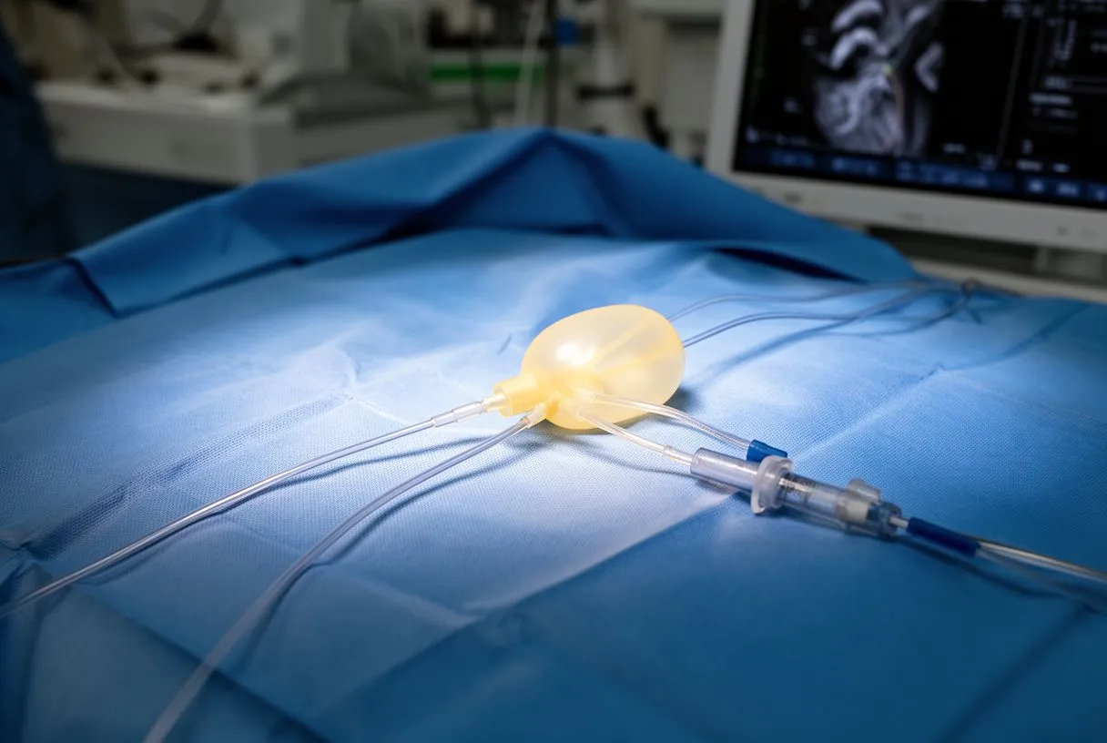
Monitoring and safety measures throughout the procedure: Continuous monitoring includes electrocardiography to detect arrhythmias, arterial blood pressure via the arterial line or the catheter itself, and pulse oximetry to track oxygen levels. If the patient develops significant rhythm disturbances such as transient heart block or ventricular ectopy, these are usually managed immediately with medications or pacing. The contrast dye used for imaging carries a small risk of kidney injury, particularly in patients with pre-existing renal impairment, so the volume is minimised and hydration is maintained intravenously. At the end of the procedure, the arterial puncture is closed with a specialised closure device or manual compression, and the patient is transferred to a recovery area for observation.
Recovery After Balloon Valvuloplasty: What to Expect
Recovery from balloon aortic valvuloplasty is generally faster than from surgical valve operations, but it still requires careful monitoring and realistic expectations. Most patients stay in hospital for one to three days after the procedure. During the first few hours, the nursing team checks the access site for bleeding or haematoma, monitors blood pressure and heart rhythm, and ensures the patient can tolerate oral fluids. Pain is usually minimal and managed with simple analgesics; many patients are up walking with assistance within six to eight hours. Medications such as aspirin are typically continued or started to reduce the risk of clot formation on the stretched valve leaflets, and any pre-existing cardiac medications are reviewed and adjusted.
Symptom improvement timeline: Many patients notice improved breathing and exercise capacity within the first week. The immediate mechanical effect of widening the valve orifice reduces the pressure load on the left ventricle, and the heart begins to function more efficiently. However, full functional recovery depends on how long the ventricle has been under strain and whether there is irreversible muscle damage. Patients who have had severe stenosis for years may have significant left ventricular hypertrophy (thickening of the heart muscle) that takes months to partially regress. Follow-up echocardiography is scheduled within a few weeks to assess the new valve area, gradient, and degree of residual regurgitation, and then at regular intervals to detect restenosis early.
Activity and lifestyle after discharge: Patients are advised to avoid heavy lifting and strenuous exercise for about one week to allow the arterial access site to heal fully. Walking and light household activities are encouraged. Driving should be avoided for several days or as advised by the treating physician. For patients travelling for treatment, the medical team will usually provide clearance for air travel after the first follow-up assessment confirms stable valve function and a well-healed access site. It is important to carry a discharge summary and procedure notes when returning home, so the local cardiologist can continue monitoring seamlessly.
Risks and Limitations of Balloon Valvuloplasty
While balloon aortic valvuloplasty is less invasive than open surgery, it is not without risks, and patients must weigh these carefully against the expected benefits. The most common complications relate to the arterial access site: bleeding, bruising, or in rare cases, pseudoaneurysm formation (a contained leak from the artery wall). Vascular complications are more frequent in elderly patients with calcified or tortuous arteries, and the choice of access route is tailored to each patient's anatomy. The interventional cardiologist reviews the iliofemoral vessels on pre-procedure CT angiography to select the safest entry point.
- Valve regurgitation: Over-aggressive balloon inflation can tear the leaflets, causing or worsening aortic regurgitation — blood leaking backwards into the ventricle with each heartbeat.
- Arrhythmias: Catheter manipulation within the heart can trigger transient arrhythmias, most of which resolve spontaneously but occasionally require medication or temporary pacing.
- Stroke risk: Dislodgement of calcium fragments during balloon inflation can travel to the brain; embolic protection devices are sometimes used to reduce this risk.
- Restenosis: The valve typically re-narrows within 6–12 months as scar tissue and calcium reform, making valvuloplasty a temporary rather than permanent solution.
Why valvuloplasty is not a standalone cure: The fundamental limitation of balloon aortic valvuloplasty is that it addresses the mechanical narrowing without removing the underlying calcium or restoring normal valve architecture. The stretched leaflets remain stiff and calcified, and the biological processes that caused the stenosis in the first place — calcium deposition, inflammation, and fibrosis — continue. This is why valvuloplasty is not recommended as a definitive treatment for adults who are candidates for valve replacement. In the elderly population, where atheromatous plaque and systemic vascular disease often coexist with aortic stenosis, the valve pathology is part of a broader cardiovascular risk profile that requires ongoing medical management, including statin therapy, blood pressure control, and attention to conditions such as atrial fibrillation. The valvuloplasty improves the valve; it does not cure the patient. Understanding this distinction helps set realistic expectations and reinforces the importance of continued cardiology follow-up.
When to Ask Your Cardiac Team About Balloon Valvuloplasty
Deciding whether balloon aortic valvuloplasty is the right intervention requires a detailed conversation with a qualified cardiac team. If you or a loved one has been diagnosed with severe aortic stenosis and are experiencing symptoms — breathlessness on exertion, chest discomfort, dizziness, or reduced exercise capacity — it is important to understand all available treatment paths. Balloon valvuloplasty may be appropriate if surgical valve replacement or TAVR is not immediately feasible due to medical conditions, if a temporary improvement is needed to bridge another procedure, or in pediatric cases of congenital valve narrowing. The only way to know with certainty is through a comprehensive evaluation including echocardiography, CT imaging, and a multidisciplinary heart team review.
Questions to bring to your consultation: When meeting with your cardiologist, consider asking: What is my estimated surgical risk score, and how does it influence my treatment options? Is my valve anatomy suitable for balloon valvuloplasty, or would TAVR be a better catheter-based approach? How long can I expect the benefit to last, and what is the plan if restenosis occurs? What are the specific risks for my case, given my other medical conditions? Are there any non-cardiac procedures I need that could be made safer by improving my valve first? These questions help frame the discussion around your individual circumstances rather than generic guidelines. The heart team should be able to explain the rationale for any recommendation in terms you can understand, including the thresholds and criteria they used to reach their conclusion.
We encourage you to discuss your specific situation with a qualified cardiac team before making any decisions. Every patient's anatomy, medical history, and personal goals are unique, and treatment recommendations should reflect that individuality. If you would like to explore whether balloon aortic valvuloplasty or another valve intervention might be appropriate for you or a family member, we invite you to book a consultation with Dr Alejandro Molina and the team at Cirugía de Corazón. With clinics in Guadalajara, Querétaro, and Hermosillo, we provide detailed evaluations, second opinions, and personalised treatment planning for patients from across Mexico and abroad. Do not self-diagnose based on this information — let an experienced cardiac specialist assess your condition and guide you toward the option with the lowest risk for your situation.






