

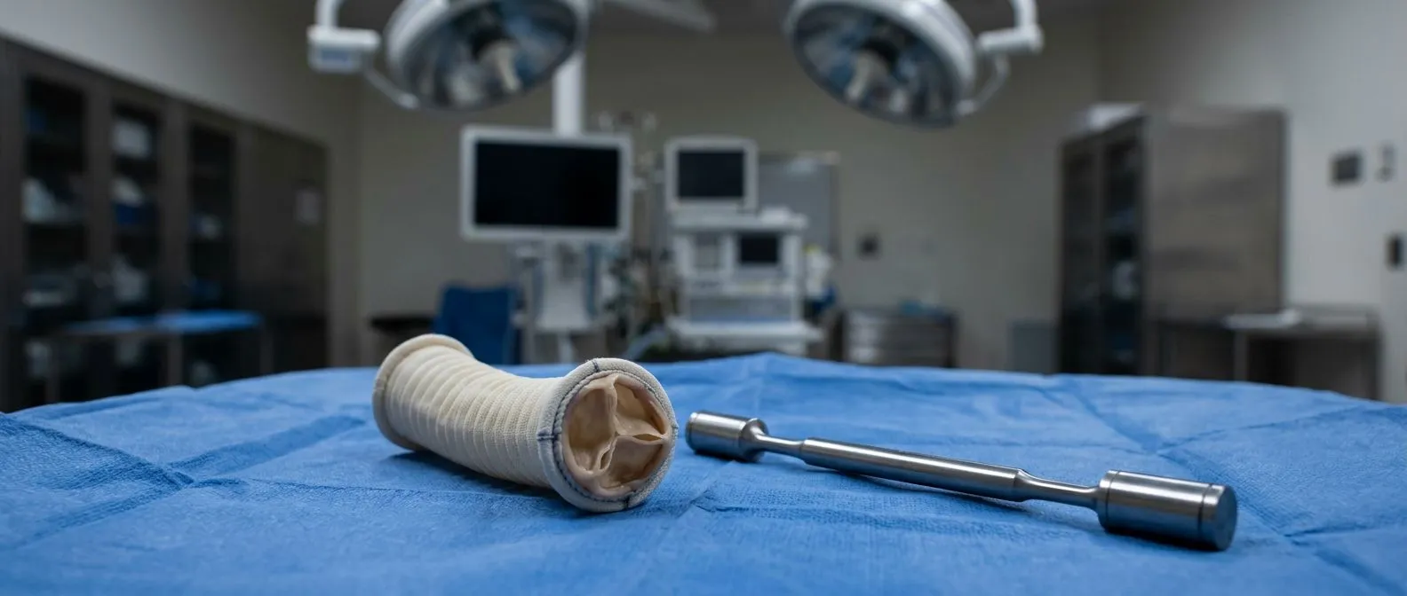
Bicuspid Aortic Valve: Monitoring and When to Operate
Bicuspid aortic valve surgery: learn when to operate versus monitor from Guadalajara heart specialists.
The bicuspid aortic valve is the most common congenital heart defect, affecting roughly 1 to 2 percent of the population. Unlike a normal aortic valve with three leaflets that open and close in precise coordination, a bicuspid valve has only two. From the outside, many people live for decades without knowing they carry this difference. Yet inside the heart, that altered architecture changes how blood flows through the ascending aorta and changes the forces acting on the valve itself. Over years, this can lead to narrowing, leaking, or dangerous enlargement of the aorta. Understanding when to choose bicuspid aortic valve surgery versus careful monitoring can shape the trajectory of your life.
This guide explains what the condition means anatomically, how we track it, and the clinical thresholds that prompt us to recommend intervention.
What Is a Bicuspid Aortic Valve and Why It Matters
A normal aortic valve sits between the left ventricle and the aorta, the body's main arterial highway. Its three leaflets, or cusps, open during systole to eject oxygenated blood upward into the ascending aorta, then snap shut during diastole to prevent backward flow. In a bicuspid aortic valve, two of these leaflets are fused from birth, leaving two functional cusps instead of three. This seemingly small change disrupts laminar flow patterns.
Fusion patterns predict your risk profile. The most common configuration fuses the right and left coronary cusps, though right-noncoronary fusion also occurs. These patterns influence not only how the valve deteriorates but also how the ascending aorta expands over time. The abnormal jet of blood striking the aortic wall creates shear stress in predictable locations, which explains why bicuspid patients develop atheromatous plaque changes and elastin degradation in their aorta at younger ages than the general population. Research published in Atlantis Press documents unique patterns of elastin breakdown in the ascending aorta specific to bicuspid patients, distinct from the degenerative changes seen with tricuspid valves.
The condition travels in families. If one parent carries a bicuspid aortic valve, first-degree relatives should undergo screening echocardiography. Early detection matters because it permits structured monitoring before complications arise.
How Bicuspid Aortic Valve Surgery Works
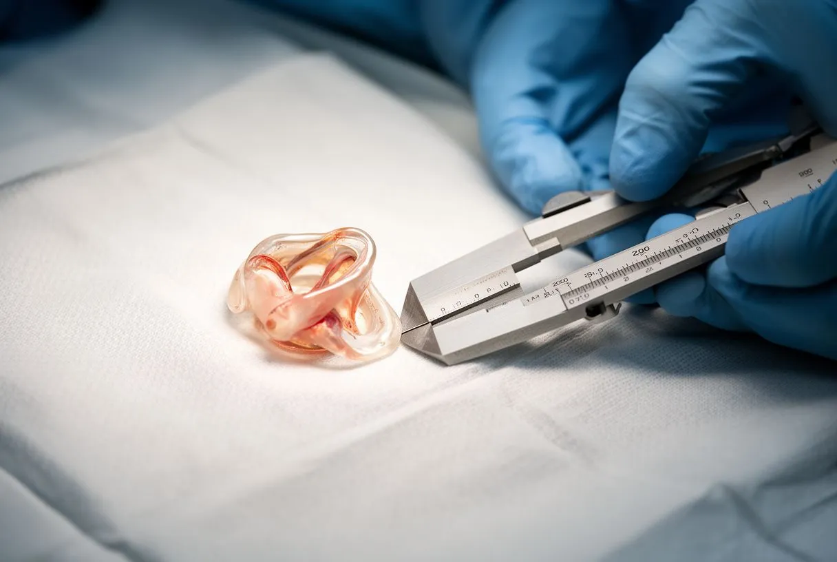
When we discuss bicuspid aortic valve surgery, we are describing procedures that either repair your native valve or replace it with a biological or mechanical substitute. The choice depends on valve anatomy, patient age, associated aortic disease, and individual priorities around anticoagulation and future reintervention.
Valve repair preserves your own tissue when feasible. Surgeons reshape the leaflets, reconstruct the commissures where the leaflets meet, and sometimes perform aortic annuloplasty to stabilize the valve ring. Repair avoids prosthetic material and its attendant risks. However, bicuspid valves present challenges: the fused leaflet often has abnormal tissue quality, and the geometric distortions may not be fully correctable. Repair rates are lower than for tricuspid regurgitation, but experienced centers achieve durable results in selected patients.
Replacement becomes necessary when repair cannot restore adequate function. Bioprosthetic valves, typically fashioned from bovine or porcine tissue mounted on a stent frame, avoid lifelong anticoagulation but wear out over 10 to 20 years. Mechanical valves offer exceptional durability but require warfarin anticoagulation with its bleeding and lifestyle implications. For younger patients, the choice involves projecting decades of cumulative risk from anticoagulation against the probability of future reoperation.
Concomitant aortic replacement adds complexity. When the ascending aorta measures above guideline thresholds or shows rapid growth, surgeons replace the diseased segment with a tubular graft, sometimes reimplanting the coronary arteries into the graft (Bentall procedure) or sparing the aortic root if tissue quality permits.
What Are the Signs You Need Bicuspid Valve Surgery
Symptoms in bicuspid aortic valve disease develop gradually, often camouflaged by slow adaptation. Patients may unconsciously restrict their activities, blaming age or fitness rather than recognizing cardiac limitation. We teach our patients to watch for specific patterns.
Exertional symptoms signal hemodynamic compromise. Shortness of breath with ordinary activity, chest pressure with effort, or unexplained fatigue during tasks previously managed easily suggests the valve has narrowed critically or begun leaking severely. Syncope, or fainting, represents advanced obstruction and warrants urgent evaluation. Nocturnal dyspnea—waking breathless after lying flat—indicates elevated left-sided pressures.
Objective findings on examination guide us before symptoms emerge. Auscultation reveals the characteristic ejection click and systolic murmur. Echocardiography quantifies gradients across the valve, measures regurgitant volume, and assesses left ventricular function. We operate based on these measurements, not symptom presence alone, because waiting for symptomatic deterioration risks irreversible myocardial damage.
The Cleveland Clinic emphasizes that asymptomatic patients with severe stenosis and reduced exercise tolerance or progressive left ventricular dysfunction should undergo intervention. Similarly, severe regurgitation with evidence of chamber enlargement or declining ejection fraction prompts surgical referral even before frank symptoms appear.

Types of Bicuspid Aortic Valve Repair and Replacement
Surgical approaches for bicuspid aortic valve disease have expanded considerably, giving patients options matched to their anatomy and life circumstances. Understanding these distinctions empowers informed shared decision-making.
Conventional open surgery through median sternotomy remains the gold standard for complex cases. This approach provides panoramic exposure of the ascending aorta, aortic root, and cardiac chambers. It permits any combination of valve replacement, root replacement, arch intervention, and coronary reimplantation. For patients with extensive aortic disease or prior cardiac surgery, this access offers the operative control that translates to safety.
Minimally invasive alternatives reduce surgical trauma in selected patients. Upper hemisternotomy, right anterior minithoracotomy, and robotic-assisted approaches all achieve valve access through smaller incisions. These techniques may reduce pain, transfusion requirements, and recovery time. However, they demand specialized expertise and exclude patients with certain anatomic variants or concomitant conditions requiring broader exposure. The decision between minimally invasive and conventional approach depends on surgeon experience, institutional volume, and individual patient factors—not on marketing claims of superiority.
Transcatheter aortic valve replacement, or TAVR, has transformed care for inoperable and high-risk patients with tricuspid stenosis. Its role in bicuspid anatomy remains more selective. The oval annulus, asymmetric leaflets, and frequent aortopathy create technical challenges. Dedicated bicuspid TAVR valves and growing operator experience are expanding indications, but younger surgical candidates generally still receive conventional repair or replacement.
Bicuspid Valve Aneurysm Risk: When to Intervene on the Aorta
The bicuspid aortic valve and aortopathy are inseparable considerations. Even when the valve functions adequately, the abnormal flow dynamics progressively damage the aortic wall. This creates a monitoring imperative separate from valve surveillance.
Guideline thresholds for aortic intervention in bicuspid patients balance rupture risk against operative risk. Current consensus recommends prophylactic ascending aortic replacement when diameter exceeds 5.5 centimeters, or 5.0 cm with additional risk factors such as family history of dissection, rapid growth exceeding 0.5 cm annually, or coarctation. Some centers use indexed thresholds adjusting for patient body size. The ARISTOTLE trial and subsequent registry data have refined our understanding of these risk strata.
The accelerated elastin degradation documented in bicuspid aortas explains why these enlargement thresholds sit lower than for tricuspid valve patients. The wall weakens structurally rather than simply stretching. When we replace the ascending aorta electively, we eliminate this catastrophic risk before it manifests. Emergency surgery for acute dissection carries mortality rates that dwarf those of scheduled intervention.
Patients should understand that their aortic surveillance continues even after valve surgery. If the aorta was not replaced and remains above 4.0 cm, periodic imaging with CT angiography or magnetic resonance angiography continues indefinitely. Coordination between your cardiologist and surgeon ensures nothing falls through follow-up gaps.
Bicuspid Aortic Valve Monitoring Schedule for Stable Patients
Not every bicuspid aortic valve requires immediate surgery. Many patients, particularly those with mild disease and normal aortic dimensions, thrive for years with structured monitoring. The key lies in appropriate surveillance frequency and knowing what triggers escalation.
Typical monitoring intervals follow valve severity and aortic size:
- Mild stenosis or regurgitation, aorta <4.0 cm: Clinical evaluation and echocardiography every 3 to 5 years.
- Moderate disease, or aorta 4.0–4.5 cm: Annual echocardiography with periodic CT or MRI aortic assessment.
- Severe disease without surgical indications, or aorta 4.5–5.0 cm: Every 6 to 12 months with multimodal imaging.
Beyond imaging, patient self-monitoring matters. Report new exercise intolerance, chest symptoms, or any neurologic events promptly. Blood pressure control represents the single most impactful medical intervention—hypertension accelerates both valve deterioration and aortic expansion. Blood pressure is usually targeted below 130/80 mmHg in most patients, though individual goals vary.
Aspirin for atheroprotection and statin therapy for lipid management are standard adjuncts, though neither reverses valvular disease. Endocarditis prophylaxis is reserved for specific high-risk circumstances rather than all bicuspid patients, per current guideline evolution.
Minimally Invasive Bicuspid Surgery Benefits and Trade-Offs
The appeal of smaller incisions is intuitive. Patients understandably prefer reduced pain, faster return to work, and smaller scars. Minimally invasive bicuspid surgery can deliver these benefits, but candidacy requires careful selection.
Technical constraints limit applicability. The smaller operative field challenges visualization of the ascending aorta and aortic root. Concomitant procedures—such as maze ablation for atrial flutter, septal myectomy, or extensive aortic work—may be unsafe or impossible through limited access. Prior cardiac surgery with adhesions typically mandates reoperative sternotomy. Body habitus, with obesity or barrel chest deformity, can further restrict instrument maneuverability.
Outcomes data support equivalence in experienced hands for straightforward cases. When valve replacement alone is needed and the aorta requires no intervention, minithoracotomy or hemisternotomy approaches achieve comparable mortality and morbidity to full sternotomy. The operative times may be longer initially during a surgeon's learning curve, but high-volume centers normalize this difference. What matters most is not incision size but the completeness and durability of the repair or replacement performed.
Patients considering minimally invasive approaches should ask specific questions: How many of these procedures has my surgeon performed annually? What is their conversion rate to full sternotomy? What happens if the aorta proves more diseased than imaging suggested? These discussions reveal whether enthusiasm for minimalism serves your interests or the marketing department's.

Bicuspid Valve Regurgitation Versus Stenosis Treatment Paths
The bicuspid valve deteriorates in two primary patterns: it narrows, it leaks, or both. Stenosis and regurgitation impose different hemodynamic burdens and sometimes favor different therapeutic strategies.
Stenosis develops from leaflet thickening and calcification, restricting forward flow. The left ventricle generates higher pressures to eject blood, leading to concentric hypertrophy—thickening of the muscular walls. This initially compensates but eventually causes diastolic dysfunction, reduced coronary perfusion during hypertrophy, and if unchecked, systolic failure. Balloon aortic valvuloplasty offers temporary palliation in children or as a bridge in adults, but restenosis is nearly universal. Definitive treatment requires surgical valve replacement or, in carefully selected cases, TAVR.
Regurgitation allows backward flow during diastole, volume-overloading the ventricle. The chamber dilates progressively, sometimes massively, while ejection fraction remains deceptively normal until late in the disease. Unlike stenosis, where symptoms correlate better with severity, regurgitation can cause irreversible ventricular damage before patients feel limited. We therefore intervene earlier in regurgitation, often when left ventricular end-systolic dimension exceeds 50 mm or ejection fraction declines below 55 percent, even without symptoms.
Mixed disease combines both lesions and requires integrated assessment. The thresholds for intervention blend considerations from both paradigms. An arterial line during catheterization, alongside echocardiographic data, clarifies hemodynamics when noninvasive studies disagree.
Heart Valve Surgery Recovery: What Patients Actually Experience
Recovery from bicuspid aortic valve surgery unfolds in phases, each with distinct milestones and frustrations. Realistic expectations prevent the discouragement that accompanies expecting linear progress.
The immediate postoperative period centers on physiologic stabilization. After median sternotomy, patients spend 24 to 48 hours in intensive care with an arterial cannula for pressure monitoring, central venous access, and often temporary pacing wires. Autologous blood recovery systems or patient-preference transfusion protocols minimize allogeneic exposure. Pain is manageable with multimodal regimens; the stereotype of unbearable thoracotomy pain reflects outdated practice. Ambulation begins within 24 hours, and most patients transfer to a step-down unit on the second or third day.
Early convalescence at home requires patience with variable energy. Sternotomy healing takes 6 to 8 weeks for bony union, though discomfort diminishes earlier. Many patients experience emotional lability, sleep disruption, and appetite changes—normal responses to physiologic stress and medication adjustments. We counsel families to expect good days and difficult days without interpreting each fluctuation as complication.
Return to function extends over months. Cardiac rehabilitation begins 2 to 4 weeks post-discharge, progressing from monitored exercise to independent activity. Driving resumption typically waits 4 to 6 weeks. Air travel is feasible after 2 to 3 weeks with proper hydration and mobilization. Full sternal healing permits unrestricted lifting at 3 months. Biological valve patients avoid anticoagulation; mechanical valve patients begin lifelong warfarin with regular INR monitoring.
Bicuspid Valve Life Expectancy After Repair or Replacement
The fundamental question patients ask—how long will I live, and how well?—deserves honest framing without false precision. Individual prognosis depends on age at surgery, ventricular function, comorbidities, and valve type.
Operative mortality for isolated aortic valve replacement in low-risk patients sits below 2 percent in experienced centers. This risk rises with emergency status, reoperation, concomitant coronary disease requiring arterial grafting, or advanced age. EuroSCORE II and similar risk models help quantify individual risk for informed consent, though no model perfectly predicts individual outcomes.
Long-term survival after successful surgery approximates age-matched populations without valve disease, particularly when intervention occurs before significant ventricular dysfunction. Atypical infarction patterns from coronary ostial abnormalities, if present, may influence late outcomes and warrant specific surveillance. Bioprosthetic valve degeneration creates a predictable need for reintervention, which increasingly occurs via transcatheter valve-in-valve procedures rather than redo surgery.
The automated external defibrillator placement for sudden death prevention is rarely needed postoperatively unless severe ventricular dysfunction persists. Most patients resume full occupational and recreational activities, with quality of life metrics normalizing within 6 to 12 months. The key variable is timing—earlier intervention, before irreversible myocardial changes, preserves the longevity that bicuspid aortic valve surgery aims to secure.
If you carry a bicuspid aortic valve, whether newly diagnosed or under years of observation, the path forward merits personalized discussion. Our cardiac teams in Guadalajara, Querétaro, and Hermosillo evaluate your valve mechanics, aortic dimensions, symptoms, and life priorities to recommend monitoring intervals or surgical timing tailored to your situation. Rather than navigating uncertain thresholds alone, schedule a consultation to review your imaging and explore what approach offers the lowest risk for your specific anatomy and circumstances.






