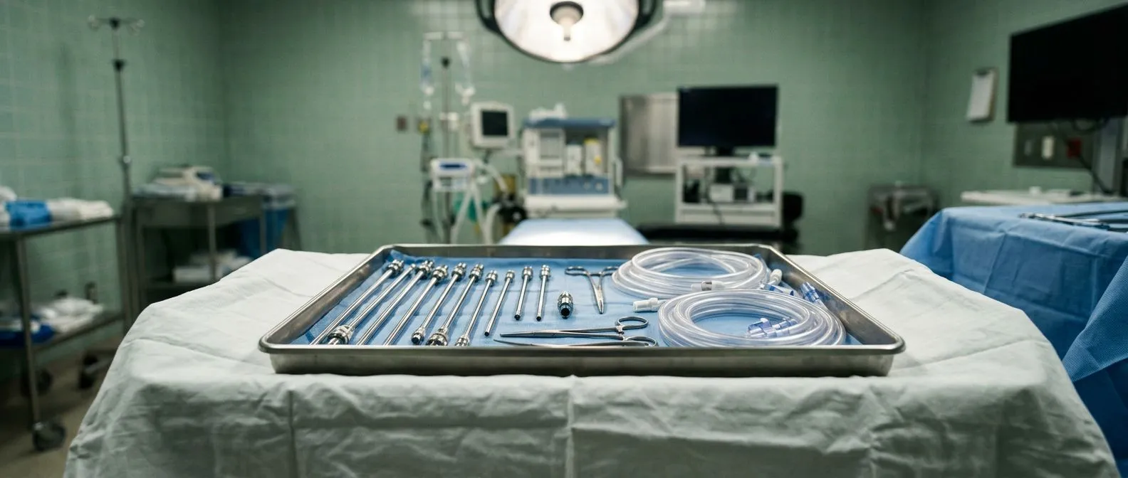
Cannulation in Heart Surgery: Connecting to Bypass
Cannulation heart surgery connects you to heart-lung bypass safely.
If you or a loved one is facing open heart surgery, one of the first steps in the operating room is something you will never feel or see: cannulation in heart surgery. It is the moment the surgical team connects your circulation to the heart-lung machine so your heart can be paused and repaired. Understanding what happens during this step, why it matters, and what the team is checking can take away much of the mystery — and much of the fear — around bypass surgery.
What Is the Cannulation Procedure in Heart Surgery?
Cannulation means placing a tube — a cannula — into a blood vessel so blood can be redirected, collected, or returned. In everyday hospital care, cannulation usually means a small intravenous line in your arm. In cardiac surgery, the word means something much bigger: large cannulae placed into the aorta and the venous system so the entire circulation can be temporarily taken over by a machine.
During cardiopulmonary bypass, the heart-lung machine does the work of both organs. It drains deoxygenated blood from your body, adds oxygen, removes carbon dioxide, and pumps the refreshed blood back into your arterial system. Cannulation is the physical connection between you and that machine. Without it, there is no bypass.
In our team, every cannulation plan is reviewed by the multidisciplinary group against international practice guidelines before the operation, and the plan is then explained to the patient in plain words as part of the surgical proposal. The cannulation strategy is tailored to your anatomy, your disease, and the operation being planned — it is never a one-size-fits-all step.
Why Do We Do Cannulation in Heart Surgery?
The reason is simple: most operations inside the heart cannot be done while the heart is beating and full of blood. To replace a valve, close a hole, or perform Coronary Artery Bypass Grafting on small vessels, the surgeon needs a still, bloodless field. Cannulation makes that possible by diverting blood away from the heart so it can be stopped safely.
It protects the brain and organs. While you are on bypass, the machine maintains blood pressure, oxygen delivery, and temperature control for the whole body. The perfusion team continuously monitors flow and pressures so your kidneys, brain, and other organs are protected throughout the operation.
It buys the surgeon time. Once on bypass, the heart can be stopped with a cardioplegia solution — a protective fluid that quiets the heart muscle. This gives the surgeon a calm, motionless field to work in, and the heart is restarted when the repair is complete, a moment known as Weaning From Bypass.
So when people ask why cannulation is done, the honest answer is: it is what makes open heart surgery safe and possible at all.
Which Sites Are Used for Cannulation in Heart Surgery?
Heart surgery cannulation sites are chosen for each operation. Most conventional On-pump Bypass surgery uses two main connections: one arterial and one or more venous.
The aorta is the usual arterial site. In standard aortic cannulation heart surgery, a tube is placed in the ascending aorta, just above where the coronary arteries branch off. This is the "outflow" cannula through which the oxygenated blood returns to your body.
The venous side has several options. Blood can be drained by gravity and suction from the right atrium through a single large cannula, or — for operations inside the heart — through two separate cannulae, one in the superior vena cava and one in the inferior vena cava. This second approach is called bicaval cannulation heart surgery and is the usual choice for valve and congenital work.
Other sites appear in specific situations: the femoral vessels at the groin (Femoral Cannulation), the axillary artery below the collarbone, or the pulmonary artery. The choice depends on the operation, the state of the aorta, and whether a minimally invasive approach is planned.
How Painful Is Cannulation in Heart Surgery?
For open heart surgery, the answer is reassuring: you are under general anesthesia and completely asleep. The surgical team places the cannulae after you are unconscious, so you feel nothing during the process. Patients often worry about this step, but it is entirely covered by anesthesia.
The discomfort you may remember afterward comes from the surgical incision, not from the cannulation itself. The cannulation sites — usually the aorta and right atrium — are inside the chest and are closed carefully at the end of the operation. Groin puncture sites, when femoral access is used, may feel bruised for a few days, like a deep injection.
We always walk patients through the sequence of the operation beforehand — including exactly when they will be asleep — because we find that knowing the order of events lowers anxiety more than any reassurance alone.
Is Cannulation the Same as Phlebotomy?
It is a common mix-up, and the two words describe very different things. Phlebotomy is simply drawing blood out of a vein, usually with a needle for a lab test or a donation. Cannulation means leaving a tube in the vessel, whether for a few hours of medication, several days of monitoring, or — in cardiac surgery — an entire operation on the heart-lung machine.
So while every cannulation involves entering a vessel like phlebotomy does, phlebotomy alone is not cannulation. An IV cannula in your forearm is a small-scale cannula; the aortic cannula used in bypass is a tube the width of a finger that carries your entire cardiac output. Same principle, completely different scale and purpose.
Understanding the difference helps when reading your operative report. If it mentions "aortic cannulation" or "bicaval venous cannulation," it refers to the connections made for the bypass machine — not to the needle used for your blood tests.
How Does the Cannulation Heart Lung Machine Connection Work?
Once the cannulae are in place and secure, they are connected to tubing that leads to the heart-lung machine. The venous line carries blood away from your body into the reservoir of the machine. From there, blood passes through the oxygenator — the artificial lung — where it takes up oxygen and releases carbon dioxide. A pump then drives it back through the arterial cannula into your aorta.
The machine takes over gradually. The perfusionist — the specialist who runs the pump — increases the bypass flow while the heart continues to beat, then the heart is slowed and stopped with cardioplegia. During this phase, the term Bypass Time begins: the clock that measures how long your circulation is supported by the machine.
When the repair is finished, the process reverses. The heart is filled, restarted, and supported as the pump flow is slowly reduced until your own heart is doing the work again. The cannulae are removed, and the entry sites are repaired. The whole circuit is a temporary bridge — your heart and lungs take over again as soon as they safely can.
Minimally Invasive Heart Surgery Cannulation Options
Not every operation needs a full sternotomy — the classic breastbone incision. When a minimally invasive approach is used, the chest is opened through smaller incisions, and cannulation is adapted to match. Often the arterial and venous cannulae are introduced through the groin or the side of the chest rather than through the main incision.
Femoral Cannulation is the workhorse here. The femoral artery and vein at the groin provide access to the circulation without touching the chest at all. Perfusion is established peripherally, and the surgeon works through a small right-chest incision, for example in robotic mitral valve repair.
Alternative arterial sites protect a diseased aorta. If the aorta is heavily calcified or diseased — a key issue in cannulation strategy for aortic surgery — the team may prefer the axillary artery, then route the flow to the whole body through a graft. This avoids squeezing debris out of a fragile aorta into the brain's circulation.
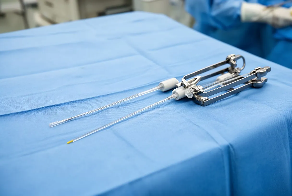
These strategies are chosen case by case. Our team performs conventional, minimally invasive, robotic and transcatheter procedures, and we recommend the less invasive route only where the evidence supports it for that specific patient — the cannulation plan follows that decision, not the other way around.
What Are the Cannulation Risks Heart Surgery Patients Should Know?
Cannulation is a routine and highly standardized part of cardiac surgery, but it carries specific risks worth understanding honestly. Being informed is not the same as being alarmed — these events are uncommon and actively managed.
- Stroke and embolism: debris from a calcified aorta can be dislodged during aortic cannulation and travel to the brain. Teams reduce this risk with imaging of the aorta, careful site selection, and alternative cannulation routes when needed.
- Bleeding and vessel injury: any large cannula in a major vessel can cause bleeding or dissection. Placement is performed under direct vision and confirmed with pressure monitoring.
- Arrhythmia after surgery: postoperative atrial fibrillation — an irregular rhythm — is a known complication after Coronary Artery Bypass Grafting, and research continues to clarify how bypass factors such as cannulation and inflammation contribute to it.
- Perfusion-related issues: malposition of a cannula can reduce flow to part of the body. Continuous monitoring of pressures and oxygen levels catches this early.
Rare local complications have also been described in the literature, such as subcutaneous emphysema — trapped air under the skin — after off-pump procedures, reported in published surgical case reports. The overall message: every element of the bypass connection is monitored continuously, precisely because the risks are known. Guidelines from organizations such as the Centers for Disease Control and Prevention frame the broader safety standards around surgical-site and device-related care that protect patients through these invasive steps.
Redo Heart Surgery Cannulation Strategies
When someone needs a second or third heart operation, everything is more delicate — including cannulation. Scar tissue can make the aorta and right atrium difficult or unsafe to touch, so the team plans a different route from the start.
Femoral access is often the answer. Establishing perfusion through the groin before opening the chest gives the team a safety net: if the heart or a great vessel is injured during re-entry, the circulation is already supported. This is one of the most important safety habits in redo surgery.
Imaging guides every decision. Before redo operations, CT scans of the aorta and chest are reviewed to map scar tissue, previous graft locations, and calcification. In our consultations we take the time to explain what these studies actually show, in plain words, so patients understand why the surgical plan — including where the cannulae will go — is what it is.
Some patients may also be candidates for an Off-pump Bypass approach, where surgery proceeds without the heart-lung machine and therefore without full cannulation, though this depends on the anatomy and the operation required.
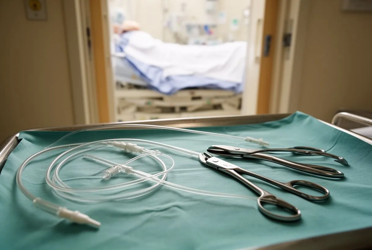
What to Expect and How to Choose Your Team
As a patient, what you will actually experience around cannulation is simple: you arrive, you go to sleep, and the next thing you know, the operation is underway. The technical work — choosing sites, placing cannulae, running the pump — happens entirely on your behalf while you rest. Recovery afterward focuses on the chest incision, breathing exercises, and gradually rebuilding strength over weeks, not on the cannulation sites themselves.
When choosing where to have heart surgery, ask questions that matter: How will you decide my cannulation strategy? Do you review the aorta before operating? What is your approach if my aorta is diseased or this is a redo operation? A good team welcomes these questions and answers them clearly.
Second opinions are a normal part of deciding. We encourage second opinions and treat seeking one as a normal, healthy step in deciding about heart surgery — a confident recommendation stands up to scrutiny. Every case deserves a proposal that explains the recommended operation, the alternatives, and the reason behind each choice.
Start with a conversation, not a commitment. Our first contact is a free video consultation: we get to know you, talk through your condition, and identify which studies are genuinely still needed, so no unnecessary tests are ordered. A face-to-face visit at our consulting rooms in Guadalajara, Querétaro or Hermosillo follows only if you decide to go ahead.

Cannulation in heart surgery is the quiet, precise step that connects you to the technology that makes your operation possible — and it reflects the care and planning of the whole team behind it. Every heart, every anatomy, and every operation deserves a plan built for that person. If you are considering heart surgery for yourself or a family member, the most valuable next step is a conversation with a qualified cardiac team who can review your specific situation. Book a consultation, bring your questions, and make the decision with clear, honest information in hand.