Cardiac Stress Test: Types, Preparation and Results
Cardiac stress test preparation guide covers types, what to expect, and how to read results.
What Is a Cardiac Stress Test?
A cardiac stress test is a non-invasive diagnostic tool that measures how well your heart performs under increased physical demand. When you exercise on a treadmill or stationary bicycle, your heart must pump harder and faster to deliver oxygen to your muscles. By monitoring your heart's electrical activity, blood pressure, and sometimes blood flow patterns during this exertion, your cardiologist can detect problems that remain hidden when you are at rest. Many heart conditions only reveal themselves under stress, which is exactly why this test exists.
Why your doctor recommends one: Your physician may suggest this test if you have been experiencing symptoms such as chest tightness, shortness of breath, unexplained Arm Pain, or fatigue during physical activity. It is also commonly used to evaluate how well a previous heart procedure is working, to assess fitness before surgery, or to monitor known coronary artery disease. The test gives your cardiac team a window into how your heart muscle responds when it is asked to work at its hardest, revealing whether blood supply through the coronary arteries is adequate or compromised.
What the test can and cannot show: A cardiac stress test is excellent at identifying whether reduced blood flow to the heart muscle is likely causing your symptoms. It can help confirm whether an Atheromatous Plaque — a fatty deposit in an artery wall — is restricting flow enough to matter. However, the test alone does not show the exact location or severity of a blockage. If results suggest a problem, your doctor may recommend further imaging or a direct look inside the arteries. It is a screening and stratification tool, not a definitive anatomical map of your coronary vessels. You can find peer-reviewed clinical literature on stress testing methodologies through the Directory of Open Access Journals.
Cardiac Stress Test Preparation Guide
Proper cardiac stress test preparation makes the difference between a smooth experience and one that needs to be rescheduled. Most of the preparation is common sense, but a few specific instructions matter greatly. You will typically be asked to avoid eating or drinking for three to four hours before the appointment, though sips of water are almost always permitted. Eating a heavy meal before the test means your body diverts blood to digestion rather than to exercising muscles, which can skew results and leave you feeling nauseated on the treadmill.
Medications to discuss with your doctor: Some medications interfere with the accuracy of a stress test. Beta-blockers, for example, keep your heart rate from rising during exercise, which can mask signs of reduced blood flow. Your cardiologist will tell you which pills to take normally and which to hold the morning of the test. Never stop any heart medication on your own. If you take Aspirin for cardiovascular protection, ask whether you should continue it that day, as instructions vary depending on the type of test planned and your overall clinical picture.
What to wear and bring: Dress as if you are going for a brisk walk. Wear comfortable, loose-fitting clothing and rubber-soled shoes suitable for exercise — running shoes are ideal. Avoid lotions or oils on your chest area, as they prevent the electrocardiogram (ECG) stickers — called electrodes — from adhering properly. Bring a list of all your medications, your insurance information, and any prior test results you may have. Plan for the appointment to take roughly one hour, even though the actual exercise portion may last only ten to fifteen minutes. If you feel anxious, tell the staff; they are accustomed to nervous patients and will explain each step as it happens.
Types of Stress Tests for Heart Evaluation
Not every cardiac stress test is the same. Your cardiologist chooses from several types based on your symptoms, physical ability, and the level of detail needed. The most common variant is the standard exercise stress test, which uses a treadmill or bike while monitoring your ECG. This is often the first step when a doctor suspects exercise-induced reduced blood flow and the patient is capable of walking comfortably. For patients who cannot exercise adequately, pharmacological alternatives use medication to simulate the effect of exertion on the heart.
- Exercise stress test: Treadmill or bike; ideal for patients who can walk and whose symptoms appear with physical effort.
- Nuclear stress test: Combines exercise or medication with a small dose of radioactive tracer to image blood flow directly.
- Dobutamine stress test: Uses medication to raise heart rate for patients with limited mobility.
- Stress echocardiogram: Ultrasound images of the heart taken before and after stress to visualize wall motion changes.
How your cardiologist chooses: The decision depends on your baseline ECG, your ability to exercise, and whether your doctor needs detailed imaging or a simple electrical tracing. If your resting ECG is abnormal in a way that would make exercise ECG hard to interpret — for instance, if you have a left bundle branch block — your doctor will likely recommend an imaging-based test such as a nuclear or echo stress test. If you have joint problems, severe lung disease, or poor balance, a pharmacological approach replaces exercise. Each method has trade-offs between sensitivity, cost, radiation exposure, and the time required.
Exercise Stress Test on a Treadmill
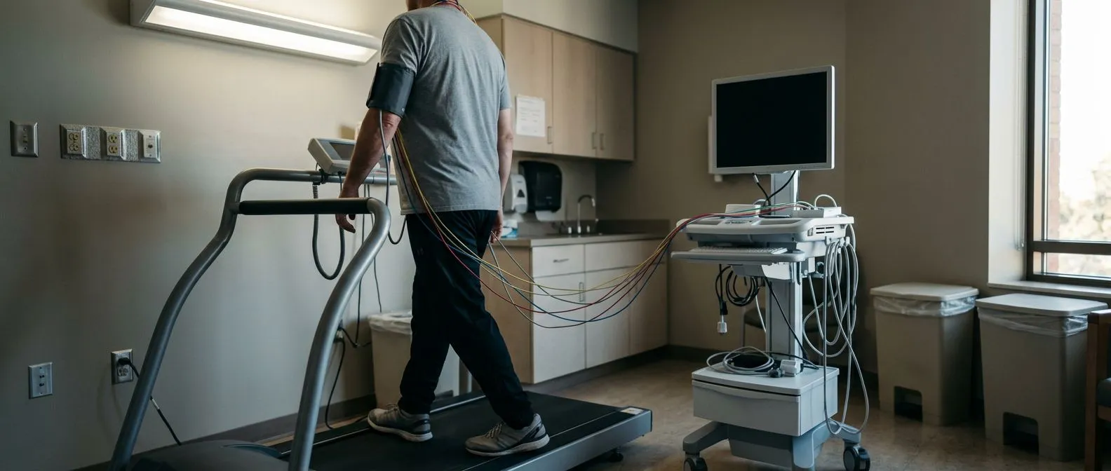
The exercise stress test on a treadmill remains the most widely performed cardiac stress test worldwide. It is straightforward, requires no radiation, and gives your cardiologist real-time data on how your heart responds to increasing effort. During the test, you walk on a motorised treadmill whose speed and incline increase at set intervals — typically every three minutes. The medical team monitors your ECG continuously and checks your blood pressure at each stage, watching for changes that suggest the heart muscle is not receiving enough blood.
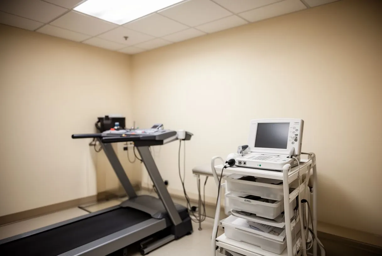
What it feels like for the patient: You start walking at a gentle, slow pace that gradually becomes brisk and uphill. The experience is comparable to a moderate hike. You are never asked to run unless a specific protocol calls for it, and you can stop at any time if you develop concerning symptoms. Most patients are surprised that the exercise portion lasts only a few minutes of actual walking. The technician will ask you to continue until you reach a target heart rate based on your age, or until symptoms prompt stopping.
Who is and isn't a good candidate: This test suits patients who can walk without severe limitation and whose resting ECG is interpretable. It is less reliable in patients who cannot achieve a high enough heart rate due to deconditioning, arthritis, neurological conditions, or medications that blunt heart rate response. If you have Atrial Flutter or other rapid arrhythmias at baseline, the treadmill ECG may be difficult to read, and your doctor may choose an imaging study instead. The exercise test is valued precisely because it reproduces the real-world conditions under which symptoms occur, giving your team clinically meaningful information about your functional capacity and your heart's supply-and-demand balance.
Nuclear Stress Test Explained
A nuclear stress test adds a layer of imaging precision to the standard exercise or pharmacological test. A small amount of radioactive tracer is injected into a vein, and a special camera captures images of blood flow to your heart muscle at rest and under stress. This allows your cardiologist to see not just whether reduced blood flow is occurring, but specifically which areas of the heart are affected and how severe the reduction is. The radiation dose is low and comparable to other common imaging studies.
Rest and stress imaging compared: By comparing images taken when your heart is at rest with images taken when your heart is working hard, the nuclear test distinguishes between fixed defects — areas of the heart muscle that have been damaged by a previous heart attack — and reversible defects, which indicate reduced blood flow that worsens under stress. This distinction matters because reversible defects point to treatable blockages, while fixed defects represent scar tissue that will not benefit from further intervention. If you have had an Atypical Infarction — a heart attack with symptoms that do not follow the classic pattern — this imaging can help clarify exactly what occurred.
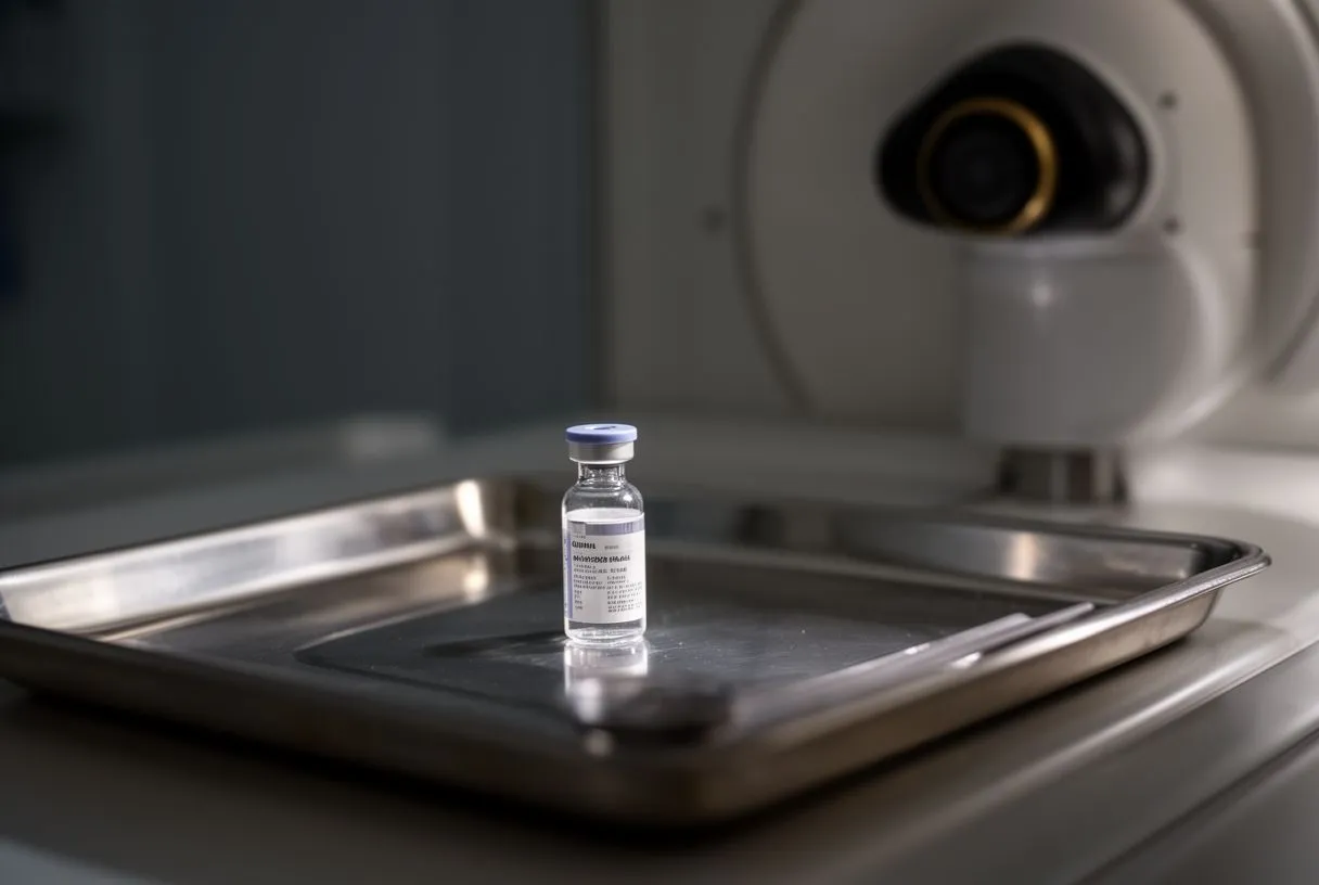
Tracers, timing, and the patient experience: Common tracers include technetium-based compounds such as sestamibi or tetrofosmin. You will receive an injection, wait for the tracer to circulate, and then lie under a gamma camera for about fifteen minutes per imaging set. The entire appointment can take two to four hours because of the need for separate rest and stress scans with a waiting period in between. Patients should know that caffeine interferes with certain tracers and must be avoided for 24 hours before the test. This test provides detailed, spatially accurate information that guides whether your cardiologist recommends medication, Balloon Angioplasty, or surgical revascularisation. Research on imaging and tissue responses in cardiovascular contexts has been documented in sources such as the Journal of Experimental Medicine.
Dobutamine Stress Test: When Exercise Isn't Possible
For patients who cannot exercise on a treadmill — due to arthritis, neurological conditions, peripheral vascular disease, or recent surgery — the dobutamine stress test offers a safe and effective alternative. Dobutamine is a medication delivered through an Arterial Line or more commonly a peripheral intravenous line that mimics the effect of exercise on the heart. It increases the force and rate of cardiac contractions, raising oxygen demand in the heart muscle just as walking on a treadmill would.
How the medication works in the body: Dobutamine acts on beta receptors in the heart, causing it to beat faster and more forcefully within minutes of starting the infusion. The dose is gradually increased, and your heart rate, blood pressure, and ECG are monitored continuously throughout. Once you reach the target heart rate or develop symptoms, the infusion is stopped and the medication clears from your system quickly. Some patients describe a sensation of warmth, a pounding heart, or mild anxiety — all expected effects of the drug. These sensations resolve within minutes of stopping the infusion.
When it is the right choice over exercise: Your cardiologist may choose a dobutamine protocol when you cannot walk adequately, when your resting ECG makes exercise ECG uninterpretable, or when a stress echocardiogram is planned and wall motion imaging is needed. It is also useful for pre-operative cardiac risk assessment before non-cardiac surgery. Patients with significant Auscultation findings suggesting valve disease may still be candidates, but the protocol is adjusted carefully. The test is generally well tolerated, and the medical team can halt the infusion instantly if any concerning sign appears. It provides reliable information about heart function when exercise is simply not practical.
Cardiac Stress Test Procedure: What to Expect Step by Step
Understanding the cardiac stress test procedure reduces anxiety and helps you arrive prepared. The process is methodical and every step serves a clinical purpose. When you arrive, a technician will ask you to change into a gown or remove clothing from the waist up. They will clean small areas of your chest with alcohol pads and attach approximately ten to twelve adhesive electrodes to specific anatomical locations. These electrodes detect the tiny electrical signals your heart produces with each beat and transmit them to an ECG monitor.
Baseline measurements before exercise: Before any walking begins, the team records your resting ECG, blood pressure, and heart rate. These baseline values are essential because they give your cardiologist a comparison point. Changes that appear only during exercise are what matter, and without a clear resting baseline, those changes are harder to interpret. The technician may also practice having you breathe deeply or stand briefly to see how your ECG responds — these manoeuvres help confirm that the electrode placement is correct and the tracing is clean.
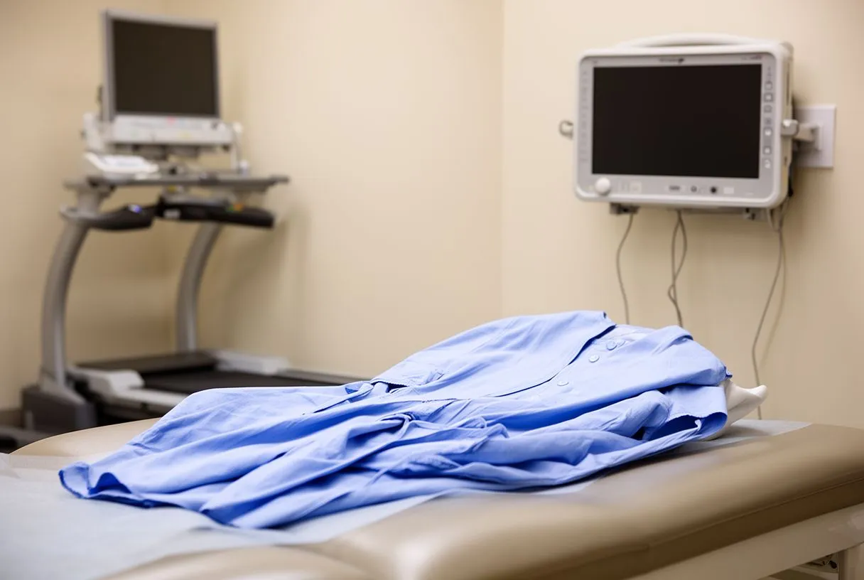
The exercise or pharmacological phase: If you are doing a treadmill test, you will begin walking at a slow, controlled pace. The treadmill gradually speeds up and tilts upward at scheduled intervals, following a standardised protocol such as the Bruce protocol. The team watches your ECG continuously, asks how you feel at each stage, and measures your blood pressure every few minutes. They are looking for ECG changes, blood pressure responses, arrhythmias, and any symptoms you report. If you are having a pharmacological test, medication is infused instead, and the monitoring is equally close. The exercise or infusion continues until you reach your target heart rate, develop symptoms, or your doctor decides sufficient information has been gathered. After stopping, you rest while monitoring continues for several minutes to capture your recovery phase, which also holds important diagnostic information.
Understanding Stress Test Results: Normal vs Abnormal
Stress test results fall broadly into three categories: normal, abnormal, or equivocal. A normal result means your heart achieved the target workload without significant ECG changes, your blood pressure rose appropriately, and you did not develop symptoms suggesting reduced blood flow. This is reassuring and significantly lowers the likelihood of severe coronary artery disease. However, a normal test does not guarantee that no plaque exists — it means that whatever is present is not currently restricting flow enough to cause problems under stress.
What an abnormal result means: Abnormal stress test results typically show one or more of the following: ST-segment depression on the ECG indicating the heart muscle is not getting enough oxygen, a drop in blood pressure during exercise, failure to reach target heart rate, or chest pain that appears during the test. In nuclear imaging, a reversible defect — an area that shows reduced tracer uptake during stress but normal uptake at rest — points to a blockage that restricts flow only when demand is high. These findings guide your cardiologist toward further evaluation, which may include coronary angiography. An abnormal result is not an emergency in itself; it is a signal that warrants a closer look. Decades of clinical research, including foundational work referenced in the clinical literature on valve and tissue outcomes, support this staged approach to diagnosis.
- Normal result: Heart handles stress well; no significant flow limitation detected. Low probability of severe blockage.
- Equivocal result: Findings are borderline or technically difficult to interpret. A repeat or alternative test may be recommended.
- Abnormal result: Evidence of reduced blood flow under stress. Further imaging or coronary angiography may be needed.
What happens after an abnormal result: Your cardiologist reviews the full picture — your symptoms, risk factors such as elevated Apolipoprotein B levels, and the specific pattern of abnormality. Some patients proceed directly to coronary angiography, which provides a direct visual of the inside of the arteries. Others may be started on or have their medication adjusted, with repeat testing scheduled to monitor for progression. The decision depends on how strongly abnormal the result is, your overall clinical context, and shared decision-making between you and your cardiac team.
Stress Test vs Angiogram: Which Do You Need?
The distinction between a stress test and an angiogram confuses many patients, yet it is one of the most important concepts to grasp when navigating cardiac care. A stress test is a functional assessment — it asks whether your heart is getting enough blood under demand. A coronary angiogram is an anatomical assessment — it shows the actual structure of your coronary arteries, revealing where blockages sit and how narrow they are. These are complementary tools, not competing alternatives.
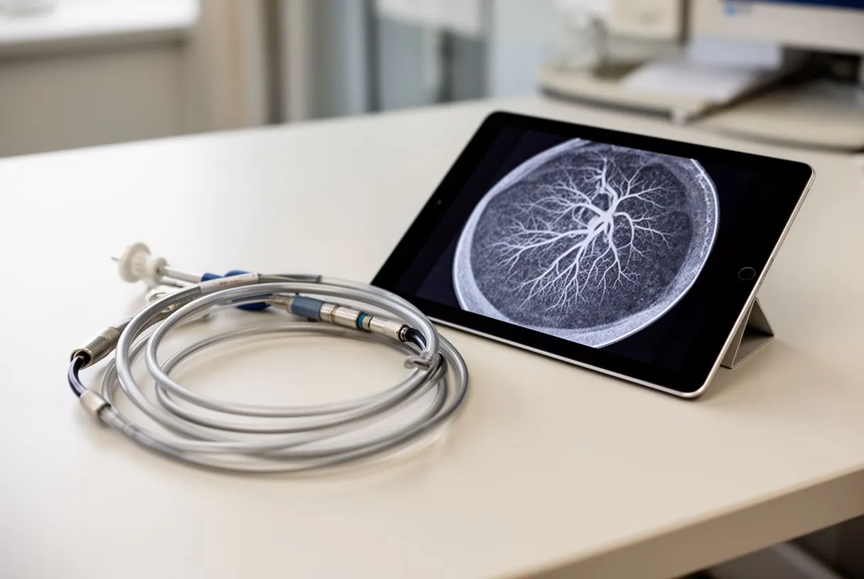
When each test is appropriate: Your doctor typically starts with a stress test because it is non-invasive, carries minimal risk, and answers the most pressing clinical question: does reduced blood flow explain your symptoms? If the stress test is normal, an angiogram is usually unnecessary. If the stress test is abnormal, the angiogram becomes the next step for patients whose findings suggest they would benefit from a procedure. During angiography, a thin tube called a catheter is guided into the coronary arteries through a small incision — sometimes near the Ascending Aorta access point — and contrast dye is injected while X-ray images are captured in real time. This reveals blockages with precision and allows immediate intervention such as stent placement if needed.
Cost, risk, and recovery compared: A stress test is performed in a clinic or hospital outpatient setting, takes about an hour, and involves essentially no recovery time. An angiogram is a minimally invasive procedure performed in a catheterisation laboratory, carries small but real risks including bleeding, infection, and contrast reactions, and requires a short observation period afterward. For patients weighing the two, it helps to understand that the stress test is the gatekeeper. It determines who proceeds to angiography and who can be managed with medication and lifestyle changes alone. This staged approach protects patients from unnecessary procedures while ensuring that those who need intervention receive it promptly.
Finding a Heart Specialist for Your Stress Test in Mexico
If you are considering a cardiac stress test and live in or travel to Mexico, accessing high-quality cardiac care is achievable without crossing borders. The team bridges advanced cardiology with patient-centred communication, ensuring that every test is explained clearly and every result is interpreted in the context of your specific situation.
What to look for in a cardiac team: Seek a specialist who takes time to understand your symptoms, reviews your complete medical history, and explains why a particular test is recommended rather than ordering studies reflexively. A good cardiac team will discuss the trade-offs of each test type, address your concerns about cost and insurance, and create a plan that fits your clinical needs and personal circumstances. International patients should ask about appointment availability, what documentation to bring, and whether results can be shared with their home-country physician. The goal is a partnership in which you feel informed and confident at every stage.
Taking the next step: If you have been experiencing symptoms that concern you, or if your primary care doctor has recommended cardiac evaluation, booking a consultation is the most important action you can take. A cardiac stress test is a powerful tool, but it is most valuable when ordered and interpreted by a specialist who understands your full clinical picture. Do not self-diagnose based on symptoms or internet research alone. Contact Cirugía de Corazón to schedule a consultation with Dr Molina and his team. They will determine whether a stress test is appropriate for you, which type suits your situation, and what the results mean for your heart health going forward. Your heart deserves expert attention — take that first step today.