Duke Criteria: How Endocarditis Is Diagnosed
Duke criteria endocarditis diagnosis uses blood cultures, echocardiograms, and clinical signs to detect heart valve infection.
What Are the Duke Criteria Endocarditis Diagnosis Tools?
When a patient arrives with persistent fever, fatigue, and a new heart murmur, the clinical team faces a critical question: is this an infection of the heart valves, or something else entirely? The Duke criteria endocarditis diagnosis framework was developed precisely to answer this question with consistency and rigor. Named after Duke University where the criteria were first proposed in 1994, this diagnostic system gives cardiologists and infectious disease specialists a structured way to classify suspected infective endocarditis as definite, possible, or rejected, based on a combination of blood culture results, imaging findings, and clinical signs.
The system matters because infective endocarditis is a condition where the inner lining of the heart—particularly the valves—becomes infected by bacteria or, less commonly, fungi. The infection can destroy valve tissue, send fragments of vegetation (infected clumps) into the bloodstream, and trigger systemic illness that, without treatment, is fatal. A delayed or missed diagnosis carries enormous consequences. The Duke criteria were designed to reduce that risk by replacing clinical guesswork with a reproducible checklist that any hospital team can apply, whether in Guadalajara, Mexico City, or anywhere else in the world.
Why the criteria were created: Before the Duke system, diagnosing endocarditis relied heavily on individual physician experience, which led to inconsistent diagnoses across hospitals. The original 1994 framework was later updated in 2000 to the modified Duke criteria, which incorporated echocardiographic imaging more formally and expanded the definition of what counted as major evidence. Today, the modified version is the global standard, used by the European Society of Cardiology and the American Heart Association in their clinical guidelines, and it remains the backbone of how cardiac infections are confirmed or ruled out before treatment begins.
How Modified Duke Criteria Work in Clinical Practice
The modified Duke criteria operate on a simple but powerful principle: endocarditis is confirmed when enough evidence accumulates from specific, defined categories. A case is classified as definite endocarditis if the patient meets two major criteria, or one major and three minor criteria, or five minor criteria. A case is classified as possible endocarditis when findings fall short of definite but do not meet the threshold for rejection. Rejected cases are those where a firm alternative diagnosis is established, or where the clinical findings resolve within four days of antibiotic therapy, or where there is no pathological evidence of endocarditis at surgery or autopsy.
In real-world hospital settings, this classification happens over the course of several days. Blood cultures are drawn before antibiotics are started—ideally three sets from different venipuncture sites spaced at least one hour apart. If the patient has already received antibiotics, cultures may be negative, which complicates the picture and often requires the team to rely more heavily on imaging and clinical signs. The modified criteria account for this reality by including a pathway for culture-negative endocarditis, where serological testing for organisms like Coxiella burnetii or Bartonella can still satisfy major criteria.
How the pathway guides treatment: Once a patient is classified as definite or possible, the team does not wait for absolute certainty to begin targeted antibiotic therapy. The Duke system is a living tool—its categories shift as new data arrive from the microbiology lab and the echocardiography suite. A patient who starts as "possible" based on fever and a murmur becomes "definite" once blood cultures grow Staphylococcus aureus and an echo reveals a vegetation on the mitral valve. The criteria help the team communicate clearly about confidence levels and make shared decisions about whether to continue medical treatment, escalate to surgical intervention, or pursue an alternative diagnosis entirely.
Duke Criteria Major and Minor Findings Explained
The architecture of the Duke criteria rests on a distinction between major findings (high-specificity evidence that directly points to endocarditis) and minor findings (supportive signs that are common but not unique to the condition). There are three categories of major criteria: positive blood cultures for typical organisms consistent with endocarditis, evidence of endocardial involvement shown by echocardiogram, and new valvular regurgitation detected on physical examination. Each of these carries significant diagnostic weight because they reflect either the organism in the bloodstream or physical damage to the heart.
Minor criteria include fever (typically above 38°C), predisposing heart conditions (such as a prosthetic valve, congenital heart disease, or a history of endocarditis), vascular phenomena (like arterial emboli, septic pulmonary infarcts, mycotic aneurysm, or Janeway lesions), immunological phenomena (such as Osler nodes, Roth spots, or a positive rheumatoid factor), and microbiological findings that do not meet the major criteria threshold. These minor findings are common in many infections, which is why they alone cannot confirm the diagnosis—but in combination with major findings, they build a compelling clinical picture.
Major versus minor in practice: A patient with a prosthetic aortic valve who presents with fever, two positive blood cultures for Enterococcus faecalis, and a transesophageal echo showing a vegetation meets two major criteria and is immediately classified as definite endocarditis. The minor criteria become less relevant in that scenario. However, when blood cultures are negative and imaging is equivocal, the minor findings carry the diagnostic burden—fever, a known predisposing valve condition, and a septic embolus to the spleen together might push the case from possible to definite.

Endocarditis Blood Cultures Diagnosis Process
Blood cultures are the single most important diagnostic test in suspected endocarditis, and their proper collection can make the difference between a swift diagnosis and days of uncertainty. The goal is to identify the microorganism growing on the valve so that antibiotic therapy can be targeted rather than empiric. Standard practice calls for three blood culture sets—each set consisting of one aerobic and one anaerobic bottle—drawn from separate venipuncture sites. If the patient has received antibiotics in the prior two weeks, cultures may be negative in up to 25% of true endocarditis cases, which is why timing and technique matter enormously.
What a positive culture tells the team: When blood cultures grow typical endocarditis organisms—such as Streptococcus viridans, Streptococcus bovis, Staphylococcus aureus, or Enterococcus—the major criteria are met. Persistent bacteremia, defined as cultures remaining positive more than 12 hours apart, is especially diagnostic because it suggests a deep-seated source rather than a transient contamination from the skin draw. According to europepmc.org, culture-positive endocarditis accounts for the majority of definite diagnoses under the Duke system, making proper microbiological technique foundational to the entire diagnostic pathway.
In culture-negative cases, the team must broaden the search. Some organisms are fastidious or require special media—HACEK organisms (Haemophilus, Aggregatibacter, Cardiobacterium, Eikenella, Kingella) grow slowly and may be missed in standard incubation periods. Others, like Coxiella burnetii (the cause of Q fever), cannot be cultured routinely and require serological diagnosis with elevated IgG titres. The modified Duke criteria explicitly recognize these pathways: a single positive blood culture for Coxiella or an anti-phase I IgG titre above 1:800 counts as a major criterion, ensuring that even hard-to-grow organisms do not escape the diagnostic net.
Transesophageal Echocardiogram Endocarditis Imaging
Echocardiography is the imaging cornerstone of the Duke criteria, and for good reason: it allows the clinician to see the valves and their surrounding structures in real time without radiation. There are two modalities—transthoracic echocardiogram (TTE) and transesophageal echocardiogram (TEE). TTE is performed by placing the probe on the chest wall; it is noninvasive, quick, and usually the first imaging test ordered. However, its sensitivity for detecting vegetations is roughly 40–60% for native valves and even lower for prosthetic valves, because ultrasound must pass through ribs, lung tissue, and body habitus.
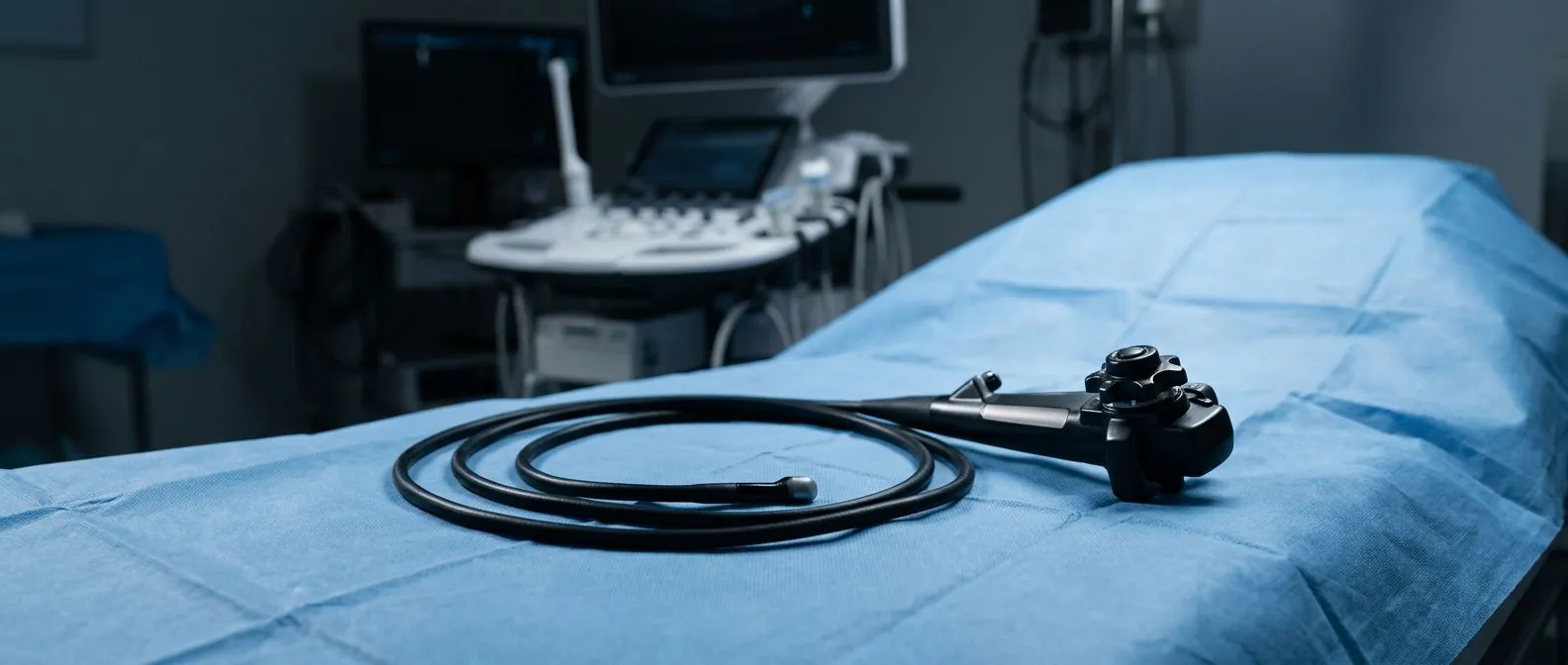
The transesophageal echocardiogram endocarditis evaluation is far more sensitive—over 90% for native valve vegetations and significantly better for prosthetic valves—because the probe sits in the esophagus directly behind the heart, giving it an unobstructed acoustic window. TEE is performed under sedation and requires the patient to fast, but the diagnostic yield justifies the additional effort. A TEE can identify vegetations as small as 2–3 mm, detect abscesses or fistula tracts around the valve annulus, and evaluate prosthetic valve function in ways that TTE simply cannot.
When TEE is mandatory: Clinical guidelines recommend TEE for any patient with suspected prosthetic valve endocarditis, any patient with a negative or inconclusive TTE, any patient with staphylococcal bacteremia, and any patient in whom complications such as abscess or extension of infection beyond the valve are suspected. The modified Duke criteria count three specific echo findings as major evidence: an oscillating intracardiac mass on the valve or supporting structures, an annular or perivalvular abscess, and new partial dehiscence of a prosthetic valve. Each of these findings, confirmed on TEE, can independently move a patient into the definite endocarditis category.
Duke Criteria Vegetation Detection on Heart Valves
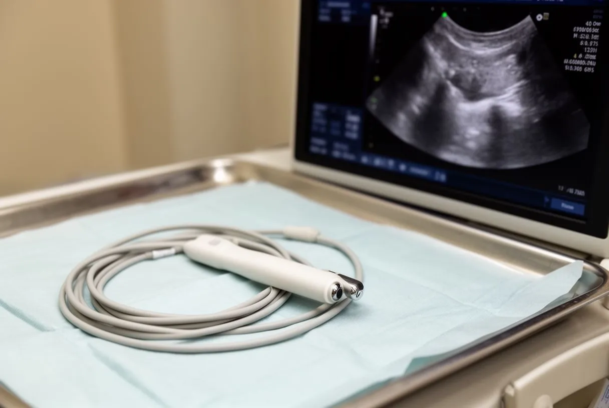
A vegetation is the pathological hallmark of infective endocarditis—a cluster of platelets, fibrin, and microorganisms that adheres to a valve leaflet or other endocardial surface. On echocardiography, a vegetation appears as a mobile, irregularly shaped mass, typically on the low-pressure side of the valve (the atrial surface of the mitral valve or the ventricular surface of the aortic valve). The Duke criteria vegetation detection standard requires that the mass be independently mobile and not part of the normal valve apparatus, which helps distinguish infection from benign structures like Lambl's excrescences or redundant leaflet tissue.
Where vegetations form and why it matters: Vegetations most commonly develop on previously damaged valves—those with degenerative changes, rheumatic scarring, or congenital abnormalities. In the left side of the heart, the mitral and aortic valves are the most frequent sites. In the right side, the tricuspid valve is often involved in patients who use intravenous drugs. The location of the vegetation matters because left-sided vegetations carry a higher risk of systemic embolism—fragments can travel to the brain, kidneys, spleen, or coronary arteries, causing strokes or organ infarcts. Tricuspid vegetations tend to shed fragments into the pulmonary circulation, producing septic pulmonary emboli rather than systemic ones.
The size and mobility of a vegetation also drive clinical decisions. Large vegetations—typically defined as greater than 10 mm in diameter—and highly mobile masses carry a higher risk of embolic events. When a vegetation is large and the organism is aggressive, the team may recommend early surgical removal of the infected valve rather than continuing antibiotics alone. The auscultation of a changing or new murmur on physical exam often prompts the echo that reveals the vegetation, underscoring how the Duke criteria integrate clinical examination with imaging findings.
Prosthetic Valve Endocarditis Criteria and Challenges
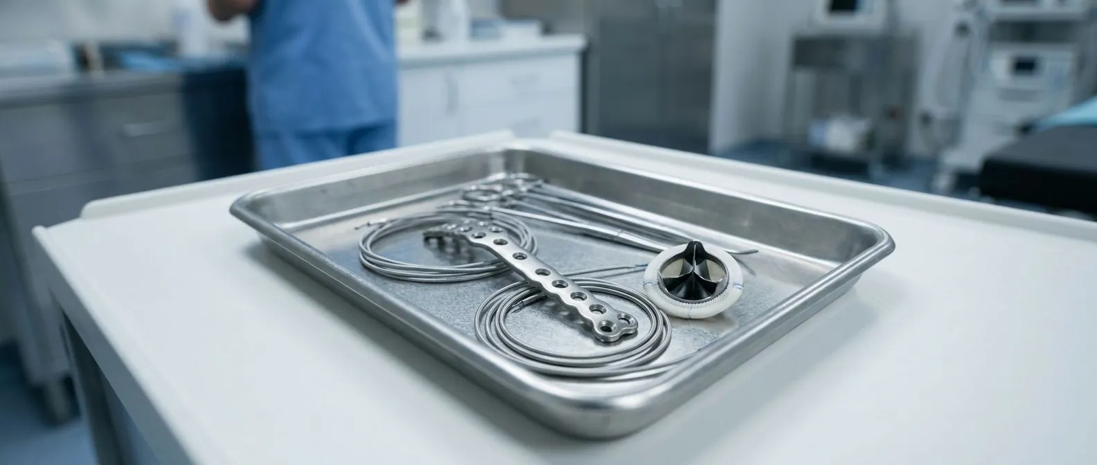
Endocarditis on a prosthetic valve—whether a mechanical or bioprosthetic replacement—presents unique diagnostic challenges that the Duke criteria address with specific modifications. Prosthetic valve endocarditis can occur early (within 60 days of surgery, often related to perioperative contamination) or late (beyond 60 days, usually from community-acquired organisms). The presence of a prosthetic valve is itself a minor criterion under the Duke system, reflecting the increased baseline risk these patients carry. However, imaging prosthetic valves is technically difficult because the synthetic materials create acoustic shadows that obscure the underlying tissue, making transthoracic echo particularly unreliable.
Why TEE is essential here: For prosthetic valves, transesophageal echocardiography is not optional—it is the standard. TEE can detect vegetations on the atrial side of a mitral prosthesis, abscesses around the sewing ring, and partial dehiscence (loosening of the valve from its annulus), all of which are major criteria findings. A paravalvular leak visible on echo, particularly when combined with fever and positive blood cultures, is highly suggestive of prosthetic valve endocarditis. The modified Duke criteria explicitly include new partial dehiscence of a prosthetic valve as a major echo finding, recognizing that this structural failure is often a direct consequence of infection loosening the valve from the surrounding annulus.
Prosthetic valve endocarditis also differs in its microbiology. Coagulase-negative staphylococci are common in early infections, while late infections often involve the same organisms that cause native valve endocarditis. The presence of a balloon-expandable valve or an autograft (a valve transplanted from the patient's own tissue, such as a pulmonary autograft in the Ross procedure) can further complicate imaging interpretation, because these structures do not always behave like traditional prostheses on echo. In these cases, the team may need to combine echo findings with other imaging modalities, such as computed tomography or fluoroscopy, to assess valve motion and detect abnormalities that echo alone might miss.
Duke Criteria Sensitivity Specificity and Diagnostic Accuracy
The Duke criteria were validated against pathological gold standards—surgical and autopsy findings—when they were first published, and they have been re-validated repeatedly since. The original 1994 criteria demonstrated a sensitivity of approximately 80% for definite endocarditis when compared against pathological confirmation. The modified 2000 version improved this figure, pushing sensitivity above 90% in most validation studies, largely because it incorporated transesophageal echocardiography more formally and expanded the definition of major microbiological findings to include serological evidence for organisms like Coxiella burnetii.
Specificity—the ability to correctly identify patients who do not have endocarditis—has historically been lower than sensitivity in some populations, particularly in the "possible" category. Many patients classified as possible endocarditis turn out to have alternative diagnoses, ranging from other bloodstream infections to non-infectious causes of fever and heart murmurs. This is why the "possible" category is not a diagnosis in itself but a prompt for further investigation: more cultures, repeat imaging, and consultation with specialists. Research compiled through Efficacy and safety of more intensive lowering of LDL cholesterol: a meta-analysis of data from 170 000 participants in 26 randomised trials underscores how large-scale, rigorously designed clinical data sets improve diagnostic and therapeutic precision across cardiovascular care.
Limitations and ongoing refinements: The Duke criteria are not perfect. They can struggle with certain scenarios: culture-negative endocarditis where serology is also negative, infections caused by unusual organisms not covered by the typical microbiological criteria, and cases where echo findings are subtle or atypical. Imaging advances continue to push diagnostic accuracy forward. For patients who develop neurological complications of endocarditis—such as embolic stroke—the diagnostic and management pathway has been further refined. According to Guidelines for the Early Management of Patients With Acute Ischemic Stroke, timely recognition of embolic stroke as a potential manifestation of endocarditis is critical, because the underlying cardiac infection must be treated alongside the neurological emergency.
Clinical Signs: Fever, Heart Murmur, and When to Act
The clinical presentation of endocarditis is often subtle at first, which is why the Duke criteria include specific physical findings as minor criteria. Fever is the most common symptom, present in over 90% of cases, but it may be low-grade and easily attributed to a minor viral illness. A heart murmur—heard through auscultation with a stethoscope—is present in most cases, but it may have been present for years if the patient has a known valve condition. What matters is a new murmur or a change in the character of an existing one, which suggests new valvular destruction.
Beyond fever and murmur, the Duke criteria recognize several other physical findings as minor criteria. Vascular phenomena include arterial emboli (sudden loss of circulation to a limb or organ), septic pulmonary infarcts (areas of lung tissue death from infected emboli in right-sided endocarditis), mycotic aneurysms (infected bulges in artery walls), intracranial hemorrhage, conjunctival hemorrhages, and Janeway lesions (painless flat red spots on the palms or soles). Immunological phenomena include Osler nodes (tender nodules on the fingers or toes), Roth spots (retinal hemorrhages with pale centers), a positive rheumatoid factor, and elevated markers of inflammation such as C-reactive protein.
- Fever above 38°C: Present in nearly all cases; may be the only early sign in prosthetic valve patients.
- New or changing heart murmur: Detected on auscultation; reflects acute valve destruction and is a major criterion if it represents new regurgitation.
- Vascular and immunological signs: Embolic events, Janeway lesions, Osler nodes, and Roth spots point to systemic infection and are minor criteria that strengthen the diagnostic case.
Additional nonspecific symptoms—fatigue, weight loss, night sweats, anorexia—often accompany the condition but are not part of the formal criteria because they overlap with many other illnesses. Arm pain from a septic embolus to a peripheral artery, or an atypical infarction pattern on an electrocardiogram if a coronary artery is occluded by an embolus, can also be presenting features. The challenge is that these signs are easily missed or attributed to other causes, which is why the Duke criteria exist: they force the clinician to systematically weigh every piece of evidence rather than relying on pattern recognition alone.

When to Seek a Cardiology Second Opinion for Endocarditis
Endocarditis is a diagnosis where the stakes are high and the pathway is complex—antibiotics may be needed for four to six weeks, surgery may be required to remove an infected valve, and the decision-making depends on interpreting blood cultures and echocardiograms that not every hospital performs with equal frequency. For patients in Mexico or those traveling for cardiac care, a second opinion from an experienced cardiac surgery team can clarify whether the Duke criteria have been applied correctly, whether imaging was adequate, and whether the proposed treatment plan aligns with current international guidelines.
- Seek a second opinion when: Blood cultures are negative but clinical suspicion remains high—specialized serological testing may identify organisms that standard cultures miss.
- Seek a second opinion when: Only a transthoracic echo was performed—TEE should be done for prosthetic valves, staphylococcal bacteremia, or any case where TTE is inconclusive.
- Seek a second opinion when: Surgery is being considered—understanding the threshold for operative intervention (such as large mobile vegetation, uncontrolled infection, or valve destruction causing heart failure) requires a team experienced in both medical and surgical management of endocarditis.
Patients with conditions like atrial flutter, those taking aspirin for other cardiovascular indications, or those who have previously undergone balloon angioplasty or have an arterial graft from coronary bypass surgery may have overlapping cardiac issues that complicate the endocarditis picture. A valve infection in a patient with an ascending aorta aneurysm or a prior autologous blood transfusion during cardiac surgery adds further layers that require subspecialist input. The modified Duke criteria provide the framework, but the interpretation of that framework—deciding what is definite, what is possible, and what the next step should be—is where experienced clinical judgment matters most.
If you or a family member are facing a suspected or confirmed diagnosis of infective endocarditis, the most important step is to ensure that the diagnostic process has been thorough: that blood cultures were properly drawn, that transesophageal echocardiography was performed when indicated, and that the Duke criteria were applied by a team experienced in their use. Dr Alejandro Molina and the team at Cirugía de Corazón in Guadalajara, Querétaro, and Hermosillo specialize in complex cardiac surgery and valve disease, offering second opinions and comprehensive evaluation for patients navigating endocarditis and its complications. We encourage you to schedule a consultation to discuss your specific situation—because every heart, every valve, and every infection presents a unique clinical picture that deserves individualized expert attention rather than a one-size-fits-all approach.