Femoral Vein Access in Heart Procedures: What to Expect
Learn what femoral vein access heart procedures involve, from sedation to recovery.
If you or a family member has been told you need a heart procedure that uses femoral vein access — reaching the heart through a vein in the groin — it is normal to feel nervous about the unknown. The good news is that femoral vein access is one of the most studied and routine routes in modern cardiology and cardiac surgery. In this guide we explain, in plain language, where the femoral vein is, how femoral vein access heart procedures are performed, what the risks really are, and what recovery actually feels like.
What Is the Femoral Vein and Where Is It Located?
Which leg is the femoral vein in? The answer is simple: you have one in each leg. The femoral vein is the large deep vein of the thigh. It runs alongside the femoral artery through the upper thigh, and at the groin crease it sits just below the skin — close enough to reach with a thin needle.
Blood from the leg travels upward through the femoral vein into the iliac veins in the pelvis, and from there into the inferior vena cava, the big vein that returns blood to the right side of the heart. Because it is a highway back to the heart, a catheter (a thin, flexible tube) placed in the femoral vein can be advanced directly into the right atrium and right ventricle without surgery.
The vein also has branches. The most famous is the great saphenous vein, the superficial leg vein that joins the femoral vein near the groin. The course of the great saphenous vein runs from the inner ankle up the inner leg to that junction — a route surgeons know well, because it is the vein most often used for coronary bypass grafts. Vascular has published case reports showing how even the saphenous system itself can be repurposed therapeutically in severe limb ischemia.
How Do Femoral Vein Access Heart Procedures Work?
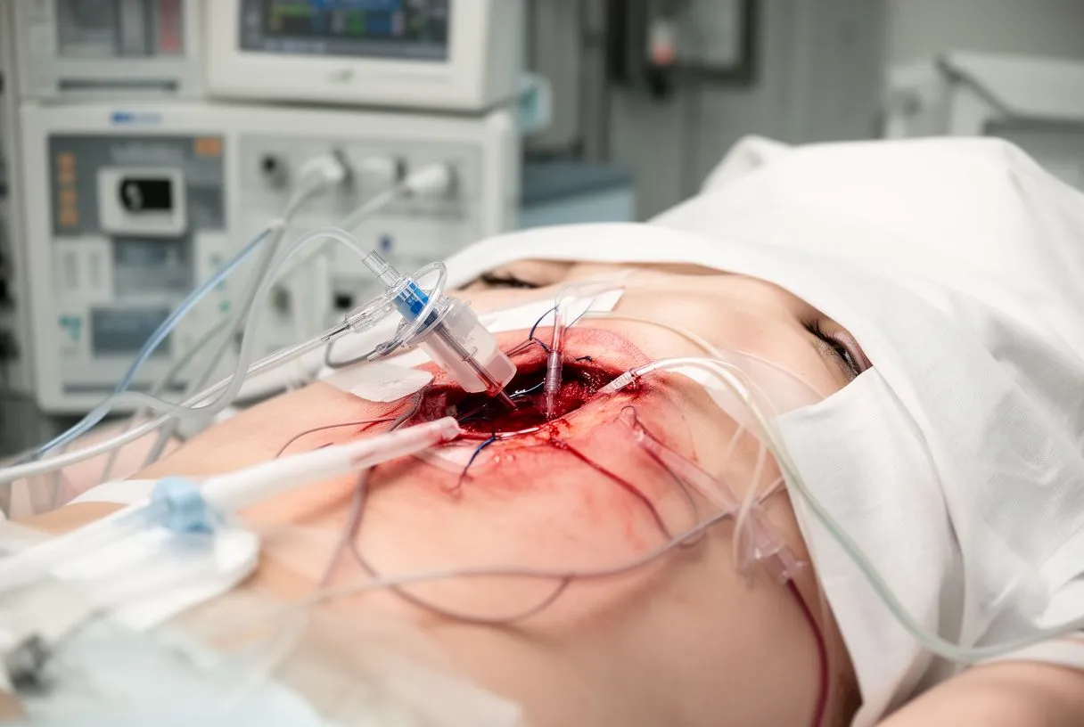
Step by step. Femoral vein access heart procedures follow a well-rehearsed sequence. First, the groin area is cleaned with antiseptic and covered with a sterile drape. Local anesthetic numbs the skin. The doctor then uses ultrasound to see the vein in real time and inserts a thin needle into it.
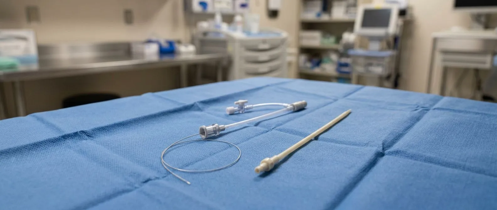
A soft guidewire passes through the needle into the vein, the needle is removed, and a short plastic tube called a sheath is slid over the wire. The sheath acts as a doorway. Through it, catheters can be exchanged freely — to measure pressures inside the heart, to study electrical signals, or to deliver a new valve — without repeatedly puncturing the vessel.
Because the femoral vein drains toward the right side of the heart, this route is the natural choice for right-sided heart catheterization, electrophysiology (rhythm) studies, ablation procedures, pacemaker or defibrillator leads, and the venous part of certain structural heart procedures. In our practice, every access plan is reviewed by the full multidisciplinary team against international guidelines before we recommend a route, and we explain the alternatives to the patient in plain words.
Femoral Artery vs Femoral Vein: What's the Difference?
Two vessels, one groove. Patients often mix up the two, and the confusion is understandable: in the groin they sit side by side, only a few millimeters apart. The femoral artery carries oxygen-rich blood away from the heart; it pulses and feels like a heartbeat under your fingers. The femoral vein carries used blood back to the heart; it does not pulse.
The difference matters clinically. Arterial access is used for procedures on the left side of the heart — the arteries, the aorta, and the aortic valve — because pressure in an artery can push a catheter upstream. Venous access through the femoral vein is used for the right side of the heart and for anything that must travel with the flow of venous blood.
Complications also differ. Arterial punctures can bleed briskly and may form a false channel in the vessel wall; venous punctures bleed less because vein pressure is low, but veins can clot. Knowing which vessel was used tells your care team exactly what to watch for afterwards.
Why Is the Femoral Vein Used for Catheterization — and When Do Doctors Avoid It?
The advantages. The femoral vein is large, reliably located, and close to the skin. It accepts large sheaths, which matters for procedures such as ablation or lead placement that need bigger catheters. Other venous routes exist — the Transfemoral Access route, the Transsubclavian Access route under the collarbone, and jugular access in the neck — but the groin vein remains the workhorse for right-heart work.
Why avoid femoral vein for catheterization? There are honest reasons to choose another route in some patients. Repeated punctures can scar the vein. Prolonged bed rest afterwards is harder on patients who cannot lie still. In patients with severe obesity, severe groin arterial disease, or a suspected clot in the leg, the team may prefer the arm (radial or brachial access) or another vein. Sometimes the answer is anatomical: vein anatomy varies from person to person, and ultrasound usually settles the question before the first needle.
The key point is that route selection is a medical decision, not a preference. When we plan a femoral vein catheterization, we check the patient's leg circulation, prior procedures, and imaging first — if the groin is not the right route for that person, we say so and offer the alternative.
What Are the Landmarks for Femoral Vein Central Line Placement?
Classic landmarks. For decades, doctors placed femoral central lines using surface anatomy alone: the femoral vein lies just medial (toward the inside of the body) to the femoral artery at the groin crease, below the inguinal ligament. Feel the arterial pulse, and the vein is a thumb's width toward the inner thigh.
Ultrasound has changed the game. Today, guidelines favor real-time ultrasound guidance for central venous access, and studies indexed on PubMed have shown it reduces failed attempts and complications compared with landmark-only techniques. The operator sees the artery, the vein, and the needle tip on screen — which is especially reassuring in patients whose anatomy differs from the textbook.
This matters to you because it means fewer needle passes, less bruising, and a lower chance of the needle going into the wrong vessel. Ask your team whether they use ultrasound guidance; at good centers it is standard, not optional.
Can You Get a Blood Clot in Your Femoral Vein?
Yes — and it has a name. A clot in the femoral vein is a form of Deep Vein Thrombosis (DVT), the condition where blood pools and solidifies in the deep veins of the leg. Femoral vein thrombosis can arise on its own (after long travel, surgery, or immobility) or as a complication of a catheter procedure.
Typical warning signs include swelling of the whole leg, a feeling of heaviness or tightness, warmth, and pain — often in the calf or thigh. The leg may look noticeably larger than the other side. If a clot breaks loose it can travel to the lungs, which is why DVT is taken seriously and treated with blood thinners.
After a femoral vein procedure, the risk is low but real, and it rises with long sheath time, prolonged bed rest, and immobility afterwards. A swollen, painful leg after a catheter procedure through the groin is never something to wait out — it needs prompt medical attention.
What Happens if the Femoral Vein Is Damaged?
Most injuries are minor. During access, the most common "damage" is a small puncture site that bruises. Because vein pressure is low, bleeding usually stops with firm pressure and time. Some patients develop a hematoma — a collection of blood under the skin — that resolves over days to weeks.
More significant problems. Rarely, the vein wall can be injured so that it narrows or blocks, or the artery and vein can form an abnormal connection called a fistula. Repair of major venous injury is a surgical matter, and surgeons have multiple tools for it, from direct suture repair to using nearby veins — including the Saphenous Vein or a Saphenous Vein Graft — as replacement conduit when a major vein must be reconstructed.
It is worth remembering the scale: serious femoral vein injury during routine catheter access is uncommon, and the problems that do occur are usually detected the same day, while you are still at the hospital.
What Does Sedation Feel Like During Femoral Vein Access Heart Procedures?
You will not feel the needle. The skin and deeper tissues at the puncture site are numbed with local anesthetic, so the access itself feels like a brief sting and then pressure. Most femoral vein procedures are done with "conscious sedation" — medication that relaxes you and makes you drowsy, while you can still respond if the team asks.
What patients most often report afterwards is not pain but memory gaps: parts of the procedure are simply not remembered. For longer procedures such as ablation, deeper sedation or light general anesthesia may be used, depending on the plan agreed beforehand.
Tell your team about any previous problems with sedation, sleep apnea, or difficult IV access — these details shape the anesthetic plan. And ask questions beforehand; in our consultations we take the time to explain what the studies actually show, in plain words, and to answer every question before any decision is made.
What Is the Recovery Time After a Femoral Vein Heart Procedure?
The first hours. When the sheath comes out, the team presses on the site or applies a closure device, and you are asked to lie still — typically with the leg straight — for a few hours. Venous access sites generally seal faster than arterial ones, so many patients are up and walking the same day or the next morning.
The first days. Expect mild bruising and a small tender lump at the groin. Avoid heavy lifting, straining, and strenuous exercise for a few days to a week, depending on the procedure. Most office work and gentle walking resume quickly. A percutaneous closure device, if used, absorbs into the body over time.
When to call. Contact your team if you notice spreading bruising, a growing painful lump, active bleeding, a leg that becomes swollen or discolored, fever, or shortness of breath. These are the signs worth a phone call, and they are almost always easier to manage early. After a groin-access procedure, it helps to have a written list of these warning signs and who to call.
Femoral Vein vs Radial Access for Heart Procedures: Which Is Better?
Radial access means the wrist. The radial artery is a common route for left-heart catheterization (angiograms of the coronary arteries). It lets patients sit up and walk sooner, and wrist bleeding is easier to control. That is why many centers prefer the wrist for arterial work.
The groin still wins for venous work. The femoral vein is larger than the arm veins, so it accepts the big catheters used for electrophysiology studies, ablation, and lead placement. It is also the natural route for procedures on the right side of the heart, where the femoral vein, the Innominate Vein, or the Pulmonary Veins may be involved in the treatment plan. Alternatives such as the Transaortic Access or Transapical Access routes — through the aorta or through the chest wall — are reserved for specific structural heart cases where the groin cannot be used.
Choosing the route. The honest answer is that the best route is the one that fits your anatomy and your procedure. For patients considering femoral vein heart valve replacement or TAVR, the femoral vein route is used for certain procedural steps while the artery handles the valve delivery itself. The right way to decide is a conversation with a team that does all of these approaches. Our team performs conventional, minimally invasive, robotic and transcatheter procedures, and we recommend the less invasive route only where the evidence supports it for that patient — the final decision is always yours. If you want a second opinion, treat it as a normal part of deciding; we encourage it in every consultation.
What to Expect and When to Seek Help
Femoral vein access has earned its place in heart medicine for good reasons: it is reliable, well tolerated, and it opens the right side of the heart to diagnosis and treatment without open surgery. For most patients, the experience is a numbed pinch, a few hours of rest, and a bruise that fades within a week or two.
- Before the procedure: you will have blood tests, an ECG, and imaging; a free video consultation is a good first step, so only the studies that are truly needed are ordered — no unnecessary ones.
- During: local anesthetic plus sedation; most people remember little of the procedure itself.
- After: a few hours of lying still, then progressive walking; a groin bruise is normal, a swollen leg is not.
- Call the team: bleeding, a growing lump, fever, breathlessness, or a swollen, painful leg deserve immediate attention.
Every heart is different, and the right access route for you depends on your anatomy, your procedure, and your overall health. If you have been told you need a femoral vein access heart procedure, the best next step is a conversation with a qualified cardiac team — ask about your options, ask about alternatives, and do not hesitate to seek a second opinion. We offer a free video consultation at our consulting rooms network in Guadalajara, Querétaro and Hermosillo, where we get to know you and your condition first, and tell you which studies are still needed before anything else is ordered.