

Inside the Catheterisation Laboratory: Your Heart Procedure
Catheterisation laboratory procedure explained step by step: prep, what you feel, equipment and recovery in Guadalajara.
What Is a Catheterisation Laboratory and What Happens There
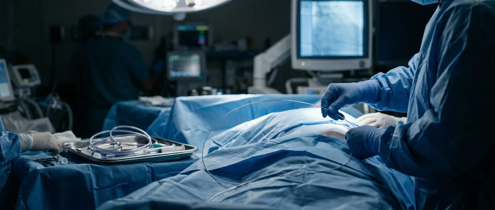
A catheterisation laboratory — often shortened to "cath lab" — is a specialised room inside a hospital where doctors use thin, flexible tubes called catheters to reach your heart and blood vessels without opening your chest. Think of it as a cross between an operating theatre and an imaging suite: the same sterile discipline applies, but instead of scalpels the team works with catheters, contrast dye and live X-ray guidance. When your cardiologist refers you for a catheterisation laboratory procedure, the goal is usually to find out why you have chest pain, shortness of breath or an abnormal stress test, and in many cases to treat the problem in the same session.
Who is in the room with you: a team of four to six people typically surrounds you. The interventional cardiologist leads, two nurses manage sedation and monitoring, a radiographer operates the X-ray machine (the angiography unit), and a cardiac physiologist records pressures and electrical signals. Everyone wears lead aprons because they stay next to you under fluoroscopy — the real-time X-ray that lets the doctor see the catheter moving through your vessels. The room is cool, brightly lit, and filled with monitors showing your heart rhythm, oxygen level and blood pressure from an arterial line.
It helps to understand why this setting exists at all. The first catheterisations were diagnostic only — a way to measure pressures inside the heart and take pictures of the coronary arteries. Over decades the same access route has become a treatment platform: balloons, stents and specialised devices can be delivered through a needle puncture no larger than a ballpoint tip. That single advance is why many patients now go home the same day instead of spending a week recovering from open surgery.
How a Heart Catheter Procedure Works Step by Step
A heart catheter procedure follows a predictable sequence, which is reassuring when you are anxious about the unknown. After you change into a gown and have a small intravenous line placed, the team cleans the access site — usually the right wrist (radial artery) or the groin (femoral artery) — with antiseptic and covers you with sterile drapes. Local anaesthetic numbs the skin so the needle puncture feels like a sharp pinch followed by pressure, not ongoing pain. A short plastic sheath then stays in the artery as a doorway through which catheters are exchanged.
How the catheter reaches your heart: the doctor slides a long, soft catheter through the sheath and threads it up your artery under X-ray guidance, watching on a large screen above your chest. Once the tip sits at the origin of a coronary artery or inside a heart chamber, a small amount of iodine-based contrast dye is injected. The dye lights up the vessel on fluoroscopy, revealing narrowings, blockages or atheromatous plaque — the fatty deposits that narrow arteries and cause angina. You may feel a brief warm flush when the dye enters; this lasts seconds and is completely expected.
Diagnostic pictures are taken from several angles so no part of the coronary tree is hidden. If the cardiologist sees a significant narrowing that matches your symptoms, they can often switch immediately to treatment — for example balloon angioplasty to stretch the vessel and a stent to keep it open. The full cath lab procedure usually takes between 30 and 90 minutes depending on how many vessels are studied and whether treatment is added.
Preparing for Your Cardiac Catheterisation in Mexico
Preparation for cardiac catheterisation in Mexico is the same as anywhere with international guidelines, but the practical details matter when you are travelling for care. You will be asked to stop eating and drinking for six to eight hours beforehand so that sedation is safe. Certain medicines need adjustment: blood thinners, diabetes tablets and drugs for kidney protection may be paused or changed, but you should never stop anything without written instructions from your cardiologist. Bring a complete list of your current medications, including doses, and any recent blood tests or echocardiogram reports.
What to pack and plan logistically: comfortable clothes you can easily step into, your identification and insurance documents, and a responsible adult who can drive or escort you home — you cannot safely drive yourself for 24 hours after sedation. If you are travelling from outside Guadalajara, Querétaro or Hermosillo, plan to stay locally overnight rather than flying the same day, because bleeding from the access site can appear hours later. Clinics accustomed to international patients will usually provide a written quote covering the facility fee, professional fees and any stent or device costs, so ask for this before confirming your date.

Kidney protection deserves special mention because contrast dye is filtered by the kidneys. Patients with diabetes, existing kidney disease or older age receive intravenous fluids before and after the procedure, and the contrast dose is kept as low as possible. This is standard, not special treatment. If you have had a reaction to contrast dye in the past — hives, swelling or difficulty breathing — tell the team so they can pre-treat you with antihistamines and steroids.
What You Feel During a Cath Lab Procedure
Patients often expect a cath lab procedure to hurt, and the honest answer is that most of it does not. You stay awake throughout, breathing on your own, with mild sedation taking the edge off anxiety without putting you to sleep. The local anaesthetic stings for a few seconds, then the area goes numb. You will feel pressure and tugging as the doctor moves catheters, but sharp pain is uncommon — if it occurs, you simply tell the nurse and more local anaesthetic is given.
The dye flush is the most noticeable sensation: when contrast enters the coronary arteries you may feel a sudden warmth spreading across your chest, sometimes down your arm or over your face, and occasionally a brief sensation that you have passed water — you have not, it is just the dye. When dye fills the left ventricle you may notice your heart beating harder for two or three beats. None of these feelings last more than a few seconds, and the team will warn you each time before an injection.
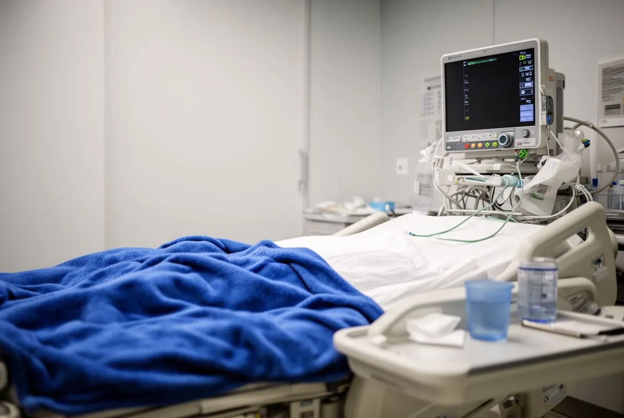
Communication is continuous and two-way. The cardiologist may ask you to hold your breath, cough, or turn your head, and the nurse checks on your comfort regularly. Many patients are surprised that the hardest part is simply lying still on a narrow, firm table for an hour. If you have back or hip problems, mention this beforehand so padding can be arranged — small comforts make a real difference when you cannot move.
Types of Coronary Catheterisation and What They Diagnose
Coronary catheterisation is the umbrella term for several related studies, and knowing which one you are having removes a layer of mystery. A diagnostic angiogram pictures the coronary arteries alone and answers one question: are there blockages, and how severe? A left-heart catheterisation additionally measures pressures inside the left ventricle and the ascending aorta, which helps assess valve disease and heart failure. A right-heart catheterisation passes a catheter through a vein into the right side and lungs to evaluate pulmonary hypertension.
What each study can reveal: coronary angiography identifies atheromatous plaque and acute blockages responsible for heart attacks; ventriculography shows how forcefully the left ventricle pumps; pressure pullbacks quantify valve narrowing such as aortic stenosis. Sometimes the combination uncovers an atypical infarction — a heart attack presenting without the classic symptoms — or atrial flutter driven by structural disease. Each piece of information changes the treatment plan, which is why the cardiologist reviews images live during the procedure.

The decision to proceed from diagnosis to treatment is usually made on the table. If a major vessel is found to be severely narrowed and you have been having symptoms, opening it with a balloon and stent in the same sitting spares you a second procedure and a second access puncture. If the disease is too extensive for stents, the angiogram data is what the surgeon uses to plan bypass grafting — including which arterial graft to use — so nothing about the study is wasted.
Catheterisation Laboratory Equipment Explained for Patients
Understanding the equipment turns an intimidating room into a logical workspace. The angiography unit is a C-shaped arm carrying an X-ray tube beneath you and an image intensifier above; it rotates around the table to capture coronary arteries from multiple angles without moving you. The monitors at the foot of the bed display fluoroscopy in real time, your electrocardiogram, oxygen saturation and the pressure tracings recorded through the catheter itself — these waveforms tell the cardiologist exactly where the tip sits.
What the catheters and wires do: the outer catheter is a stable pathway; a thinner guidewire is advanced first across a narrowing, and then a balloon catheter slides over the wire to compress the plaque. When a stent is needed, it is mounted on a balloon and expanded against the vessel wall, where it stays permanently as a scaffold. For valve work, a balloon-expandable valve can be delivered through the same femoral access route in procedures such as transcatheter aortic valve replacement (TAVR), performed in a specialised cath lab with a larger team.
Support equipment matters too. An automated external defibrillator is always nearby in case of dangerous rhythm disturbances, and a crash cart holds the drugs to manage allergic reactions or vasovagal episodes. The contrast injector delivers precise dye volumes at controlled pressure, and the pressure transducer converts fluid pressure inside the catheter into the electrical signal you see on screen. None of this should feel alarming — it is the safety net that makes same-day cardiac procedures routine.
Cardiac Catheterisation Risks and How They Are Minimised
Honesty about risk is part of informed consent, and the overall numbers are reassuring for a procedure this invasive. Major complications — heart attack, stroke, serious bleeding or kidney injury — occur in well under one percent of diagnostic catheterisations in stable patients, and somewhat more during emergency procedures for an evolving heart attack. Your individual risk depends on age, kidney function, diabetes, the extent of disease and whether treatment is performed at the same time.
The most common issues and how they are prevented: bleeding or bruising at the access site is by far the most frequent problem and is managed with firm pressure, sometimes a closure device, and a short period of bed rest. Contrast-related kidney injury is reduced with hydration and the lowest possible dye volume; allergic reactions are pre-treated when known; and dangerous arrhythmias are caught instantly by the continuous monitoring. Europe PMC catalogues much of the published evidence on these complication rates, which your cardiologist can reference for your specific risk profile.
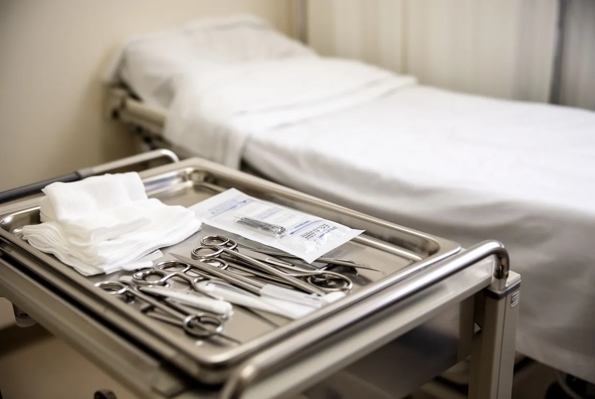
Two larger concerns deserve specific mention. Stroke during catheterisation is rare but real, because catheters and wires can dislodge plaque or clot from the aorta; meticulous technique and, where appropriate, filter devices lower this risk. Kidney injury matters most for diabetic patients and older adults, and large analyses such as the meta-analysis of intensive LDL lowering remind us that controlling vascular risk factors beforehand is itself protective. For acute stroke care specifically, the early management guidelines define time windows that also govern how urgently catheter-based treatments must be delivered.
Recovery After Your Heart Catheterisation Experience
Recovery is shorter than most patients expect. After the sheath is removed, pressure is held at the access site until bleeding stops, and a tight bandage or wrist compression device keeps it secure. If the radial artery in your wrist was used, you can usually sit up and walk within an hour; if the groin was used, you may need to lie flat for two to four hours. Most patients are discharged the same day, four to six hours after the procedure, with clear written instructions.
What to watch for at home: a small bruise and mild soreness at the puncture site are normal for a week. You should seek immediate help if you notice expanding redness, increasing swelling, persistent bleeding, numbness or coldness in the hand or foot, or a growing lump under the skin — these can signal a pseudoaneurysm or arterial leak. Avoid heavy lifting with that arm for several days, keep the site clean and dry, and take only the pain relief your doctor recommends, since some over-the-counter drugs can interact with the antiplatelet therapy started after stenting.
- First 24 hours: rest, no driving, no strenuous activity, monitor the access site.
- First week: bruise fades, light walks encouraged, keep the puncture dry.
- Follow-up: usually a clinic visit or telemedicine check within one to two weeks, plus medication review.
Medication after a stent is not optional. Aspirin and a second antiplatelet drug are typically prescribed together to prevent clot forming inside the new stent — this combination is supported by trials such as ARISTOTLE and similar studies that define optimal duration. Stopping these medicines early is one of the most common reasons stents block again, so never discontinue them without your cardiologist's explicit instruction, even if you feel perfectly well.
When a Cath Lab Procedure Leads to Further Treatment
The angiogram is only the beginning of the decision tree. When one or two vessels are severely narrowed, stenting in the same session is usually the right answer. When all three major coronary arteries are diseased, particularly if the left main artery is involved or the heart muscle is weak, coronary bypass surgery tends to give better long-term outcomes, and the cath lab data is handed directly to the surgical team. Sometimes the angiogram is normal but the pressures reveal valve disease, redirecting you toward a surgical or transcatheter valve solution instead.
How the findings shape your next steps: a stent resolves a focal narrowing and often relieves angina the same day; bypass surgery is reserved for diffuse disease and offers a more durable fix for complex anatomy; valve procedures — including TAVR for severe aortic stenosis — are planned from the same measurements. In some cases the catheterisation confirms that symptoms come from a rhythm problem such as atrial flutter rather than blocked arteries, and the treatment shifts to electrophysiology. The key point is that the cath lab is a diagnostic and staging tool, not an end in itself.
For patients travelling internationally, the workflow matters. We will always explain which steps can be combined and which must be staged for safety, and we will not recommend a combined procedure simply for convenience if it raises your risk. The right sequence depends on your anatomy and your overall health, not on a standard package.
Choosing a Catheterisation Laboratory Specialist in Guadalajara
Choosing where to have your catheterisation laboratory procedure is a decision that deserves more than a website search. The factors that matter most are the operator's experience and case volume, the availability of on-site surgical backup, the quality of the imaging equipment, and a transparent conversation about what your specific situation requires. A high-volume lab with surgical support on the same campus offers the lowest risk for complex or unexpected findings, and that safety net should not be underestimated.
Questions worth asking before you book: how many procedures like yours does the cardiologist perform each year, what is the lab's complication rate, is cardiac surgery available in the same hospital if needed, and what does the quoted price actually include? A clinic that cannot answer these questions clearly is one to approach with caution. International patients should also confirm how post-procedure communication will work once they return home — who reviews your medications, who reads your follow-up tests, and how quickly can you reach someone if a question arises.
- On-site surgical backup — essential if a complication or complex anatomy requires immediate surgery.
- Transparent quoting — facility fee, professional fees, stent or device costs, and follow-up visits listed separately.
- Post-care continuity — a named contact and a clear plan for review once you are back home.
If you are considering cardiac catheterisation in Guadalajara, Querétaro or Hermosillo, we encourage you to discuss your specific symptoms and test results with our team rather than self-diagnosing from this guide. Every heart is different, and the right plan comes from matching your anatomy, your symptoms and your goals with a qualified cardiac team. Book a consultation with Dr Alejandro Molina and the Cirugía de Corazón team to understand your options and the lowest-risk pathway for your individual situation.






