

Mechanical Ventilation After Heart Surgery: Weaning Off
Mechanical ventilation after heart surgery—learn safe weaning protocols, what families can expect in ICU recovery, and when breathing tube removal happens.
What is Mechanical Ventilation After Heart Surgery?
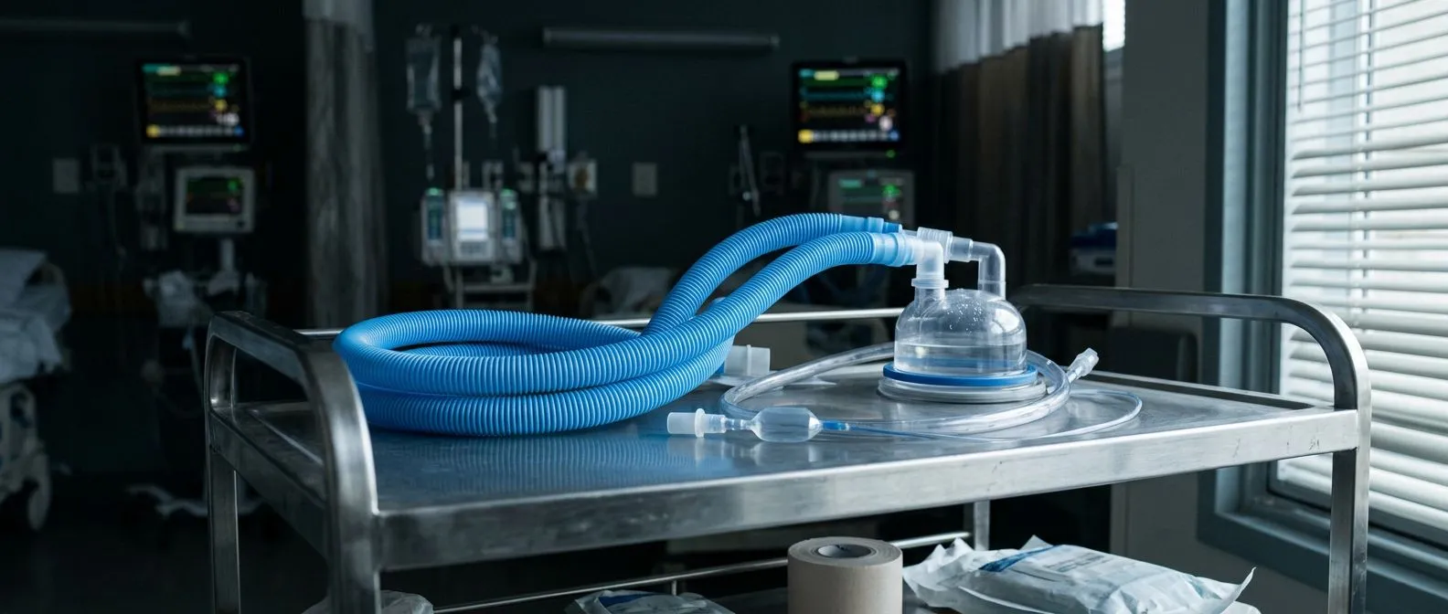
Mechanical ventilation after heart surgery is a standard and critical part of your recovery journey. When you undergo a procedure involving the Ascending Aorta or other major cardiac structures, your body needs time to wake up from anesthesia. The breathing tube connected to the ventilator ensures your lungs continue to deliver oxygen while your heart and body stabilize. It is entirely normal to feel anxious about this, but understanding the process helps demystify the intensive care unit (ICU) experience.
Why the ventilator is not a setback: Many families mistakenly believe that waking up with a breathing tube means something went wrong. In reality, mechanical ventilation is planned for every patient undergoing major cardiac surgery. The heart and lungs share a close physiological relationship, and temporarily supporting lung function allows the heart to rest and heal without the strain of independent breathing.
How you will feel during this time: Patients are typically heavily sedated or in a twilight state while intubated. You will not be able to speak because the tube passes through your vocal cords, but the nursing team will use alternative communication methods once you are awake enough. The goal is always to remove the tube as soon as your own respiratory drive and cardiac function are strong enough to sustain you safely.
How Long is a Breathing Tube Needed Post-Cardiac Surgery?
For most routine cardiac surgeries, the breathing tube remains in place for only a few hours. The surgical team aims to extubate, or remove the tube, within two to six hours after the operation to reduce the risk of ventilator-associated issues. The exact timing depends on how quickly you wake up from anesthesia and how well your heart is pumping. Your blood pressure, oxygen levels, and overall stability dictate when weaning can safely begin.
Factors that extend ventilation time: Certain complexities can require a longer duration of mechanical ventilation after heart surgery. If the operation was an emergency, involved extensive repair of the Atrium, or if you have pre-existing lung conditions, the medical team will keep the tube in longer to ensure total stability. According to Johns Hopkins Medicine, prolonged ventilation may be necessary for patients who experience significant fluid shifts or require advanced hemodynamic support in the immediate postoperative period.
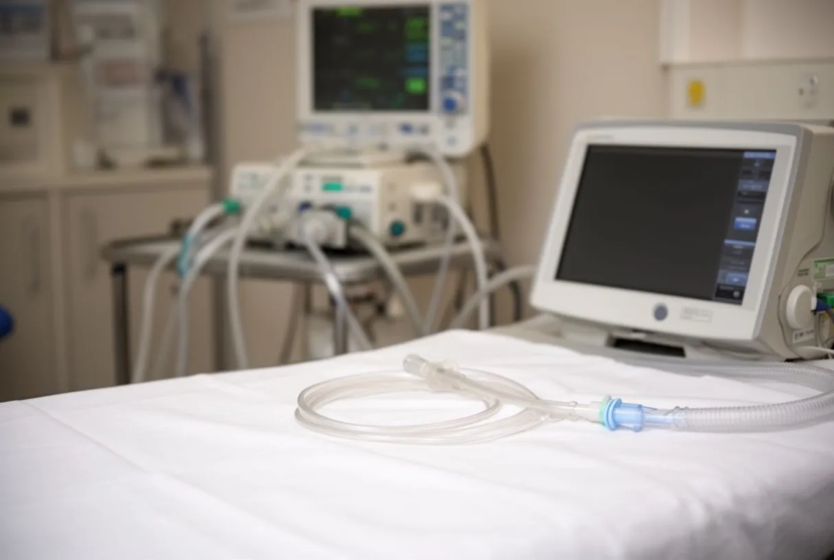
Setting realistic expectations for families: Seeing a loved one with a breathing tube can be distressing, especially when the timeline extends beyond the standard few hours. It is helpful to remember that the ICU team continuously evaluates readiness for extubation. If the tube needs to stay overnight, it is a precautionary measure to protect your airway and ensure your cardiac recovery is not compromised by respiratory fatigue.
What is the Ventilator Weaning Protocol?
Weaning from mechanical ventilation is the gradual process of reducing ventilator support until you can breathe entirely on your own. The clinical team follows a strict protocol to assess your readiness, looking at your ability to trigger breaths independently and maintain adequate oxygen levels. This involves lowering the pressure support and allowing your respiratory muscles to take over the work of breathing. It is a delicate balance between supporting your lungs and encouraging them to function unassisted.
Spontaneous breathing trials: The cornerstone of the weaning protocol is the spontaneous breathing trial (SBT). During an SBT, the ventilator is adjusted to provide minimal support, mimicking normal breathing conditions. If you can maintain your oxygenation and carbon dioxide levels without respiratory distress for 30 to 120 minutes, the team will typically proceed with extubation.
Monitoring during the transition: Throughout the weaning process, clinicians rely on continuous Auscultation of your lungs and vital sign monitoring. They listen for clear breath sounds and watch for signs of labor in your breathing. If your heart rate spikes or your oxygen drops, the trial is paused, and full ventilator support is resumed to let you rest before trying again.
Recognizing Weaning Failure in Cardiac Surgery
Weaning failure occurs when the heart and lungs cannot sustain independent breathing after the ventilator support is reduced. This does not mean your surgery was unsuccessful, but rather that your body needs more time to recover from the physiological stress of the operation. Recognizing the signs of weaning failure early allows the medical team to step in before respiratory exhaustion sets in, protecting your heart from additional strain.
Cardiac causes of weaning difficulty: The heart and lungs are deeply interconnected. If your heart is not pumping with enough force, fluid can back up into the lungs, making it incredibly difficult to breathe without assistance. This is often managed with medication rather than rushing another extubation attempt. A study highlighted in a Successful treatment of challenging case of infective endocarditis demonstrates how complex cardiac dynamics directly impact respiratory weaning timelines in critically ill patients.
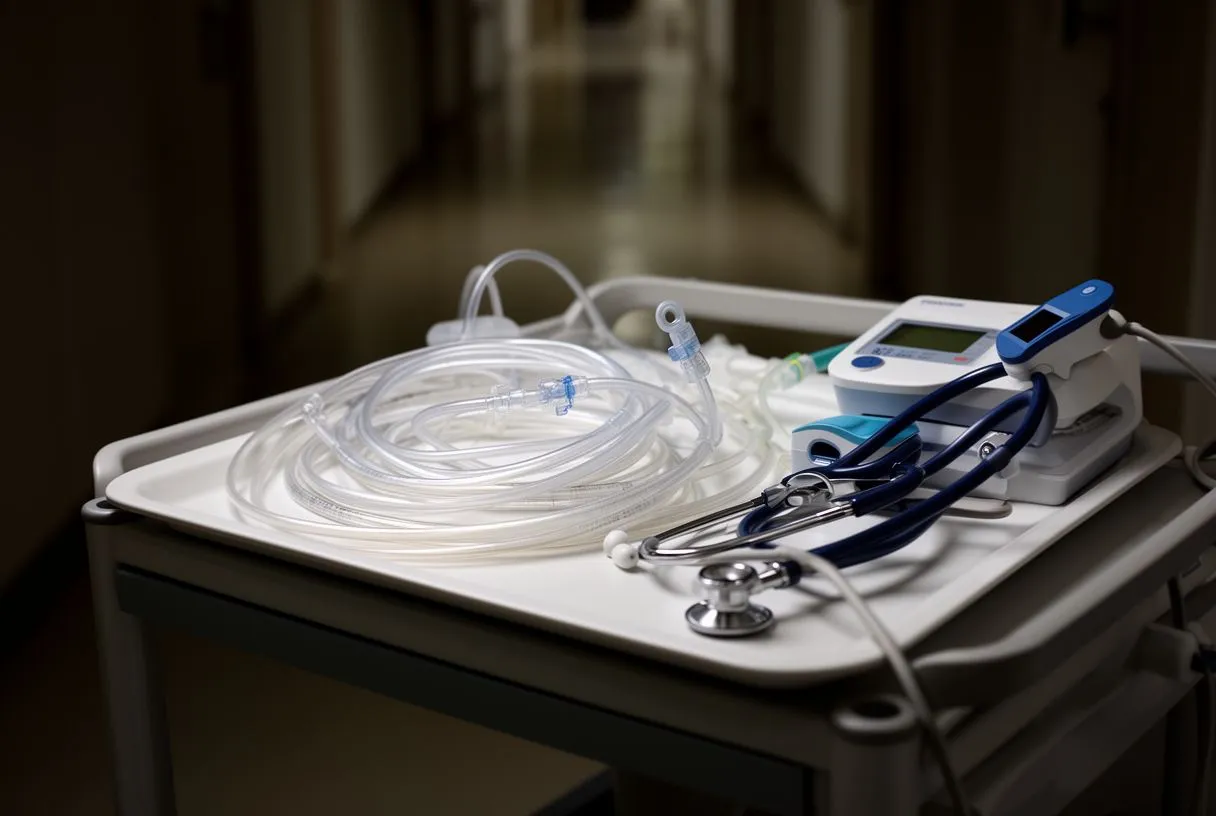
What happens if extubation fails: If the breathing tube needs to be reinserted, it is done swiftly and safely to protect your airway. The team will then investigate the underlying cause, such as fluid overload or weakened respiratory muscles, and adjust your treatment plan. Patience is essential; allowing the heart to recover fully often resolves the respiratory issues without further intervention.
What Causes Postoperative Respiratory Failure?
Postoperative respiratory failure is a broader term for the inability to maintain adequate oxygenation or ventilation after surgery. It is a recognized complication for patients requiring mechanical ventilation after heart surgery, driven by the effects of cardiopulmonary bypass, anesthesia, and the physical manipulation of the chest cavity. Understanding the causes helps patients and families grasp why the ICU stay is structured the way it is.
Fluid shifts and lung mechanics: During cardiac surgery, the body experiences significant fluid shifts. This fluid can accumulate in the spaces around the lungs, decreasing their ability to expand fully. Additionally, the surgical incision through the sternum makes deep breathing painful, leading patients to take shallow breaths, which can cause the small air sacs in the lungs to collapse.
- Fluid overload: Excess fluid in the lungs impairs gas exchange and is managed with diuretics.
- Atelectasis: Collapsed lung tissue caused by shallow breathing, treated with breathing exercises.
- Sedation effects: Lingering anesthesia can depress the respiratory drive temporarily.
Reversing anesthesia effects: The lingering effects of anesthesia can also depress the brain's respiratory drive. Until the medication clears your system, your brain may not send strong enough signals to breathe deeply. This is why patients are closely monitored and why weaning only begins once you are awake enough to participate in your own breathing.
How Arterial Lines and Advanced Support Aid Recovery
While mechanical ventilation is the most visible life-support machine in the ICU, it works in tandem with other monitoring and support systems. An Arterial Line, for instance, is a thin catheter placed in an artery to provide beat-to-beat blood pressure readings. This real-time data is crucial when adjusting medications that support the heart while the lungs are weaning from the ventilator.
Advanced oxygenation strategies: In rare cases where the lungs are severely compromised, the team may use Awake ECMO (Extracorporeal Membrane Oxygenation). This advanced technology acts as an artificial lung, oxygenating the blood outside the body so the heart and lungs can rest entirely. The decision to use such aggressive support is based on specific clinical thresholds and is reserved for the most critical scenarios.
Blood conservation and stability: Maintaining the right blood volume is essential for both heart function and lung recovery. Whenever possible, the surgical team utilizes Autologous Blood—your own blood collected before or during surgery—to minimize transfusion risks. This careful management ensures your heart is not overworked, giving you the best chance at a smooth and rapid weaning process.
Breathing Exercises for Post Heart Surgery Recovery
Once the breathing tube is removed, your active participation becomes critical. The nursing and respiratory therapy teams will teach you specific breathing exercises to expand your lungs and clear out any lingering secretions. The most common tool is an incentive spirometer, a simple device that encourages you to take slow, deep breaths to keep your airways open.
The importance of coughing safely: Coughing is necessary to clear mucus from your lungs, but it hurts after chest surgery. The team will show you how to splint your chest with a pillow to minimize pain while coughing. Effective coughing prevents pneumonia, a risk that increases the longer you rely on mechanical ventilation.
Mobilization as respiratory therapy: Sitting up in a chair and walking are powerful respiratory therapies. Moving helps expand the lower lobes of your lungs and promotes better circulation. The faster you can begin gentle mobilization with assistance, the quicker your respiratory function will return to its baseline, helping you transition out of the ICU.
Mechanical Ventilation Complications to Watch For
While mechanical ventilation after heart surgery is lifesaving, it is not without risks. The longer the breathing tube remains in place, the higher the risk of certain complications. The clinical team weighs the benefits of continued ventilation against these risks daily, deciding when the safest time to extubate is.
Infection and airway damage: The breathing tube bypasses the natural defenses of your nose and throat, creating a direct pathway for bacteria to enter the lungs. Ventilator-associated pneumonia is a serious risk, which is why the team prioritizes early extubation and rigorous oral care. Additionally, prolonged pressure from the tube can cause vocal cord irritation or damage, leading to temporary hoarseness after removal.
Muscle deconditioning: When a machine breathes for you, the diaphragm and chest muscles can weaken. This is why weaning protocols emphasize spontaneous breathing trials early on. A documented Eyes wide shut - unusual two stage repair of pectus excavatum and annuloaortic ectasia in a 37 year old marfan patient: case report illustrates how complex structural repairs demand careful prolonged ventilation, but the medical team balances this against the inevitable muscle deconditioning that occurs. Overcoming this deconditioning requires dedicated physical therapy after extubation.
Heart Surgery ICU Recovery: What Families Can Expect
The ICU environment is often overwhelming, filled with monitors, alarms, and specialized equipment. Understanding what you are seeing and hearing can reduce anxiety. The ventilator will beep if it detects a disconnected circuit or a change in pressure, but these alarms often trigger for minor adjustments and do not always indicate an emergency. The nursing staff is constantly monitoring these systems and will intervene if necessary.
Communication while intubated: Because the breathing tube prevents speech, the ICU team relies on sedation breaks to assess your neurological status. During these breaks, you may be asked to follow simple commands like squeezing a hand or wiggling your toes. If you are awake enough, the nurses may provide a clipboard and pen or picture boards to help you communicate basic needs.
- Daily rounds: The multidisciplinary team meets at your bedside to discuss your progress and weaning goals.
- Sleep preservation: Lights are dimmed at night, and care is clustered to allow for rest, which is vital for weaning success.
- Family involvement: Families are encouraged to participate in non-clinical care, like holding hands and providing orientation.
Transitioning out of the ICU: Once the breathing tube is removed and your respiratory status is stable, the team prepares to move you to a lower level of care. This transition happens when you no longer require the minute-by-minute monitoring of the ICU. It is a major milestone in your recovery, signaling that your heart and lungs are functioning well enough to continue healing on the standard ward.
When to Seek Help After Breathing Tube Removal
After successful extubation, your recovery transitions to the step-down unit or standard ward. You will still be monitored, but the focus shifts to independent breathing and physical recovery. It is completely normal to feel short of breath when walking or to have a persistent cough for a few weeks as your airways recover from the intubation.
Warning signs to report: While mild discomfort is expected, certain symptoms require immediate medical attention. If you experience sudden severe shortness of breath that does not improve with rest, a high fever, or an inability to cough up secretions, you must alert the nursing staff immediately. These could be signs of a developing respiratory complication that requires prompt intervention.

In conclusion, mechanical ventilation after heart surgery is a carefully orchestrated bridge to recovery, not a sign of failure. The weaning process is tailored to your specific cardiac and respiratory needs, ensuring the lowest risk for your situation. Every step, from the initial intubation to the final breathing exercises, is designed to protect your heart and lungs. We encourage you to discuss your specific risk factors and recovery timeline with a qualified cardiac team. If you or a loved one are preparing for heart surgery, book a consultation with our practice to understand how we individualize your postoperative care and ensure a safe, comfortable recovery.






